천마 추출액이 Scopolamine으로 유발된 기억력 감퇴 흰쥐에 미치는 영향
김진호1․추한나1․박은혜1․정종길2․김경옥3․김정상1†
1
동신대학교 한의과대학 해부학교실
2
동신대학교 한의과대학 본초학교실
3
동신대학교 한의과대학 한방신경정신과학교실
Effects of Gastrodia elata Extracts on Scopolamine-induced Memory Impairment in Rats
Jin-Ho Kim1, Han-na Choo1, Eun-hye Park1, Jong-Kil Jeong2, Kyeong-Ok Kim3, and Jeong-Sang Kim1†
1Dept. of Anatomy, 2Dept. of Herbology, and 3Dept. of Neuropsychiatry, College of Oriental Medicine, Dongshin University, Jeonnam 520-714, Korea
Abstract
Alzheimer's disease is a progressive neurodegenerative disorder characterized by a gradual decline in memory associated with shrinkage of brain tissue, with a localized loss of neurons mainly in the hippocampus and basal forebrain. This study investigated the neuroprotective effect of Gastrodia elata aqueous extracts against scopol- amine-induced neurotoxicity in the hippocampus of male Sprague-Dawley rats. The animals (n=25) were divided into five different groups with five animals per each group. The normal group (Nor) was administered with saline, while the control (Con) group was administered saline after scopolamine treatment. The experimental group (Exp) was administered Gastrodia elata aqueous extracts (200 mg/kg body weight) for 20 or 30 days after scopolamine treatment. From a light microscopy study, the nuclei of neurons in the hippocampus were more shrunken or condensed in the 20 or 30 day control groups compared to experimental groups. The densities of neurons from the CA1 and CA3 area of the hippocampus in the Exp increased compared with the Con. Amyloid β protein, containing PAS-positive materials, was lower in the Exp compared with the Con. The present study demonstrates that Gastrodia elata aqueous extracts possess neuroprotective potential, thus validating its use in alleviating the toxic effects of scopolamine.
Key words: Gastrodia elata, hippocampus, scopolamine
†
Corresponding author. E-mail: [email protected]
†
Phone: 82-61-330-3512, Fax: 82-61-330-3519
서 론
알츠하이머병(Alzheimer's disease, AD)은 나이든 사람 들에서 나타나는 만성적인 신경퇴행성 치매로 기억력 손실 로 인한 인지기능에 현저한 감소와 행동장애를 일으킨다(1).
AD 환자의 주요 병리학적 특징은 amyloid 전구체 단백질 (amyloid precursor protein, APP)로부터 발생된 amyloid-β (Aβ)가 광범위하게 뇌 조직에 축적된다(2). 인지기능이 저 하되면 중추신경계의 포도당 대사가 감소하며, 신경조직에 서는 β-amyloid가 축적된 노화반(senile plaque)이 나타난 다(2,3). 주요 우울장애에서 보이는 감정표현과 기억과정의 조절 이상은 해마와 편도가 관련된 기능적 회로가 연관이 있다고 알려져 있고, 성인과 노년기 우울장애 환자들에서 이 구조들의 이상이 관찰된다고 하였다(4,5). 또한 우울증 환자에서는 해마의 용적이 줄어든다고 하였다(6,7).
해마는 학습에 의한 기억의 형성에 관여하며, 해마의 신경 세포는 신경의 발생기 동안뿐만 아니라 그 이후에도 생성되 는 특징이 있다(8,9). 그러므로 해마 신경세포들은 유효한 약물을 투여하면 새로운 신경연접을 형성하여 기능을 보다 활성화하며(10), 지속적인 운동은 해마의 줄기세포(stem cells) 증식을 유도하여 새로운 신경세포로의 분화도 가능하 다고 하였다(11).
Scopolamine은 무스카린성 콜린 수용체(muscarinic cholinergic receptor) 길항제로서 실험동물의 인지능력 결 핍연구에 널리 사용되고 있다. 흰쥐에 scopolamine을 복강 투여하면 콜린성 신경전달물질이 차단되어 콜린성 기능장 애(cholinergic dysfunction)와 인지 능력의 손상이 온다. 그 러므로 scopolamine으로 유발된 기억력 결핍 흰쥐는 항치매 약물의 효능을 조사할 목적으로 많이 사용되고 있다(12-14).
천마(
Gastrodia elataBlume)는 난초과(Orchidaceae)에
속한 다년생 기생초본의 근경을 건조한 것이다. 주요 성분은 vanillyl alcohol, vanillin 및 gastrodin, phenolic compounds, hydroxybenzaldehyde, p-hydroxybenzyl alcohol 등이 있다 (15-17). 천마에 대한 연구로는 중추신경계의 혈류 개선(18, 19), 뇌조직의 출혈이나 부종(20), 뇌허혈 시 신경보호 효과 (21)에 대한 연구가 이루어져 왔으나, 천마의 추출액이 기억 의 형성과 저장과정에 관여하는 해마의 신경세포에 미치는 영향에 관한 연구는 미흡한 실정이다. 따라서 본 연구에서는 국내에서 생산되는 천마의 물추출물을 scopolamine을 투여 하여 해마의 신경세포를 손상시킨 흰쥐에 투여하여 그 효과 를 규명하고자 하였다.
재료 및 방법
실험동물
체중 200±10 g 내외의 7주령 흰쥐(Sprague-Dawley)를 샘타코(주)(Osan, Korea)로부터 구입하였다. 실험동물은 동 신대학교 한의과대학 동물사육실에서 일정한 조건(온도, 21±2
oC; 습도, 50~60%; 12시간 주기 명/암) 하에서 일반 고형사료(샘타코(주), 흰쥐 용)와 물을 충분히 공급하면서 1주 동안 적응시킨 후 실험에 사용하였다. 모든 실험은 동신 대학교 동물실험윤리위원회의 허락을 얻은 다음 수행하였다.
시료 준비
천마를 수증기로 삶고서 70
oC에서 120시간 숙성시킨 다음 40 g을 물 900 mL에 넣고 약탕기로 3시간 동안 가열하였다.
얻은 추출액은 3,000 rpm으로 원심분리 하여 상층액을 감압 농축(CCA-1100, Eyela, Tokyo, Japan)한 다음 저온순환수 조(COOL ACE CA-1500, Eyela)에서 1차 동결 후 동결건조 기(FD8508, Ilshin, Dongducheon, Korea)로 동결건조 하여 5.5 g의 분말을 얻었다.
기억력 감퇴 동물모델 구축
정상군을 제외한 모든 실험동물은 scopolamine(Sigma- Aldrich, St. Louis, MO, USA) 1 mg/kg을 0.9% 식염수 1 mL에 용해하여 7일 동안 1일 1회 복강 주사하여 동물모델을 구축하였다.
실험군 분류와 처치
실험군은 기억력 감퇴를 유발시키지 않는 정상군(Nor), scopolamine를 7일간 복강 주사하여 기억력 감퇴를 유발시 킨 다음 음용수만 20일과 30일 동안 공급한 20일 대조군(C- 20)과 30일 대조군(C-30), scopolamine을 대조군과 동일한 방법으로 투여한 다음 성인의 1일 복용량에 해당하는 천마 추출물(200 mg/kg)을 동일기간 동안 투여한 20일 실험군 (G-20)과 30일 실험군(G-30)으로 구분하였다. 실험동물은 각 군마다 5마리씩 사용하였다.
뇌조직의 적출 및 조직 처리
모든 실험이 끝난 직후 실험동물을 urethane(75 mg/kg)
으로 마취시킨 다음 심장을 통하여 phosphate buffered sal- ine(PBS) 100 mL에 이어 4% paraformaldehyde 용액으로 관류고정 하였다. 그 다음 뇌를 꺼내 같은 고정액에 넣어 4
oC에서 보관하였다.
광학현미경 관찰
흰쥐를 도살한 후 각각 뇌를 적출하여 4% paraformalde- hyde에 24시간 고정시킨 다음, 30, 50, 70, 80, 90, 95, 100Ⅰ, 100Ⅱ와 같이 알코올 농도를 상승시켜 조직 속의 수분을 제 거한 후 xylene으로 투명화 과정을 거친 다음 paraffin으로 포매하였고, 포매된 조직을 microtome을 사용하여 5 μm 두 께로 절편하였다. 절편한 조직을 slide glass 위에 부착시키 고 xylene으로 paraffin을 제거한 다음 100%, 90%, 80%
ethanol과 같이 농도가 낮아지는 순으로 5분씩 담그어 함수 과정을 거치게 하였다. Cresyl violet 염색을 한 다음 탈수 후 Canada balsam으로 봉입하고 카메라 부착 광학현미경 (Nikon Eclipse 80i, Tokyo, Japan)으로 관찰한 후 사진을 촬영하였다. 또한 염색이 끝난 조직은 광학현미경을 사용하 여 저배율(×100)에서 신경세포의 밀도를 Scion image pro- gram(Scion Corp., Frederick, MD, USA)을 이용하여 측정 하였다.
Periodic acid Schiff(PAS) 염색
뇌 조직 절편을 PBS로 수세한 후 0.5% periodic acid와 함께 10분간 상온에서 배양하였다. Schiff's reagent로 5분 씩 2회 처리한 후 sulfurous rinse 용액으로 각각 2분씩 3회 에 걸쳐 씻어내고 이어서 흐르는 물에 10분간 수세하였다.
Harri's hematoxylin으로 1분간 염색한 후 1% acid alcohol 에 2~3회 담그는 방법으로 탈수시키고 다시 흐르는 물에 수세 과정을 거친 후 ammonia액에 5~10회 담가 염색하였 다. Alcohol을 이용한 탈수 과정을 거친 후 Canada balsam 으로 봉입하였다.
통계처리
실험 결과는 mean±SE로 나타냈으며, 대조군과 실험군 사이의 평균 차이를 검정할 때에는 Student's t-test로 검정 하여 p 값이 0.05 미만일 때 통계적으로 유의한 차이가 있는 것으로 판정하였다.
결과 및 고찰
Hippocampus의 광학현미경 관찰
Scopolamine은 무스카린성 콜린 수용체(muscarinic
cholinergic receptor) 길항제로서 작용을 하기 때문에 흰쥐
에 투여하면 뇌신경뿐만 아니라 해마의 신경세포들이 손상
되거나 위축이 된다고 하였다(12-14). 본 연구에서도 실험동
물에 7일간 scopolamine을 투여한 다음 20일 동안 음용수를
공급한 대조군(Fig. 1B)에서는 정상군(Fig. 1A)에 비하여 해
마의 신경세포는 매우 응축되어 있었다. 천마의 효능을 검증
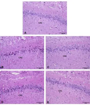
Fig. 1. The light micrographs of hippocampus. A, normal group; B, C-20 control group administered saline for 20 days after scopolamine treatment; C, G-20 group administered Gastrodia elata extract (200 mg/kg/day) for 20 days after scopolamine treatment; D, C-30 control group administered saline for 30 days after scopolamine treatment; E, G-30 group administered Gastro- dia elata extract (200 mg/kg/day) for 30 days after scopolamine treatment. PAS stain. ×400.
Fig. 2. The light micrographs of hippocampus. A, normal group;
B, C-20 control group administered saline for 20 days after sco- polamine treatment; C, G-20 group administered Gastrodia elata extract (200 mg/kg/day) for 20 days after scopolamine treatment;
D, C-30 control group administered saline for 30 days after sco- polamine treatment; E, G-30 group administered Gastrodia elata extract (200 mg/kg/day) for 30 days after scopolamine treatment.
Cresyl violet stain. ×100.
Table 1. The densities of neurons from CA1 and CA2 areas of hippocampus
Groups Area
CA1 CA3
C-20 Nor G-20 C-30 G-30
140.6±7.17 96.0±1.38 130.3±5.37
*102.8±3.48 134.6±7.09
*161.6±12.66 113.6±5.84 137.8±6.26
*126.2±11.12 134.0±7.95 Nor, normal group; C-20, control group administered saline for 20 days after scopolamine treatment; G-20, group administered Gastrodia elata extract (200 mg/kg/day) for 20 days after scopol- amine treatment; C-30, control group administered saline for 30 days after scopolamine treatment; G-30, group administered Gastrodia elata extract (200 mg/kg/day) for 30 days after scopol- amine treatment. Significant differences were compared with control at
*p<0.05. (n=5).
하고자 성인의 1일 복용량(200 mg/kg)에 해당하는 물추출 물을 실험동물에 투여한 결과, 실험군(Fig. 1C)에서는 대조 군에 비하여 손상이나 응축 정도가 미약하였다. 30일군에서 는 대조군(Fig. 1D) 해마의 신경세포의 손상 정도가 보다 현저하여 그 수가 감소하였으나, 실험군(Fig. 1E)에서는 손 상정도가 미약하였다. 이와 같은 결과로 보아 scopolamine 에 의하여 손상된 흰쥐에 투여된 천마 추출액이 해마의 신경 세포 손상을 억제하는 것으로 보인다.
Hippocampus의 신경세포 밀도
Scopolamine을 투여한 흰쥐의 해마를 AChE 염색을 하여 밀도를 분석한 결과 40~50% 감소하였다고 하였다(22). 또 한 Probst 등(23)에 의하면 해마의 피라미드 세포 밀도는 정상인에 비하여 치매 환자에서 매우 감소한다고 하였다.
본 연구에서는 뇌조직을 cresyl violet으로 염색한 후 Scion image program(Scion Corp.)을 이용하여 세포 밀도를 측정 하였다. 그 결과 해마의 CA1 영역의 신경세포 밀도는 정상 군(140.0 density)에 비하여 C-20군(96.0 density)은 약 32%
감소하였고 실험군인 G-20군(130.5 denstiy)과 G-30군 (134.6 density)은 정상군에 비하여 다소 감소하였으나, 대조 군에 비하여 각각 통계적으로 유의성 있게 증가하였다(Fig.
2, Table 1). 해마의 CA3 영역의 신경세포 밀도 또한 정상군 (161.6 density)이 가장 높았으며, C-20군(113.6 density)은
정상군에 비하여 약 30% 감소하였다. 실험군에서는 G-20군 (137.8 denstiy)과 G-30군(134.0 denstiy) 밀도가 대조군에 비하여 높았으며, G-20군은 통계적으로 유의성 있게 증가하 였다(Fig. 2. Table 1). 이와 같은 결과는 위 연구자의 결과와 일치한다고 할 수 있을 것이다.
뇌조직의 PAS 염색
인지기능이 저하되면 중추신경계의 포도당 대사가 감소
하고 신경조직에서는 β-amyloid가 축적된 노화반(senile
Fig. 3. The light micrographs of CA1 area from hippocampus.
A, normal group; B, C-20 control group administered saline for 20 days after scopolamine treatment; C, G-20 group administered Gastrodia elata extract (200 mg/kg/day) for 20 days after scopol- amine treatment; D, C-30 control group administered saline for 30 days after scopolamine treatment; E, G-30 group administered Gastrodia elata extract (200 mg/kg/day) for 30 days after scopol- amine treatment. PAS stain. ×200.
plaque)이 나타난다(2,3)고 하였으며, scopolamine으로 유발 된 알츠하이머병 생쥐모델의 해마를 Congo Red 염색을 한 결과 노화반이 관찰되었다고 보고하였다(24). 알츠하이머병 에서 노화반은 PAS 염색을 통하여 확인할 수 있으며(25), 본 실험에서도 scopolamine으로 유도된 동물 모델의 뇌조직 을 PAS로 염색한 결과 정상군(Fig. 3A)에 비하여 C-20군 (Fig. 3B)의 신경세포는 핵들이 응축되어 있었고 신경섬유는 PAS-양성반응이 보다 높게 관찰되었으며, 실험군인 G-20 군(Fig. 3C)에서는 응축된 세포의 수가 현저히 감소하였을 뿐만 아니라 PAS-양성반응도 대조군에 비하여 낮았다. 30 일군에서는 C-30군(Fig. 3D)에서는 여전히 신경세포의 핵 이 응축되어 있었으며 신경섬유는 PAS-양성반응이 높았고, 실험군인 G-30군(Fig. 3E)에서는 신경세포의 핵이 뚜렷이 관찰되었으며 신경섬유에서 관찰되는 PAS-양성반응은 낮 았다.
위와 같은 결과로 보아 천마 물추출액은 scopolamine으로 유발된 신경 독성을 개선하는 효과가 있는 것으로 보인다.
그러나 해마의 신경세포를 독성으로부터 보호 또는 회복 효 과를 나타내는 성분이나 기전에 관한 연구가 지속되어야 할 것으로 사료되었다.
요 약
알츠하이머병은 신경세포가 점차적으로 퇴화되는 질환으 로 특히 해마와 기저쪽 앞뇌의 뇌조직이 위축되어 점진적으 로 기억력을 잃어간다. 본 연구는 scopolamine(1 mg/kg, 7일 동안 1일 1회)으로 유발된 수컷 Sprague-Dawley 흰쥐 해마 의 신경독성에 대한 천마 물 추출액의 효과를 규명하기 위하 여 수행하였다. 실험동물(n=25)은 5군으로 나누었으며, 각 군마다 5마리씩 사용하였다. 실험군은 생리식염수를 투여한 정상군(Nor), scopolamine으로 유발한 다음 생리식염수를 20일(C-20)과 30일(C-30) 동안 투여한 대조군, scopolamine 으로 유발한 다음 천마 추출액(200 mg/kg)을 20일(G-20)과 30일(G-30) 동안 투여한 실험군으로 구분하였다. 광학현미 경으로 관찰한 결과 해마의 신경세포 핵은 G-20과 G-30에 비하여 C-20과 C-30에서 보다 응축되었거나 위축되어 있었 다. 해마의 CA1과 CA3의 세포밀도를 조사한 결과 실험군이 대조군보다 높게 나타났다. 해마 주변의 뇌세포를 PAS로 염색한 결과 amyloid β 단백질을 함유한 PAS-양성물질이 대조군에 비하여 실험군에서 감소하였다. 이상의 결과로 보 아 천마 물추출액은 scopolamine으로 유발된 신경 독성을 개선하는 효과가 있는 것으로 사료되었다.
문 헌