177
<원례보저
>
Protective effect of the methanol extract of Polyopes lancifolia (Harvey) kawaguchi et wang against ionizing radiation-induced
mouse gastrointestinal injury
Jinwoo Jeong
1,2, Wonjun Yang
1,2, Meejung Ahn
2,3, Ki Cheon Kim
2,4, Jin Won Hyun
2,4, Sung-Ho Kim
5, Changjong Moon
5, Taekyun Shin
1,2,*
1Department of Veterinary Anatomy, College of Veterinary Medicine and Veterinary Medical Research Institute, Jeju National University, Jeju 690-756, Korea
2Applied Radiological Science Research Institute, Jeju National University, Jeju 690-756, Korea Departments of 3Anatomy and 4Biochemistry, College of Medicine, Jeju National University, Jeju 690-756, Korea
5Department of Veterinary Anatomy, College of Veterinary Medicine, Chonnam National University, Gwangju 500-757, Korea
(Received: June 02, 2011; Revised: July 21, 2011; Accepted: July 21, 2011)
Abstract :The radioprotective efficacy of a methanol extract of the red algae Polyopes lancifolia (Harvey) kawaguchi et wang (mPL) was evaluated in mice subjected to total-body gamma irradiation. mPL protection against radiation-induced oxidative stress was examined by histological evaluation of intestinal crypt-cell survival and liver activities of the antioxidant enzymes superoxide dismutase (SOD) and catalase (CAT). mPL (100 mg/kg body weight) administered intraperitoneally at 24 h and 1 h prior to irradiation protected jejunal crypt cells from radiation-induced apoptosis (p< 0.01). The pretreatment of mPL attenuated a radiation-induced decrease in villous height (p< 0.05), and improved jejunal crypt survival (p< 0.05). The dose reduction factor was 1.14 at 3.5 days after irradiation. Treatment with mPL prior to irradiation resulted in significantly higher (p< 0.01) levels of SOD and CAT activities, compared to those levels of irradiated control mice with vehicle treatment. These results suggest that mPL is a useful radioprotective agent capable of defending intestinal progenitor cells against total-body irradiation, at least in part through mPL antioxidative activity.
Keywords :antioxidation,intestinal progenitor cells, irradiation, Polyopes lancifolia (Harvey) kawaguchi et wang, red seaweed
Introduction
Radiation exposure triggers the generation of intracellular free radicals and related reactive oxygen species, which subsequently damage vital cellular targets such as DNA, membrane lipids, and proteins [14, 23]. Rapidly proliferating epithelial progenitor cells in small intestinal crypts are highly vulnerable to radiation-induced cell damage, leading to the development of gastrointestinal syndrome [5, 20]. The fundamental requirements for putative radioprotective substances include prevention of apoptosis and protection of intestinal crypt cells and architecture following radiation exposure. Although chemicals such as amifostine have been developed as
radioprotective agents, many agents induce dose-limiting side effects [1]. Certain naturally occurring antioxidants have also been shown to be effective radioprotectors due to their ability to scavenge free radicals or neutralize free radical reactions [2, 4].
Many single compounds and plant extracts with antioxidative and anti-inflammatory activities have been associated with radioprotection [12, 18, 21, 24]. Natural products derived from seaweeds have been shown to exert antioxidant effects with minimal toxicity, making them attractive potential radioprotective agents. These seaweed derivatives include fucoidan [10], eckol [19, 27], and phloroglucinol [17] from brown algae.
Callophyllis japonica, a red seaweed, was also found to
*Corresponding author
Tel: +82-64-754-3363, Fax: +82-64-756-3354 E-mail: [email protected]
have antioxidant [6] and radioprotective [8] activities. In addition, Palmaria palmata, another red seaweed, is reported to contain high concentrations of antioxidant polyphenolic compounds, which exert a number of biological effects [11, 26].
In line with previous antioxidant and radioprotection investigations using red seaweeds, Polyopes lancifolia (Harvey) kawaguchi et wang (PL), a red algae, was selected for evaluation of its in vivo radioprotective effects. To determine whether the methanolic extract of PL (mPL) offers protection against radiation-induced injury, mPL were administered to mice prior to acute gamma irradiation exposure and subsequent analysis of antioxidant enzymes in the liver and intestinal crypt-cell survival was conducted.
Materials and Methods
Preparation of mPL
The Polyopes lancifolia (Harvey) kawaguchi et wang (AR309; Jeju Hi-Tech Industry Development Institute, Korea), a red alga, was collected from the sea surrounding Jeju island at 2010. The wet leaves and twigs of the plant were dried, and extracted twice with methanol to produce a crude extract. The extract mPL was filtered, evaporated to dryness under reduced pressure and then concentrated in vacuum. The mPL, dissolved in phosphate buffered saline, was intraperitoneally injected in mice with a single dose of 100 mg/kg twice prior to the irradiation.
Animals and experimental groups
Eight-week-old male BALB/c mice were obtained from OrientBio (Korea). All experimental procedures were conducted in accordance with the Guidelines for the Care and Use of Laboratory Animals at Jeju National University, Korea.
Total of 60 male BALB/c mice were divided into the following four groups (5 mice/each group): Vehicle treated control group (vehicle), the mPL (100 mg/kg) only treated group (mPL100), the vehicle-treated irradiation group (vehicle + IR), and the mPL (100 mg/
kg) treated irradiation group (mPL100 + IR).
Irradiation
All irradiation and evaluation protocols were conducted as previously described [16, 17]. In brief, mice were placed in a specially designed, well-ventilated acrylic
container and subjected to whole-body irradiation. Mice were irradiated with either 2 gray (Gy) for the apoptosis assay in the intestines, 9 Gy for the intestinal crypt assay or 9, 10 and 11 Gy for dose reduction factor (DRF) analysis using 60Co gamma rays (Nordion Gammacell 3000 Elan; Nordion International, Canada), which is installed in the Applied Radiological Science Research Institute, Jeju National University at a dose-rate of 3.1 Gy/min.
Apoptosis assay
To determine the protection of jejunal crypt apoptosis against irradiation, the mice four groups (4 mice/each group) were killed 12 h after irradiation (2 Gy). The small intestines were fixed in 10% buffered formalin, and embedded in paraffin. 5µm-thick sections were cut and stained using a H&E. The apoptotic cells in the jejunal cross sections were counted using an optical microscope as shown in our previous paper [17]. The cells were recorded as a single cell based on their size and clustering when several apoptotic fragments were believed to represent the remains of a single cell. Forty crypt sections were recorded for each mouse.
Intestinal crypt assay
Jejunal crypt stem-cell survival was determined using the microcolony technique reported by Withers and Elkind [25]. Briefly, each mouse jejunum was removed at 3.5 days post-irradiation, fixed in 10% buffered formalin, and embedded in paraffin. Paraffin blocks were cut into 5µm-thick sections andstained with H&E.
Two sections each from four different regions of the jejunum from each mouse were prepared for histological examination. For 10 histological jejunal cross-sections per mouse, the number of regenerating crypts and the total number of crypts per transverse circumference were counted under a microscope. The average number of crypt cells was plotted against the radiation dose.
Sections stained with H&E were used for the determination of villous height. Five non-overlapping field in each section were examined at ×100 optical magnification. The villous height was measured from the tip of the villi to the crypt.
DRF analysis
The protective capacity of an agent can be expressed as the DRF [10, 15]. Animals with or without mPL pretreatment (100 mg/kg body weight/day, 24 h and 1 h
prior to irradiation) were exposed to 9, 10, and 11 Gy (4 mice/per dosage group). Mice were sacrificed at 3.5 days after radiation exposure. D10 represented the dose at which the curve intersected 10 crypt cells. The DRF was calculated using the formula D10 with mPL/D10 with vehicle.
Detection of liver superoxide dismutase and cata- lase activities
BALB/c mice (5 mice/each group) with or without mPL pretreatment (100 mg/kg) at 24 h and 1 h prior to irradiation were sacrificed 3.5 days after irradiation (9 Gy). The absolute and relative (organ-to-body weight ratio) weights of the liver were measured for all mice immediately following sacrifice. The excised livers were then immediately frozen, and the frozen liver tissue was homogenized in a glass-Teflon homogenizer with 50 mM phosphate buffer (pH 7.4) to obtain a 1 : 9 (w/v) whole homogenate. The homogenates were then centrifuged at 11,000 g for 10 min at 4oC to remove cellular debris. The protein content of the supernatant was determined using the Bradford method.
For detection of superoxide dismutase (SOD) activity, 50 µg liver protein was added to 500 mM phosphate
buffer (pH 10.2) and 1 mM epinephrine. Epinephrine rapidly undergoes auto-oxidation at pH10 to produce the pink-colored product adrenochrome, which was assayed at 480 nm using a UV/VIS spectrophotometer operating in the kinetic mode. SOD inhibits the auto-oxidation of epinephrine. The rate of inhibition was monitored at 480 nm, and the amount of enzymerequired to produce 50%
inhibition was defined as one unit of enzyme activity.
The total SOD activity was expressed as units/mg protein [13].
For detection of catalase (CAT) activity, 50 µg of liver protein was added to 50 mM phosphate buffer (pH 7.0) and 100 mM H2O2, and the mixture was incubated for 2 min at 37oC. Following incubation, the absorbance of the mixture at 240 nm was monitored for 5 min. The change in absorbance wasproportional to the breakdown of H2O2 and the CAT activity present in the sample.
CAT activity was expressed as units/mg protein [3].
Statistical analysis
Data are presented as the mean±SE. All data were analyzed using one-way analysis of variance (ANOVA) followed by the Tukey’s test for multiple comparisons.
In all cases, p< 0.05 was considered significant.
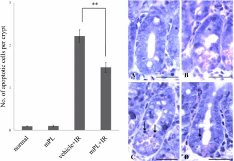
Fig. 1. Analysis of crypt apoptotic cells. Bar graphs showing the number of apoptotic cells per crypt in mouse jejunum (sections stained with H&E stain) from the vehicle, 100 mg/kg methanol extracts of Polyopes lancifolia (Harvey) kawaguchi et wang (mPL100), vehicle + irradiation (2Gy) (Vehicle + IR), and mPL100 + IR mice. Mice were pretreated with vehicle or mPL100 at 24 h and 1 h prior to irradiation. (A~D) Representative images showing apoptotic cells in H&E-stained jejunal sections from vehicle (A), mPL100 (B), vehicle + IR (C), and mPL100 + IR (D) mice. The arrows indicate apoptotic cells.
Values are the mean±SE of four mice in each group. **p< 0.01 compared with the vehicle-treated irradiation controls.
Scale bars = 20µm.
Results
mPL reduces mouse intestinal cell apoptosis fol- lowing total body irradiation
Irradiation induced apoptosis of intestinal crypt cells in BALB/c mice (Fig. 1). Pretreatment with mPL100 group at 24 h and 1 h prior to irradiation was significantly reduced the number of apoptotic cells (1.48
±0.12) as compared with vehicle treated irradiation group (2.21±0.15) (p< 0.01) (Fig. 1). This result suggests that mPL inhibits apoptosis of immature progenitor cells in jejunal crypts following irradiation.
mPL protection of intestinal crypt cells from total body irradiation
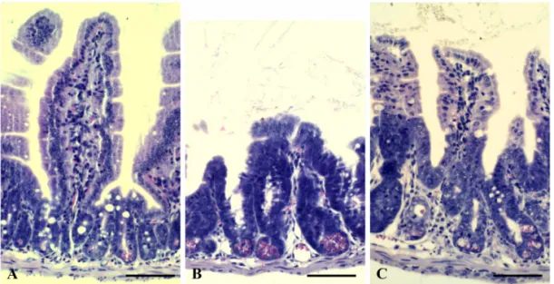
To evaluate the gastroprotective effects of mPL against mouse whole-body irradiation, the mean height of jejunal villi was compared in irradiated mice with or without mPL pretreatment. Mean jejunal villus height in the mPL-pretreated, irradiated mice (306.02±7.39 µm) was markedly higher than that in vehicle-treated, irradiated mice (235.64±5.03 µm) at 3.5 days after 9 Gy irradiation (Figs. 2A~C). In line with the taller villi in mPL-pretreated mice, preservation of the number of jejunal crypts (93.69±0.97) was significantly increased (p< 0.05) compared with vehicle-treated mice (74.73±
6.38) (Table 1). These results suggested that mPL significantly protected the intestinal crypt cells in irradiated mice against radiation-induced crypt cell damage.
Dose reduction factor
The dose reduction factor is one indicator of radioprotective activity. As shown in Table 2, the D10 values of mice pretreated with mPL 100 and vehicle were 14.02 and 12.34 Gy, respectively. The dose reduction factor of the mPL-pretreated mice for gamma irradiation was 1.14, suggesting that mPL has a radioprotective activity in this model system.
Changes of antioxidant enzymes
To investigate mPL antioxidant effects, we evaluated the SOD and CAT antioxidant activities in liver tissues treated with mPL prior to irradiation. As presented in Fig. 2. Representative images of H&E-stained jejunal sections showing the jejunal villus height (A~C, cross section of jejunum) from the vehicle (A), vehicle + IR (B), and mPL100 + IR (C) mice at 3.5 days after 9 Gy irradiation. Scale bars = 100µm.
Table 1. Effect of mPL on intestinal crypt survival in irradiated mice
Group Crypts per circumference
Vehicle 146.58±1.65
mPL100a 145.88±3.91
Vehicle + Irradiation 574.73±6.38**
mPL 100 a+ Irradiation 593.69±0.97*
aBALB/c mice were pretreated with intraperitoneal injection of mPL (100 mg/kg) at 24 h and 1 h prior to sham irradia- tion or 9 Gy irradiation.
**Significant difference (p< 0.01) compared with the vehi- cle control and mPL100 only group.
*Significant difference (p< 0.05) compared with the vehi- cle-treated irradiation group.
All data represent mean±SE.
Fig. 3, mean SOD activity with mPL100 pretreatment prior to irradiation group (302.02±33.94 unit/mg) was significantly higher (p< 0.01) than the activity in the vehicle-treated irradiation group (79.33±11.15 unit/mg) (Fig. 3A). Likewise, CAT activity in the mPL100 pretreated irradiation group (1,060.48±110.05 unit/mg) was significantly higher (p< 0.01) than in the vehicle- treated irradiation group (642.88±4.35 unit/mg) (Fig.
3B). Collectively, both SOD and CAT activity were greater in the mPL-treated, irradiated group, implying that mPL exerts anti-oxidant activity leading to radioprotection.
Discussion
It has been well established that single compounds, including phlorotannins and fucoidan from brown seaweeds, have radioprotective effects in both in vitro and in vivo systems [7, 10, 17, 19]. However, less is known about the protective effects of red seaweed other
than Callophyllis japonica [8], which also has antioxidant effects [6].
In line with our previous investigations demonstrating that red seaweed collected around the island of Jeju has radioprotective effects, we first demonstrated that mPL, a methanol extract of red seaweed, has a radioprotective effect in mice exposed to total-body irradiation. DRF is one indicator used in the evaluation of radioprotective effects. The DRF of amifostine (WR-2721), an established radioprotector, is reported to be 1.51 (200 mg/kg), while that of cystamine is reported as 1.36 (1.36 mg/kg) [9].
In the present study, we have confirmed that the DRF of mPL is 1.14, suggesting that mPL has radioprotective efficacy in mice exposed to total-body irradiation.
To gain insight into the mechanism of mPL protection, we evaluated liver SOD and CAT activities with and without mPL pretreatment of mice exposed to irradiation.
As has been reported for the single compound phloroglucinol, a component of brown seaweed [7, 17], and eutigoside C [16] from Eurya emarginata (Thumb) Table 2. D10 estimation using regression analysis of intestinal crypt assay results
Group Intercept (b) Slope (m) y = mx + b D10 DRF
Vehicle + IR 193.50 −14.87 y = –14.867x + 193.50 12.34 1.14
mPL100 + IR 219.31 −14.93 y = –14.929x + 219.31 14.02
DRF: dose reduction factor, mPL 100: methanol extracts of Polyopes lancifolia (Harvey) kawaguchi et wang 100 mg/kg, IR:
irradiation.
Fig. 3. Analysis of superoxide dismutase (SOD) and catalase (CAT) activities. Bar graphs showing the activities of SOD and CAT in hepatic tissues from the vehicle, 100 mg/kg methanol extracts of Polyopes lancifolia (Harvey) kawaguchi et wang (mPL100), vehicle + irradiation (9 Gy) (Vehicle + IR), and mPL100 + IR mice. The values are the mean±SE of four mice in each group. **p< 0.01 compared with the vehicle-treated irradiated mice.
Makino, irradiated mice pretreated with mPL had higher levels of SOD and CAT activity compared with mice in vehicle treatment groups. This finding suggests that mPL radioprotective efficacy (as shown in DRF; > 1) might be mediated through antioxidative effects.
Intestinal injury is one of the serious side effects of radiation therapy. Identification of bioactive compounds derived from seaweed was the main aim of the present study, with preservation of intestinal crypt cells after irradiation as a primary endpoint. mPL pretreatment of mice exposed to total-body irradiation was shown to protect jejunal crypt cells against apoptosis and to preserve crypt villus height. These protective effects were similar to the protection reported for other radio- protective resources, including phloroglucinol [7] and an ethyl acetate extract of Callophyllis japonica [22].
Based on these results, we postulate that mPL, a red seaweed, exerts radioprotective efficacy in a mouse model through the preservation of intestinal progenitor cells through a radioprotective mechanism at least partially associated with preservation of cellular antio- xidative capacity.
Acknowledgments
This work was supported by a program of the Basic Atomic Energy Research Institute (BAERI), which is part of the Nuclear R&D Programs funded by the Ministry of Education, Science, and Technology of Korea, and this Project was also supported by Honam Sea Grant R&D Program fund (2009).
References
1.Andreassen CN, Grau C, Lindegaard JC. Chemical radioprotection: a critical review of amifostine as a cytoprotector in radiotherapy. Semin Radiat Oncol 2003, 13, 62-72.
2. Arora R, Gupta D, Chawla R, Sagar R, Sharma A, Kumar R, Prasad J, Singh S, Samanta N, Sharma RK. Radioprotection by plant products: present status and future prospects. Phytother Res 2005, 19, 1-22.
3.Carrillo MC, Kanai S, Nokubo M, Kitani K. (−) Deprenyl induces activities of both superoxide dismutase and catalase but not of glutathione peroxidase in the striatum of young male rats. Life Sci 1991, 48, 517- 521.
4. Hosseinimehr SJ. Trends in the development of radioprotective agents. Drug Discov Today 2007, 12, 794-805.
5. Ijiri K, Potten CS. Radiation-hypersensitive cells in small intestinal crypts; their relationships to clonogenic cells. Br J Cancer Suppl 1986, 7, 20-22.
6. Kang KA, Bu HD, Park DS, Go GM, Jee Y, Shin T, Hyun JW. Antioxidant activity of ethanol extract of Callophyllis japonica. Phytother Res 2005, 19, 506-510.
7. Kang KA, Zhang R, Chae S, Lee SJ, Kim J, Kim J, Jeong J, Lee J, Shin T, Lee NH, Hyun JW.
Phloroglucinol (1,3,5-trihydroxybenzene) protects against ionizing radiation-induced cell damage through inhibition of oxidative stress in vitro and in vivo. Chem Biol Interact 2010, 185, 215-226.
8. Kim J, Moon C, Kim H, Jeong J, Lee J, Kim J, Hyun JW, Park JW, Moon MY, Lee NH, Kim SH, Jee Y, Shin T. The radioprotective effects of the hexane and ethyl acetate extracts of Callophyllis japonica in mice that undergo whole body irradiation. J Vet Sci 2008, 9, 281-284.
9. Kuna P, Volenec K, Vodicka I, Dostál M.
Radioprotective and hemodynamic effects of WR-2721 and cystamine in rats: time course studies. Neoplasma 1983, 30, 349-357.
10. Lee J, Kim J, Moon C, Kim SH, Hyun JW, Park JW, Shin T. Radioprotective effects of fucoidan in mice treated with total body irradiation. Phytother Res 2008, 22, 1677-1681.
11. Londhe JS, Devasagayam TP, Foo LY, Ghaskadbj SS. Radioprotective properties of polyphenols from phyllanthus amarus Linn. J Radiat Res (Tokyo) 2009, 50, 303-309.
12. Maurya DK, Devasagayam TP, Nair CK. Some novel approaches for radioprotection and the beneficial effect of natural products. Indian J Exp Biol 2006, 44, 93-114.
13. Misra HP, Fridovich I. The role of superoxide anion in the autoxidation of epinephrine and a simple assay for superoxide dismutase. J Biol Chem 1972, 247, 3170-3175.
14. Miura Y. Oxidative stress, radiation-adaptive responses, and aging. J Radiat Res (Tokyo) 2004, 45, 357-372.
15. Monobe M, Hamano N, Sumi M, Mukai K, Moritake T, Anzai K, Uzawa A, Ando K. Effects of glycine betaine on bone marrow death and intestinal damage by gamma rays and carbon ions. Radiat Prot
Dosimetry 2006, 122, 494-497.
16. Moon C, Ahn K, Kim J, Kim J, Kim SH, Oh TH, Lee NH, Jee Y, Hyun JW, Park JW, Shin T.
Eutigoside C attenuates radiation-induced crypt injury in the mouse intestine. Phytother Res 2010, 24, 840- 17. 845.Moon C, Kim SH, Kim JC, Hyun JW, Lee NH, Park JW, Shin T. Protective effect of phlorotannin components phloroglucinol and eckol on radiation- induced intestinal injury in mice. Phytother Res 2008, 22, 238-242.
18. Nair CKK, Parida DK, Nomura T. Radioprotectors in radiotherapy. J Radiat Res 2001, 42, 21-37.
19. Park E, Lee NH, Joo HG, Jee Y. Modulation of apoptosis of eckol against ionizing radiation in mice.
Biochem Biophys Res Commun 2008, 372, 792-797.
20. Potten CS, Merritt A, Hickman J, Hall P, Faranda A. Characterization of radiation-induced apoptosis in the small intestine and its biological implications. Int J Radiat Biol 1994, 65, 71-78.
21. Rao BSS, Shanbhoge R, Rao BN, Adiga SK, Upadhya D, Aithal BK, Kumar MRS. Preventive efficacy of hydroalcoholic extract of Cymbopogon citratus against radiation-induced DNA damage on V79 cells and free radical scavenging ability against radicals generated in vitro. Hum Exp Toxicol 2009, 28, 195- 202.
22. Shin T, Kim H, Kim J, Ahn M, Moon C, Hyun JW, Jee Y, Lee NH, Park JW. A comparative study of radioprotection with Callophyllis japonica extract and amifostine against lethal whole body gamma irradiation in mice. Orient Pharm Exp Med 2010, 10, 1-6.
23. Shirazi A, Ghobadi G, Ghazi-Khansari M. A radiobiological review on melatonin: a novel radioprotector. J Radiat Res 2007, 48, 263-272.
24. Wang Y, Liu L, Pazhanisamy SK, Li H, Meng A, Zhou D. Total body irradiation causes residual bone marrow injury by induction of persistent oxidative stress in murine hematopoietic stem cells. Free Radic Biol Med 2010, 48, 348-356.
25. Withers HR, Elkind MM. Microcolony survival assay for cells of mouse intestinal mucosa exposed to radiation. Int J Radiat Biol Relat Stud Phys Chem Med 1970, 17, 261-267.
26. Yuan YV, Walsh NA. Antioxidant and antiproliferative activities of extracts from a variety of edible seaweeds.
Food Chem Toxicol 2006, 44, 1144-1150.
27. Zhang R, Kang KA, Piao MJ, Ko DO, Wang ZH, Lee IK, Kim BJ, Jeong IY, Shin T, Park JW, Lee NH, Hyun JW. Eckol protects V79-4 lung fibroblast cells against γ-ray radiation-induced apoptosis via the scavenging of reactive oxygen species and inhibiting of the c-Jun NH2-terminal kinase pathway. Eur J Pharmacol 2008, 591, 114-123.