http://www.medicinalcrop.org http://dx.doi.org/10.7783/KJMCS.2014.22.1.17
파종 방법에 따른 고려인삼의 대사체 비교
양승옥*1·이성우*1·김영옥*·이상원*·김나현**·최형균***·정주연****·이동호**†·신유수*†
*농촌진흥청 국립원예특작과학원 인삼특작부 **고려대학교 생명과학대학 생명공학부
***중앙대학교 약학대학 ****오레곤 주립대 식품공학과
Comparative Analysis of Metabolites in Roots of Panax ginseng Obtained from Different Sowing Methods
Seung Ok Yang∗1, Sung Woo Lee∗1, Young Ock Kim∗, Sang Won Lee∗, Na Hyun Kim∗∗, Hyung Kyoon Choi∗∗∗, Joo Yeoun Jung∗∗∗∗, Dong Ho Lee∗∗† and Yu Su Shin∗†
*Department of Medicinal Crop Research, NIHHS, Rural Development Administration, Eumseong 369-873, Korea.
**Division of Biotechnology, College of Life Sciences and Biotechnology, Korea University, Seoul 136-701, Korea.
***College of Pharmacy, Chung-Ang University, Seoul 156-756, Korea.
****Department of Food Science and Technology, Oregon State University, Corvallis, OR 97330, USA.
ABSTRACT : Ginsenosides of roots in Panax ginseng were analyzed by metabolic-targeting HPLC using the partial least squares discriminant analysis (PLS-DA) and compared depending on sowing methods between direct seeding and transplanting method. Score plots derived from PLS-DA could identify the sowing method between the direct seeding and transplanting method in P. ginseng roots. The ginsenoside compounds were assigned as Rg1, Re, Rf, Rg2, Rb1, Rc, Rb2, Rb3, and Rd. Contents of Re, Rf, Rg2, Rb1, Rc, Rb3, and Rd of main roots produced from the transplanting method were relatively higher than those of samples produced from direct seeding method. Also, contents of Rg1, Re, Rf, Rg2, Rb1, Rc, Rb2, Rb3, and Rd of lateral roots from the transplanted samples were relatively higher than those of samples produced from direct seeding method. Therefore, HPLC with PLS-DA analysis can be a straightforward tool for identification of ginsenosides in main or lateral roots of P. ginseng obtained from two different seeding methods between direct and transplanting methods.
Key Words : Panax ginseng Root, Sowing Method, Metabolic Targeting, HPLC
INTRODUCTION
Panax ginseng C. A. Meyer (Araliaceae) is a medicinal herb around the world. The ginseng products have been rapidly increased and expanded in the commercial market.
Ginseng has been known to have ginsenoside compounds including protopanaxadiols and potopanaxatriols. Rb1, Rb2, Rb3, Rc, Rd, Rg3, and Rh2, were classified in the group of 20-(s)-protopanaxadiols, and Re, Rf, Rg1, Rg2, and Rh1 were identified as the group of 2-(s)-protopanaxatriols
(Lü et al., 2009; Cho et al., 2010; Han et al., 2013).
Ginsenosides are the major components having biological activities and pharmacological effects in ginsengs (Keith and Mark, 2003; Nah et al., 2007; Carlini, 2003; Xie et al., 2005; Yun, 2003; Choi, 2008; Kim et al., 1992, 2011; Lim et al., 2010; Attele et al., 2002).
Commonly, the shape and weight of ginseng roots are important properties evaluating the quality of ginseng. The shape of roots can determine the quality and cost of fresh ginseng in commercial markets. There are two methods
1
SO Yang and SW Lee contributed equally to this work.
†
Corresponding author: (Tel) +82-43-871-5584, +82-2-3290-3017 (E-mail) [email protected], [email protected]
Received 2013 October 23 / 1st Revised 2013 November 19 / 2nd Revised 2013 December 23 / 3nd Revised 2013 December 31 / Accepted 2014 January 14
This is an open access article distributed under the terms of the Creative Commons Attribution Non-Commercial License (http://creativecom-
cultivating ginseng, either direct seeding or transplanting method. The direct seeding can define the plant grown from seeds directly in the ground. Direct seeding cultivation can omit the step raising seeds, sorting, digging of young ginseng, and transplanting seeds (Lee et al., 2005). On the other hand, the transplanting method was to demonstrate the effect of growing condition on mature ginseng roots in the first year. In addition, it was expected to explore opportunities to manage beds and manipulate the shape of root in transplanted ginseng, which may be a common practice in future (Roy et al., 2008).
The metabolomics using target analysis can be an effective tool for performing metabolites analysis and profiling medicinal plants. Also, it has been used in the agriculture fields including the quality control of crops and the assessment of plant breeding (Okada et al., 2010). The typical equipment includes chromatography and spectroscopy, such as liquid chromatography (LC) and gas chromatography (GC) coupled with mass spectrometry (MS) and nuclear magnetic resonance (NMR), which are recommended for quality control in medicinal plant (Lan et al., 2010; Sangster et al., 2006; Van et al., 2009). LC- based equipment has higher resolution and sensitivity than NMR, and its use has been more various and convenient than GC. High performance liquid chromatography (HPLC) can be used for analyzing a wide range of metabolites, polar, thermo stable or non-derivatization samples (Wilson et al., 2005). It can be applied in the fields of plant, clinical, and biomarker discovery (Warwick and David, 2005). There have been a few previous studies regarding to investigation of metabolomics in ginseng roots, such as the identification of different origins (Kang et al., 2008), the differentiation of cultivation ages (Yang et al., 2011), and the quality control of ginsengs (Yang et al., 2006).
However, there are no report regarding to the differentiation of the metabolic compounds and the prediction of different sowing method in main or lateral roots of P. ginseng obtained from the same region.
Therefore, this study compared the metabolic targeting and prediction of main or lateral roots of P. ginseng obtained from different cultivation methods between direct seeding and transplanting method using an HPLC coupled with multivariate statistical.
MATERIALS AND METHODS
1. Solvents and Chemicals
The standards of ginsenoside, Rb1, Rb2, Rb3, Rc, Rd, Re, Rf, Rg1, and Rg2
,were purchased from Sigma- Aldrich (St. Louis, MO, USA). HPLC grade acetonitrile and methanol were obtained from J. T. Baker (Phillipsburg, NJ, USA) and S.K Chemicals (Ulsan, Korea), respectively.
2. Plant Materials
P. ginseng of 5 year-old ginseng root were collected from Yeongju province (GPS: E 128
o62'53''N 36
o81'15'') in Korea during October in 2007 (Fig. 1). Main and lateral roots (each 10 roots) of 5 year-old ginseng was obtained from direct and transplanting seeding method. The main and lateral part of ginseng root was divided as following Fig. 1. The root samples were freeze-dried and stored in a −70℃ frseezer prior to analysis.
3. Sample Preparation and Extraction
The main and lateral roots of ginseng were put in 25 mL centrifuge tubes (Corning, Union City, California, USA) and extracted with 15 mL of 99.8% methanol for 24 h at room temperature under the continuous shaking.
Each sample was analyzed by HPLC after filtering with polyvinylidene fluoride syringe filter (0.45 ㎛, Whatman, Piscataway, NJ, USA).
Fig. 1. Geographic location of Yeongju in Korea and parts of ginseng root.
4. HPLC Analysis
Methanol extracts of main and lateral roots in P. ginseng were separated and identified using HPLC equipped with an ultraviolet detector (Agilent 1100 series, Santa Clara, CA, USA). Sample separation was achieved using a 5 ㎛ Capcell pak C
18MGII column (150 ㎜ × 3.0 ㎜ I.D., Shiseido, Co. LTD, Tokyo, Japan). Injection volume was 20 µL, and UV absorbance was at 203 ㎚. The flow rate of mobile phase was 0.8 mL/min
−1and column oven temperature was 30 ℃. The Mobile phase was composed of a 100% acetonitrile (solvent A) and 100% water (solvent B). The gradient program of mobile phase was a linear gradient from 30% solvent A and 70% solvent B (t = 0 min) to 100% solvent A at t = 70 min.
5. Statistical Analysis
The 9 variables were normalized for principle component analysis (PCA) and partial least squares discriminant analy- sis (PLS-DA). PLS-DA was performed with SIMCA-P software (version 13.0, Umetrics, Umea, Sweden). Data were analyzed statistically by t-test using SPSS software program (SPSS Inc., Chicago, IL, USA). A P-value < 0.05 was considered to be significant difference.
RESULTS AND DISCUSSION
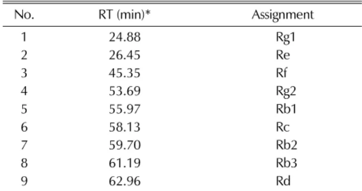
1. Assignment of the Peaks in P. ginseng Root Samples HPLC analysis of P. ginseng was carried for comparing ginsenosides of methanol extracts in main and lateral roots obtained from different cultivation methods between direct seeding and transplanting method. Nine peaks were assigned in main and lateral roots of P. ginseng, indicating ginsenosides, Rg1, Re, Rf, Rg2, Rb1, Rc, Rb2, Rb3, and each Rd was identified using ginsenoside standards. The nine peaks of HPLC were quantified depending on a retention time/assignment (Table 1).
2. Difference of Metabolic Compounds between Direct Seeding and Transplanting Method of P. ginseng Roots
To investigate the differences of metabolites in the main or lateral root of P. ginseng between samples produced from direct seeding and transplanting cultivation, P. gin- seng root samples were analyzed using HPLC-based meta- bolic targeting technique. PLS-DA is a PLS regression
mation to maximize the group classification and/or biomar- ker selections (Eriksson et al., 2006). We conducted PLS- DA using the processed HPLC chromatogram data to differentiate cultivation method of P. ginseng root sam- ples by using metabolic targeting. The chromatogram data were scaled to Pareto and mean-centered by SIMCA-P 13.0 software.
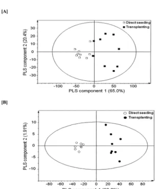
We excluded 1 and 2 outliers in the preliminary PCA of 10 samples of each P. ginseng main and lateral root samples, respectively (data not shown), and PLS-DA was thus performed using 9 and 8 samples for each P.
ginseng main and lateral, respectively. Figure 2 (A) and (B) show the score plots derived from the PLS-DA of P.
ginseng main and lateral roots obtained from direct seeding and transplanting method, respectively. Direct seeding and transplanting samples of P. ginseng main and lateral root could be clearly separated by using PLS component 1 and 2. The two PLS components accounted for 85.4% and 94.1% of the total variance in main and lateral roots of P. ginseng, respectively.
Figure 2 (A and B) show the PLS-DA score plots (PLS component 1 and PLS component 2) derived from the HPLC chromatogram data. Parameter in PLS-DA including R
2and Q
2could evaluate the models, and demonstrate the goodness of fit and ability of prediction.
When the values of R
2and Q
2are close to 1.0, the model has good capacity and prediction (Eriksson et al., 2006). PLS-DA modeling between direct seeding and transplanting revealed R
X2, R
Y2, and Q
2value of 0.95, 0.76, and 0.65 for main root and 0.96, 0.96, and 0.93 for
Table 1. Retention time and assignment of nine peaks of HPLC chromatogram of methanol extracts main and lateral of P.ginseng roots.
No. RT (min)* Assignment
1 24.88 Rg1
2 26.45 Re
3 45.35 Rf
4 53.69 Rg2
5 55.97 Rb1
6 58.13 Rc
7 59.70 Rb2
8 61.19 Rb3
9 62.96 Rd
*RT; Retention Time.
The PLS-DA score plot between direct seeding and transplanting in main and lateral roots of P. ginseng showed clear separation by PLS component 1 (Fig. 2A and B). The loading plot of P. ginseng main and lateral root from direct seeding and transplanting method showed that the main root of transplanting method contained higher amount of Rg1, Re, Rf, Rg2, Rb1, Rc, Rb2, Rb3, and Rd compounds than that of direct seeding cultivation.
Variable influence on projection (VIP) is a weighed sum of squares of the PLS taking into account the amount of Y-variance in each dimension. VIP values of 0.7-0.8 could be considered as important value for separation of each sample through a PLS model (Eriksson et al., 2006). As shown in Table 2, VIP values of the major contributing compounds in the score plots derived from PLS-DA (Fig. 2 (A and B)) were like follows; Re: 1.62, Rb1: 1.35, Rc: 1.31, Rb2: 0.84, Rf: 0.73, Rg1: 0.72, Rd:
0.70, in main root of P. ginseng samples, and Rg1: 2.01, Re: 1.23, Rb1: 1.15, Rf: 0.96, Rc: 0.72 in lateral root of P.ginseng samples.
To obtain clear information of the relative levels in
each compound based upon the VIP analysis, t-test was performed and shown in Fig. 3. The levels of Re, Rf, Rg2, Rb1, Rc, Rb3, and Rd were significantly (p < 0.05 in all cases) higher in main root of transplanting method than direct seeding samples. The levels of Rg1, Re, Rf, Rg2, Rb1, Rc, Rb2, Rb3, and Rd were also significantly (p < 0.05 in all cases) higher in lateral root of transplanting cultivation than direct seeding samples.
This study found higher amount of Re representing protopanaxatriol and Rb1 and Rc representing proto panaxadiol in main root of P. ginseng, and Rg1, Re, and Rb1 representing protopanaxatriol in lateral roots of P. ginseng. Total amount of ginsenoside from main and lateral root was 1.9 times and 3.3 times higher in transplanting cultivation samples than direct seeding samples, respectively. According to the result comparing ginsenoside between direct seeding and transplanting method in the study of Li et al., total amount of ginsenoside in transplanting cultivation samples was 56.8% higher than direct seeding cultivation samples. The reason was
Fig. 2. PLS-DA derived score plots (A and B) obtainedusing HPLC chromatogram data of P. ginseng roots from different cultivation method demonstrating the separation between direct seeding and transplanting cultivation from main (A) and lateral (B). □; Main root by direct seeding, ■; Main root by transplanting,
○;Lateral root by direct seeding, ●; Lateral root by transplanting.
Table 2. VIP of various peaks of HPLC chromatogram of methanol extracts main and lateral of P. ginseng roots.
(A) Main root of P. ginseng
RT (min)* VIP** Ginsenosides
26.45 1.62 Re
55.97 1.35 Rb1
58.13 1.31 Rc
59.70 0.84 Rb2
45.35 0.73 Rf
24.88 0.72 Rg1
62.96 0.70 Rd
53.69 0.59 Rg2
61.16 0.38 Rb3
(B) Lateral root of P. ginseng
RT (min)* VIP** Ginsenosides
24.88 2.01 Rg1
26.45 1.23 Re
55.97 1.15 Rb1
45.35 0.96 Rf
58.13 0.72 Rc
55.97 0.61 Rb1
53.69 0.32 Rg2
61.19 0.27 Rb3
62.96 0.26 Rd
*RT; Retention Time, **VIP; Variable Influence on Projection.
demonstrated that the weight per individual ginseng obtained from samples of transplanting cultivation was heavier than that from direct seeding samples; especially lateral root in ginseng from transplanting cultivation was much more mature in comparison with that from direct seeding (Li et al., 2009). This result is consistent with Qu et al. (2009), showing higher amount of Re and Rb1 in main roots of American ginseng than those in root-hair of 5-years-old American ginseng. However, the amount of Re, Rb1, and Rc of root-hair in American ginseng was higher in order.
Also, the amount of Rb1 and Rg1 of main roots in and Rb1 and Re of lateral root in P. ginseng was higher in Yunpoong ginseng cultivated in Daejeon, Korea (Li et al., 2009). Shi et al. (2007) compared the amount of ginsenoside in roots of 5-years-old P. ginseng, and confirmed that the amount of ginsenoside including Rg1, Rb1, and Re of main roots and Re, Rb1, and Rc of root-hair were higher in order.
This study investigated the amount of ginsenoside of
different methods, such as direct seeding and transplanting method, and newly found that there was significant difference of ginsenoside between different cultivation methods according to multivariate statistical. The amount of ginsenoside of roots in P. ginseng was higher in transplanting method than direct seeding cultivation method.
ACKNOWLEDGEMENTS
This study was supported by 2014 Post Doctoral Course Program of Department of Medicinal Crop Research(PJ 00935501), Rural Development Administration, Republic of Korea.
REFERENCES
Attele AS, Zhou YP, Xie JT, Wu JA, Zhang L, Dey L, Pugh W, Rue PA, Polonsky KS and Yuan CS. (2002). Antidiabetic effects of Panax ginseng berry extract and the identification of an effective component. Diabetes. 51:1851-1858.
Carlini EA. (2003). Plants and the central nervous system.
Pharmacology Biochemistry and Behavior. 75:501-512.
Cho JG, Lee MK, Lee JW, Park HJ, Lee DY, Lee YH, Yang DC and Baek NI. (2010). Physicochemical characterization and NMR assignments of ginsenosides Rb1, Rb2, Rc, and Rd isolated from Panax ginseng. Journal of Ginseng Research.
34:113-121.
Choi KT. (2008). Botanical characteristics, pharmacological effects and medicinal components of Korean Panax ginseng C. A.
Meyer. Acta Pharmacologica Sinica. 29:1109-1118.
Eriksson L, Johansson E, Kettaneh-Wold N and Wold S.
(2006). Multi-and megavariate data analysis. MKS Umetrics AB. Umea. Sweden, p.99-108.
Han JS, Tak HS, Lee GS, Kim JS and Choi JE. (2013).
Comparison of ginsenoside content according to age and diameterin Panax ginseng C. A. Meyer cultivated by direct seeding. Korean Journal of Medicinal Crop Science.
21:184-190.
Kang JH, Lee SY, Kang SM, Kwon HN, Park JH, Kwon SW and Park SH. (2008). NMR-based metabolomics approach for the differentiation of ginseng(Panax ginseng) roots from different origins. Archives of Pharmacal Research. 31:330-336.
Keith IB and Mark NM. (2003). Immune system effects of echinacea, ginseng, and astragalus. Integrative Cancer Therapies.
2:247-267.
Kim HY, Chen X and Gillis CN. (1992). Ginsenosides protect pulmonary vascular endothelium against free radical-induced injury. Biochemical and Biophysical Research Communications.
189:670-676.
Kim HJ, Lee SG, Chae IG, Kim MJ, Im NK, Yu MH, Lee EJ and Lee IS. (2011). Antioxidant effects of fermented red ginseng extracts in streptozotocin-induced diabetic rats. Journal Fig. 3. Peak area of Rg1, Re, Rf, Rg2, Rb1, Rc, Rb2, Rb3, and
Rd in P. ginseng root samples. t-test was performed to compare and assess statistical significance (p< 0.05).
The error bars are expressed as the standard deviation.
(A); Main root of P. ginseng, (B); Lateral root of P. ginseng.
Lan K, Zhang Y, Yang J and Xu L. (2010). Simple quality assessment approach for herbal extracts using high performance liquid chromatography-UV based metabolomics platform.
Journal of Chromatography A. 1217:1414-1418.
Lee SW, Cha SW, Hyun DY, Kim YC, Kang SW and Seong NS. (2005). Comparison of growth characteristics, and extract and crude saponin contents in 4-year-old ginseng cultured by direct seeding and transplanting cultivation. Korean Journal of Medicinal Crop Science. 13:241-244.
Li XG, Kang SJ, Han JS, Kim JS and Choi JE. (2009). Effects of root diameter within different root parts on ginsenoside composition of yunpoong cultivar in Panax ginseng C. A.
meyer. Korean Journal Medicinal Crop Science. 17:452-457.
Li XG, Kang SJ, Han JS, Kim JS and Choi JE. (2010).
Comparison of growth increment and ginsenoside content in different parts of ginseng cultivated. Korean Journal of Medicinal Crop Science. 18:70-73.
Lim SI, Cho CW, Choi UK and Kim YC. (2010). Antioxidant activity and ginsenoside pattern of fermented white ginseng.
Journal of Ginseng Research. 34:168-174.
Lü JM, Yao Q and Chen C. (2009). Ginseng compounds: An update on their molecular mechanisms and medical applications.
Current Vascular Pharmacology. 7:293-302.
Nah SY, Kim DH and Rhim H. (2007). Ginsenosides: Are any of them candidates for drugs acting on the central nervous system.
CNS Drug Reviews. 13:381-404.
Okada T, Mochamad AF, Altaf-Ul-Amin M, Takahashi H, Nakamura K and Kanaya S. (2010). Metabolomics of medicinal plants: The importance of multivariate analysis of analytical chemistry data. Current Computer Aided Drug Design. 6:179-196.
Qu C, Bai Y, Jin X, Wang Y, Zhang K, You J and Zhang H.
(2009). Study on ginsenosides in different parts and ages of Panax quinquefolius L. Food Chemistry. 115:340-346.
Roy R, Coelho BRB, Reeleder R, Bruin A, Grohs R, White P and Capell B. (2008). Effect of planting bed shape, mulch and
soil density on root yield and shape in north American ginseng(Panax quinquefolius L.). Canadian Journal of Plant Science. 88:937-949.
Sangster T, Major H, Plumb R, Wilson AJ and Wilson ID.
(2006). A pragmatic and readily implemented quality control strategy for HPLC-MS and GC-MS-based metabonomic analysis. Analyst. 131:1075-1078.
Shi W, Wang Y, Li J, Zhang H and Ding L. (2007).
Investigation of ginsenosides in different parts and ages of Panax ginseng. Food Chemistry. 102:664-668.
Van der Kooy F, Maltese F, Choi YH, Kim HK and Verpoorte R. (2009). Quality control of herbal material and phytopharmaceuticals with MS and NMR based metabolic fingerprinting. Planta Medica. 75:763-775.
Warwick B. Dunn and David IE. (2005). Metabolomics: Current analytical platforms and methodologies. Trends in Analytical Chemistry. 24:285-294.
Wilson ID, Plumb R, Granger J, Major H, Williams R and Lenz EM. (2005). HPLC-MS-based methods for the study of metabonomics. Journal of Chromatography B. 817:67-76.
Xie JT, Mchendale S and Yuan CS. (2005). Ginseng and diabetes. The American Journal of Chinese Medicine.
33:397-404.
Yang SY, Kim HK, Lefeber AW, Erkelens C, Angelova N, Choi YH and Verpoorte R. (2006). Application of two-dimensional nuclear magnetic resonance spectroscopy to quality control of ginseng commercial products. Planta Medica. 72:364-369.
Yang SO, Shin YS, Hyun SH, Cho S, Bang KH, Lee D, Choi SP and Choi HK. (2011). NMR-based metabolic profiling and differentiation of ginseng roots according to cultivation ages.
Journal Pharmaceutical and Biomedical Analysis. 25:19-26.
Yun TK. (2003). Experimental and epidemiologic evidence on non-organ specific cancer preventive effect of Korean red ginseng and identification of active compounds. Mutation Research. 523-524:63-74.