저작자표시-동일조건변경허락 2.0 대한민국 이용자는 아래의 조건을 따르는 경우에 한하여 자유롭게 l 이 저작물을 복제, 배포, 전송, 전시, 공연 및 방송할 수 있습니다. l 이차적 저작물을 작성할 수 있습니다. l 이 저작물을 영리 목적으로 이용할 수 있습니다. 다음과 같은 조건을 따라야 합니다: l 귀하는, 이 저작물의 재이용이나 배포의 경우, 이 저작물에 적용된 이용허락조건 을 명확하게 나타내어야 합니다. l 저작권자로부터 별도의 허가를 받으면 이러한 조건들은 적용되지 않습니다. 저작권법에 따른 이용자의 권리는 위의 내용에 의하여 영향을 받지 않습니다. 이것은 이용허락규약(Legal Code)을 이해하기 쉽게 요약한 것입니다. Disclaimer 저작자표시. 귀하는 원저작자를 표시하여야 합니다. 동일조건변경허락. 귀하가 이 저작물을 개작, 변형 또는 가공했을 경우 에는, 이 저작물과 동일한 이용허락조건하에서만 배포할 수 있습니다.
A THESIS
FOR THE DEGREE OF DOCTOR OF PHILOSOPHY
Improving ginsenoside content in Korean wild
ginseng (Panax ginseng Meyer) through
different biotechnological approaches
Jun-Ying Zhang
Department of Biotechnology
GRADUATE SCHOOL
JEJU NATIONAL UNIVERSITY
A THESIS
FOR THE DEGREE OF DOCTOR OF PHILOSOPHY
Improving ginsenoside content in Korean wild
ginseng (Panax ginseng Meyer) through
different biotechnological approaches
Jun-Ying Zhang
(Supervised by Professor Hyo-Yeon Lee)
Department of Biotechnology
GRADUATE SCHOOL
JEJU NATIONAL UNIVERSITY
Improving ginsenoside content in Korean wild ginseng (Panax
ginseng Meyer) through different biotechnological approaches
Jun-Ying Zhang
(Supervised by Professor Hyo-Yeon Lee)
A thesis submitted in partial fulfillment of the requirement for the degree of
Doctor of Philosophy
2014. 08
This thesis has been examined and approved by
김인중
……… Chairperson of the supervising committees
Professor In-Jung Kim, Faculty of Biotechnology, Jeju National University
이효연
……… Professor Hyo-Yeon Lee, Faculty of Biotechnology, Jeju National University
선현진
………
Professor Hyeon-Jin Sun, Subtropical Horticulture Research Institute, Jeju National University
김혜란
………
Professor Hye-Ran Kim, Korea Research Institute of Bioscience and Biotechnology
송인자
………
Professor In-Ja Song, Subtropical Horticulture Research Institute, Jeju National University
Department of Biotechnology
GRADUATE SCHOOL
CONTENTS
CONTENTS………...1 요약문……….5 ABBREVIATIONS………10 LIST OF FIGURES………...12 LIST OF TABLES……….16 SUMMARY………17Chapter I. Improving ginsenoside content in Korean wild ginseng (Panax ginseng Meyer) mutant lines induced by γ-irradiation (60Co) of adventitious roots………...23
INTRODUCTION………...25
MATERIALS AND METHODS………..31
Plant materials……….31
Establishment of adventitious root cultures………...32
Petridish culture condition………...32
Flask culture condition………...32
Bioreactor culture condition………...32
Gamma irradiation………...34
Selection of cell lines from suspension culture………...34
Determination of root weight and growth ratio………...34
Extraction of crude saponin……….35
Method validation and statistical analysis………...39
RESULTS AND DISCUSSION………40
Effects of gamma irradiation on survival rate of adventitious roots ………..40
Effects of gamma irradiation on growth rate of cell lines ………..40
Effects of gamma irradiation on ginsenoside production………...41
CONCLUSIONS………42
REFERENCES………...54
Chapter ІI. Plant regeneration of Korean wild ginseng (Panax ginseng Meyer) mutant lines induced by γ-irradiation (60Co) of adventitious roots………66
INTRODUCTION……….67
MATERIALS AND METHODS………..70
Callus induction and proliferation………...70
Induction of somatic embryos……….70
Maturation and germination of somatic embryos………...71
Development of plantlets and acclimatization………...72
Data analysis………...73
RESULTS AND DISCUSSION………...74
Induction of callus from adventitious roots……….74
Induction of somatic embryos……….75
REFERENCES……….90
Chapter III. Impr oving gins enos ide cont ent in Kor ea n wild gins eng b y Agrobacterium-mediated transformation with a squalene synthase gene………..95
INTRODUCTION………...96
MATERIALS AND METHODS………99
Plant materials………...99
Total RNA extraction and cDNA synthesis………...100
TA Cloning of squalene synthase gene……….101
Vector construction………...101
Agrobacterium-mediated transformation protocol of Panax ginseng…………...102
Adventitious root induction from transgenic Panax ginseng………...104
Molecular characterization of transgenic plants………105
Extraction of crude saponin………...109
Determination of ginsenoside contents by HPLC…………..……...…………....110
RESULTS AND DISCUSSION………...112
Isolation of PgSS2 and vector construction for Panax ginseng transformation…112 Establishment of an effective in vitro transformation system………...112
Production of transgenic adventitious roots………...113
Molecular analysis of putative transgenic adventitious root………....113
Accumulation of ginsenoside content (Rg1, Re, Rf, Rh1, Rb1, Rc, Rb2 and Rd) in Panax ginseng overexpressing squalene synthase (PgSS2) gene……….114
REFERENCES………136 ACKNOWLEDGMENT………....142 PUBLICATIONS………....143
요약문
인삼은 동양사람들에게 널리 이용되어온 가장 오래 된 약초 중의 하나이고, 이 인삼의 주요 성분인 진세노사이드는 30 여종이 알려져 있다. 감마선(γ-ray)이 산삼 부정근의 성장 및 진세노사이드 함량에 미치는 영향을 평가하기 위해, 산삼 뿌리 유래의 부정근에 25, 50, 75, 100, 125Gy 의 감마선을 조사하여 돌연변이를 유도하였다. 감마선(60Co) 조사는 제주대학교 원자력과학기술연구소에서 수행하였다. 감마선을 조사한 부정근은 30 일간 플라스크 배양한 후, 형성된 secondary root 의 수를 조사하여 선발하였다. 감마선을 조사하지 않은 야생형과 선발된 변이 부정근의 진세노사이드 (Rg1, Re, Rf, Rh1, Rb1, Rc, Rb2 와 Rd) 함량은 HPLC 와 PDA 검출기를 이용하여 측정하였으며, Digoxin 을 internal standard 로 사용하였다. 그 결과, 50Gy 이상의 방사선을 조사한 처리구에서는 secondary root 의 수가 야생형에 비해 적었고, 50Gy 이하의 방사선을 조사한 처리구에서 야생형보다 secondary root 의 수가 많은 변이 부정근을 선발할 수 있었다. 따라서 진세노사이드의 생산성을 높이기 위한 방사선량으로는 50Gy 이하가 적합함을 알 수 있었다. Secondary root 의 수가 야생형보다 많은 5 개의 계통을 선발한 후, 한달 간 플라스크에 배양하여 건조중량을 측정한 결과, 야생형은 0.31g 이었고, 선발한 1, 3, 9, 13 및 14 번 계통들은 각각 0.46g, 0.45g, 0.29g, 0.34g, 0.48g 이었다. 생장율은 야생형이 4.42 였으며, 선발한 계통들인 1, 3, 9, 13, 14 번은 각각 6.57, 6.48, 4.10, 4.90, 6.90 이었다. 선발한 1, 3, 14 번 계통들의 생장율은 야생형에 비해 각각 49%, 47%, 57% 증가하였다.총 진세노사이드 함량은 1 번 계통에서 가장 크게 증가하였고, 1 번 계통의 총 진세노사이드 (Rg1, Re, Rf, Rh1, Rb1, Rc, Rb2, Rd) 함량은 야생형과 비교하여 약 1.6 배 증가하였다. 또한, 이 계통의 진세노사이드 중 Rg1, Re, Rh1 과 Rb2 의 함량은 야생형에 비해 2-3 배 증가하였음을 알 수 있었다. 3 번 계통의 진세노사이드 Re, Rh1, Rb2, Rc 와 Rd 함량은 야생형에 비해 1.6-2.3 배 증가하였으며, 9 번 계통에서는 진세노사이드 Rg1, Re 와 Rh1 함량이 야생형에 비해 1.7-2 배 증가하였다. 4 번 계통의 진세노사이드 함량은 야생형과 비슷하였고, 13 번 계통의 진세노사이드 함량은 야생형에 비해 감소하였음을 알 수 있었다. 이들 결과로부터, 선발한 변이 계통들 중 1 번 계통이 가장 높은 진세노사이드 함량을 가지고 있음을 알 수 있었다. 본 연구결과 우리는 biomass 생산성과 진세노사이드 함량이 크게 증가된 돌연변이 세포주를 선발할 수 있었으며, 감마선 조사는 2 차 대사산물 특히 진세노사이드의 생산량 증대에 있어 강력하고 유용한 수단임을 확인할 수 있었다. 이 결과는 감마선 조사가 진세노사이드 합성에 필요한 효소의 활성 등에 영향을 미쳤음을 시사한다. 인삼은 성장이 느리고 긴 생산주기 (4-6 년)를 가지고 있는 다년생 식물이며, 종자를 생산하기까지 3 년이상의 유년기가 필요하다. 이러한 특성 때문에 전통육종에 의한 우수 유전자형의 창출은 매우 어려운 실정이다. 또한, 지금까지 산삼의 부정근에 방사선을 조사한 후 그 돌연변이 부정근을 재분화한 보고는 없다. 따라서, 본 연구에서는 인삼의 신품종 육성을 위해 진세노사이드 함량이
재분화 시스템은 인삼식물의 형질전환에도 활용될 수 있을 것이다. 한국 산삼의 체세포배발생과 식물체 재분화를 위하여 부정근 유래의 캘러스로부터 체세포배를 유도한 결과, 0.5 mg/L 2,4-D 가 포함된 배지에서 가장 높은 빈도로 유도되었고, 야생형과 돌연변이 부정근의 체세포배 형성 빈도는 각각 15.3% 와 14.7% 였다. 야생형과 돌연변이 부정근의 체세포배 형성수는 각 캘러스 당 각각 25.6 개와 23.7 개였으며, 야생형과 돌연변이 부정근의 체세포배 형성 빈도에는 유의 차가 없었다. Globular 형태의 체세포배는 배발생 캘러스의 표면에 형성되었다. 체세포배발생으로부터 식물체 재분화에 미치는 GA3 의 영향을 평가한 결과, GA3 무처리구에서는 36%의 재분화효율을 보였으나, 5 mg/L 의 GA3처리구에서는 85% 로 가장 높은 재분화효율을 보였다. 배의 성숙과 재분화는 GA3 의 농도에 크게 영향을 받았으며, 발아한 배는 5 mg/L GA3가 포함된MS 배지에서 식물체로 발달하여 성장하였다. 뿌리가 형성되지 않은 식물체는 MS 또는 SH 기본 배지에 0.25 mg/L NAA 혹은 0.5% 활성탄이 포함된 배지에 옮겨 약 1 달후 뿌리유도 효율을 평가하였다. 그 결과, 유도된 뿌리의 질은, 0.25 mg/L NAA 와 1%의 sucurose 가 첨가된 1/3 SH 배지에서 빠르고 두껍게 성장하여 가장 좋은 결과를 보였다. 0.5%의 활성탄과 2% sucrose 가 첨가된 1/3 SH 배지에서 뿌리 유도 효율이 가장 높았으나, 이 배지에서 유도된 뿌리는 잘 자라는 반면 약한 단점이 있었다. 따라서 뿌리유도를 위한 최적 배지는 0.25 mg/L NAA 와 1%의 설탕을 첨가한 1/3 SH 배지였다. 뿌리가 잘 발달한 산삼 재분화 식물체는 인공 혼합토 [peatmoss:vermiculite:
perlite=2:3:1(v/v)]를 넣은 플라스틱 화분 (10x18cm)에 이식하여 생장실에서 순화하였다. 순화 3 개월 후 신초가 형성되어 성장하기 시작하였으며 생존율은 약 30% 였다. 본 연구에서는 중요한 약용식물인 산삼의 효율적인 재분화계를 확립하였고, 이 프로토콜은 한국 산삼의 빠른 대량 생산을 가능하게 해줄 것으로 기대된다. 산삼 부정근으로부터 캘러스를 유도하여 재분화 식물을 얻기까지는 약 6-8 개월이 소요되었다. 본 연구에서는 방사선조사를 통해 얻은 돌연변이 부정근으로부터 식물체를 재분화하였고, 감마선을 이용한 돌연변이 기술과 조직배양에 의한 식물재분화 기술의 융합은 인삼 품종개량에 있어서 효과적인 방법의 하나가 될 수 있을 것으로 생각된다. 산삼 돌연변이 부정근의 진세노사이드 생산성을 개량하기 위하여 합성 스쿠알렌 신타아제(PgSS2) 유전자를 35S 프로모터하에 도입한 후 산삼의 돌연변이 부정근에 형질전환을 수행하였다. 형질전환은 돌연변이 산삼 부정근 계통에서 선발한 배발생캘러스를 PgSS2 유전자와 bar 유전자를 포함하는 아그로박테리움 EHA105 에 감염하여 수행하였다. 형질전환체는 phosphinothricin (PPT) 3mg/L 를 첨가한 MS 배지에서 선발하였다. 그 결과, 10 개체의 PPT-저항성 식물을 선발하였고, 이 식물체들의 유전자 도입은 PAT strip test 와 Southern blot 분석을 통하여 확인하였다. 유전자의 도입이 확인된 산삼 형질전환 식물을 이용하여 스쿠알렌 신타아제(PgSS2) 유전자가 진세노사이드의 생합성에서 어떤 역할을 하는지를 조사하였다. 형질전환 산삼 식물 계통들에서 재분화시킨
계통 1, 2, 5, 7, 8 번은 비형질전환 돌연변이 부정근보다 진세노사이드 함량이 약 1.3-1.6 배 증가하였음을 확인하였다. 또한, 이들 형질전환 계통들 중 1 번과 8 번의 진세노사이드 함량이 야생형 및 공벡터가 도입된 계통들보다 약 1.6 배로 가장 높게 나타났다. 이들 결과로부터 형질전환 기술은 진세노사이드 축적이라는 관점에서 인삼의 개량을 용이하게 해 줄 것으로 생각된다. 감마선을 이용한 돌연변이 기술, 식물재분화 및 형질전환 기술의 융합은 새로운 인삼 품종의 개발에 매우 효과적인 방법의 하나가 될 것이다.
ABBREVIATIONS
NAA Naphthalene acetic acid IAA IBA GA Indole-3-acetic acid Indole-3-butyric acid Giberellic acid
HPLC-PDA High performance liquid chromatography-photodiode array detector
ACN Acetonitrile MS FW DW PD PT MJ UV Gy Bar PPT MCL1 CL
Murashige & Skoog Fresh weight Dry weight Panaxadiol Panaxatriol Methyl jasmonate Ultraviolet Gray Phosphinothricin acetyltransferase Phosphinothricin
Mutant control line Cell line
LB RB p35S RT-PCR 2,4-D DEPC SEM SH LOD LOQ LD SPSS S/N Left border Right border CaMV 35S promoter
Reverse transcriptase polymerase chain reaction 2,4-dichlorophenoxy-acetic acid
Diethylpyrocarbonate Standard error of mean
Schenk & Hidebrandt Medium Limit of detection
Limit of quantification Lethal dose
Statistical Package for the Social Sciences Signal-to-noise
LIST OF FIGURES
Figure 1. Structures of ginsenosides isolated from Panax ginseng. Figure 2. Korean wild ginseng (Panax ginseng Meyer).
Figure 3. Culture system of Panax ginseng.
Figure 4. Extraction procedures of crude saponin in Panax ginseng.
Figure 5. Survival rate among gamma-ray irradiated adventitious roots of Panax ginseng. Figure 6. . Phenotypic characteristics of secondary roots in the gamma-irradiated cell lines of
Panax ginseng (scale bar = 1 cm).
Figure 7. Growth ratio of Panax ginseng adventitious root in the control and gamma-irradiated cell lines.
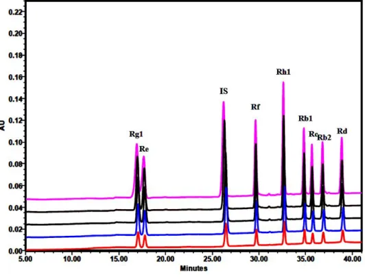
Figure 8. Typical chromatograms obtained from the standard solution by UV detection at 203 nm. IS: internal standard (digoxin).
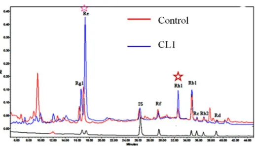
Figure 9. Typical chromatograms and ginsenoside content obtained from the standard, control and cell line CL1.
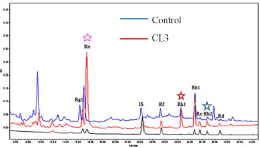
Figure 10. Typical chromatograms and ginsenoside content obtained from the standard, control and cell line CL3.
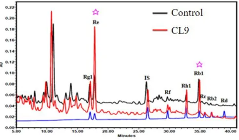
Figure 11. Typical chromatograms and ginsenoside content obtained from the standard, control and cell line CL9.
Figure 13. Typical chromatograms and ginsenoside content obtained from the standard, control and cell line CL14.
Figure 14. Total ginsenoside content of the control and mutant cell lines of Panax ginseng adventitious root.
Figure 15. Effects of different concentration of 2,4-D and Kinetin on the frequency of callus formation from ginseng adventitious roots after 6 weeks of a culture (scale bar = 1 cm). Figure 16. Somatic embryogenesis and regeneration of plantlet from adventitious roots of
Panax ginseng.
Figure 17. Effect of 2,4-D on somatic embryogenesis from embryogenic callus of control and mutant cell line.
Figure 18. Effects of GA3 concentration on somatic embryos germination frequency from
150 somatic embryos.
Figure 19. Development of ginseng plant and transplantation into soil. Figure 20. Transplantation of regenerated ginseng plants into soil. Figure 21. Total RNA extraction.
Figure 22. Construct of binary vector plasmid pCAMBIA3301-PgSS2.
Figure 23. Extraction of genomic DNA. DNA from adventitious root of Panax ginseng extracted by CTAB method.
Figure 24. Scheme of Southern hybridization.
Figure 25. TA cloning of squalene synthase gene from Panax ginseng adventitious root and T-DNA region of binary vector (pCAMBIA3301-PgSS2) construct used for
Figure 26. Transformation procedure of Panax ginseng adventitious root.
Figure 27. Phosphinothricin-resistant plant lines generated from Panax ginseng adventitious roots.
Figure 28. PAT test strip analysis of transformants.
Figure 29. Adventitious root induction from transgenic Panax ginseng shoot on MS medium supplemented with 3 mg/L IBA.
Figure 30. Adventitious root proliferation from transgenic Panax ginseng shoot on MS medium supplemented with 2 mg/L NAA and 0.25 mg/L IAA.
Figure 31. Transgene expression in wild type and transgenic plant lines of P. ginseng.
Figure 32. Southern blot analysis of transgenic P. ginseng. Genomic DNAs extracted from ten different transgenic P. ginseng lines.
Figure 33. Southern blot analysis of transgenic P. ginseng. Genomic DNAs extracted from ten different transgenic P. ginseng lines.
Figure 34. Typical chromatograms and ginsenoside content obtained from the standard solution, mutant control (MCL1) and transgenic Panax ginseng line SS-1. The ginsenosides were extracted from adventitious roots cultured in the flask.
Figure 35. Typical chromatograms and ginsenoside content obtained from the standard solution, mutant control (MCL1) and transgenic Panax ginseng line SS-2. The ginsenosides were extracted from adventitious roots cultured in the flask
.
were extracted from adventitious roots cultured in the flask.
Figure 37. Typical chromatograms and ginsenoside content obtained from the standard solution, mutant control (MCL1) and transgenic Panax ginseng line SS-4. The ginsenosides were extracted from adventitious roots cultured in the flask.
Figure 38. Typical chromatograms and ginsenoside content obtained from the standard solution, mutant control (MCL1) and transgenic Panax ginseng line SS-5. The ginsenosides were extracted from adventitious roots cultured in the flask.
Figure 39. Typical chromatograms and ginsenoside content obtained from the standard solution, mutant control (MCL1) and transgenic Panax ginseng line SS-6. The ginsenosides were extracted from adventitious roots cultured in the flask.
Figure 40. Typical chromatograms and ginsenoside content obtained from the standard solution, mutant control (MCL1) and transgenic Panax ginseng line SS-7. The ginsenosides were extracted from adventitious roots cultured in the flask.
Figure 41. Typical chromatograms and ginsenoside content obtained from the standard solution, mutant control (MCL1) and transgenic Panax ginseng line SS-8. The ginsenosides were extracted from adventitious roots cultured in the flask.
Figure 42. Typical chromatograms and ginsenoside content obtained from the standard solution, mutant control (MCL1) and transgenic Panax ginseng line SS-9. The ginsenosides were extracted from adventitious roots cultured in the flask.
Figure 43. Total ginsenoside content of the mutant control (MCL1) and transgenic lines of
LIST OF TABLES
Table 1. HPLC conditions for ginsenosides analysis.
Table 2. Mobile phase of HPLC gradient condition for ginsenosides analysis.
Table 3. Calibration curve, limit of detection (LOD) and limit of quantification (LOQ) for eight ginsenosides.
Table 4. Effects of 2,4-D and kinetin on the frequency of callus formation from ginseng adventitious roots.
Table 5. Effects of 2,4-D on somatic embryo formation from embryogenic callus of wild-type and mutant adventitious roots.
Table 6. Effects of GA3 on germination of somatic embryos.
Table 7. Comparison of rooting media for ginseng root development.
Table 8. Media used in the Panax ginseng transformation system. Table 9. Set of primers used for PgSS2 gene cloning and PCR.
SUMMARY
Panax ginseng (Korean wild ginseng) is one of the oldest and the most widely used
herbal medicines for the oriental people. Ginsenosides are the major active components in the ginseng and more than 30 ginsenosides were identified from the plant. In order to evaluate effects of γ-irradiation on adventitious root growth and ginsenoside content, adventitious roots of Panax ginseng were treated with γ-ray of 25, 50, 75, 100 and 125Gy to induce mutation. The γ-irradiation was performed from 60Co source at the Applied
Radiological Science Research Institute, Jeju National University. Cell lines irradiated by
γ-ray
were cultured in flask for 30 days and selected based on the secondary root numbers of the irradiated adventitious roots. The contents of ginsenosides Rg1, Re, Rf, Rh1, Rb1, Rc, Rb2 and Rd in wild type and the selected cell lines were evaluated by HPLC-PDA detector with digoxin as the internal standard. After irradiation, below 50Gy, secondary root number of irradiated adventitious root was more than that of non-irradiated adventitious root. Otherwise, over 50Gy, the secondary root number was less than that of non-irradiated control. Therefore, the dosage below 50Gy was considered as adequate for the induction of mutant cell lines of Panax ginseng. Five cell lines, CL1, 3, 9, 13, and 14, were selected according to the secondary root number. After 30 days culture in flask, dry weight of control was 0.31g and the selected cell lines CL1, 3, 9, 13 and 14 were 0.46g, 0.45g, 0.29g, 0.34 and 0.48g respectively. The value of growth ratio of control was 4.42 and the selected cell lines CL1, 3, 9, 13, 14 were 6.57, 6.48, 4.1, 4.9 and 6.9, respectively. The values of growth ratio in celllines CL1, CL3 and CL14 were increased 49%, 47% and 57%, respectively, compared to the control.
The contents of the eight ginsenosides were significantly enriched, especially in the cell line CL1. The content of total ginsenosides (Rg1, Re, Rf, Rh1, Rb1, Rc, Rb2 and Rd) in the cell line CL1 was 1.6-fold higher than that of the control. In the cell line CL1, the level of ginsenoside Rg1, Re, Rh1and Rb2 contents were increased by 2-3 times than that of the control. In the cell line CL3, level of ginsenoside Re, Rh1, Rb2, Rc and Rd contents were increased by 1.6-2.3 times than that of the control. In the cell line CL9, level of ginsenoside Rg1, Re and Rh1 contents were increased by 1.7-2 times than that of the control. In the cell line CL14, level of ginsenoside contents were similar with that of the control. In case of the cell line CL13, the ginsenoside contents were affected negatively in ginseng adventitious root cultures. Overall, the highest ginsenoside contents were obtained in cell line CL1.
In this study, I selected cell lines with a significantly increased biomass productivity and ginsenoside content. Based on the results obtained in this report, gamma irradiation is powerful and useful tool for the enhancement of production of secondary metabolites, especially for ginsenoside. This result suggests that gamma irradiation might have triggered the enzyme activities for the synthesis of ginsenosides.
Ginseng is a perennial plant that grows slowly and has a long production cycle (4-6 years). And juvenile period is required longer than three years for producing seeds. Thus, it is very difficult to make superior genotypes by conventional breeding. So, I used the mutant
new cultivar of Panax ginseng. At present no information is available on the regeneration of mutant adventitious root line that has been selected from γ-irradiated P. ginseng adventitious roots. I report on an efficient procedure for the regeneration of wild-type and mutant cell lines of P. ginseng adventitious roots through somatic embryogenesis. This regeneration system was also used for genetic transformation.
I established an efficient in vitro protocol for somatic embryogenesis and plantlet conversion of Korean wild ginseng (Panax ginseng Meyer).The highest induction frequency of somatic embryos was observed on the medium supplemented with 0.5 mg/L 2, 4-D. The frequency of somatic embryo formation in wild-type and the mutant cell line was15.3% and 14.7%, respectively. The number of somatic embryos per callus was 25.6 and 23.7 in wild-type and the mutant cell line, respectively. There was no significant difference in somatic embryo formation frequency between wild-type and the mutant cell line. Globular shaped somatic embryos formed on the surfaces of embryogenic callus.
The optimal concentration of GA3 in germination medium was 5 mg/L, yielding the
highest germination frequency of 85%. Without GA3 treatment, the germination frequency
was lowest at 36%. Maturation and germination of embryos were strongly influenced by the GA3 concentration. The germinated embryos were developed to shoots and elongated on MS
medium with 5 mg/L GA3.
The shoots without roots were excised and transferred to different rooting media, half or one-third strength MS, or SH basal medium supplemented with 0.25 mg/L NAA or with 0.5% activated charcoal. Adventitious roots formed from the excised regions of the shoots.
After 1 month, the rate of root formation from the shoots was examined. As far as root quality is concerned, 1/3 SH medium with 0.25 mg/L NAA and 1% sucrose showed the best result among the tested rooting media; the roots grew fast and thickened on the medium. Although 1/3 SH medium with 2% sucrose and 0.5% activated charcoal was most effective in inducing roots, the roots grew well but weak. The optimal medium for rooting is therefore 1/3 SH medium supplemented with 0.25 mg/L NAA and 1% sucrose among the tested rooting media in this study. In my comparative studies, SH medium was more effective than MS medium in root induction and proliferation. So, SH medium, especially 1/3 strength SH medium is suitable for root induction and growth of regenerated ginseng plants.
Well-developed plantlets with both shoots and roots derived from adventitious roots were transferred to plastic pots (10×18cm) containing an artificial soil mixture of peatmoss, vermiculite and perlite (2:3:1 v/v) in a growth room. The survival rate of the plantlets was about 30% after 3 months of culture and new leaf began growing.
I developed an efficient in vitro regeneration protocol for an important medicinal plant of
P. ginseng. The protocol described here will allow a relatively rapid mass production of
Korean wild ginseng plants. It takes 6-8 months from the callus induction of adventitious roots to the plantation of plants. In the present study, I also produced the regenerated plants from the mutant adventitious roots which were obtained by γ-irradiation. The combination of mutation technique by γ-irradiation and plant regeneration by tissue cultures may be an effective way to ginseng improvement. The protocol established in this study was used for
With the purpose of improving ginsenoside production in Panax ginseng (Panax ginseng Meyer) mutant adventitious root lines, a synthetic gene encoding squalene synthase (PgSS2) was placed under the control of 35S promoter and transferred to Panax ginseng. The mutant lines used in this study generated by γ-irradiation (60Co) of ginseng adventitious roots.
Transgenic plants were generated by Agrobacterium-mediated transformation. Embryogenic callus obtained from ginseng adventitious root lines were transformed by infection with A.
tumefaciens strain EHA105 containing the PgSS2 gene and the phosphinothricin
acetyltransferase (bar) gene as a selectable marker. Transformants were selected on Murashige Skoog medium containing 3mg/L phosphinothricin. Ten phosphinothricin-resistant plant lines were generated on selective medium, and the integration of the transgene in these plants was confirmed by PAT test strip, RT-PCR and Southern hybridization. Southern hybridization analysis suggested the insertion of one or multiple copies of T-DNA into ginseng genome. I am investigating the regulatory role of
Panax ginseng squalene synthase (PgSS2) on the biosynthesis of ginsenoside by using the
transgenic ginseng plant lines. The total contents of the 8 ginsenoside types were significantly enriched in the 5 transgenic lines compared to the mutant control (MCL1). The line SS 1 showed a 1.6-fold increase than the MCL1. The line SS 2 showed a 1.5-fold increase than the MCL1, and the line SS 5, SS 7 showed a 1.3-fold increase than the MCL1. The total contents of the 8 ginsenoside types (Rg1, Re, Rf, Rh1, Rb1, Rc, Rb2 and Rd) obtained from the mutant control (MCL1), empty vector and the SS lines were 6.14 mg g-1,
mg g-1, 9.75 mg g-1, 4.56 mg g-1, respectively. Overall, the highest ginsenoside content was
obtained with SS line 1 and 8.
This transformation method may facilitate the improvement of Panax ginseng in terms of the accumulation levels of ginsenoside. The combination of mutation technique by γ-irradiation, plant regeneration by tissue cultures and genetic transformation may be an effective way to develop new ginseng cultivars.
Chapter I
Improving ginsenoside content in Korean wild ginseng (Panax
ginseng Meyer) mutant lines induced by γ-irradiation (
60Co) of
adventitious roots
INTRODUCTION
There are several medicinal Panax species identified from all over the world. From a number of speices, Panax ginseng (Korean wild ginseng) is one of the oldest and the most widely used herbal medicines and this species is almost extinct in wild habitats. Fortunately,
Panax ginseng is widely cultivated in Korea, China, Japan, and several countries in North
America and Europe under special shade conditions (Hobbs 1996, Mabberley 1987, Toda et
al. 2003, Nam et al. 2002). Depending on the species, growth condition and location etc, the
ginsenoside contents vary widely which act on the central nervous system, cardiovascular system, endocrine system, and immune system. These active ingredients also increase endocrine secretion, promote immune function, and have anti-aging and stress relieving effects (Briskin 2000, Shibata 2001, Vogler et al. 1999). Panax ginseng (Korean ginseng) and Panax quinquefolius (American ginseng) are characterized well by phytochemistry (Mallol et al. 2001, Ngan et al. 1999) and more than 30 ginsenosides were identified from this genus. They can be classified into three groups based on their aglycones (Figure 1): the protopanaxadiol-type, protopanaxatriol-type, and oleanane type saponins (Jung et al. 2003, Nah 1997).
Figure 1. Structures of ginsenosides isolated from Panax ginseng.
The both species are used for preparation of tonic, prophylactic and anti-ageing agents (Chang and But 1986). Some ginsenosides are used widely, such as Rg1, Re, Rb1 are used to determinate ginseng quality (Wu et al. 2007). Some ginsenosides, like Rg3, Rh2 and Rh1 are rarely detected in cultured ginseng. Ginsenoside Rb1, Re, Rg1 and Rb2 were the precursors
to form other ginsenosides by Human Intestinal Microflora and steaming. Different forms of ginsenosides showed several different functions.
Ginsenoside Rg1: Ginsenoside Rg1 inhibits the rat brain cAMP phosphodiesterase
activity, excitatory amino acids are involved in neuronal survival, synaptogenesis, neuronal plasticity, learning and memory processes, estrogen-like activity, and promoted functional neovascularization into a polymer scaffold in vivo (Stancheva et al. 1993, Balazs et al. 1998, Muller et al. 1998, Chan et al. 2002, Sengupta et al. 2004).
Ginsenoside Rb1: Ginsenoside Rb1 successfully improved homocysteine-induced
reduction of endothelial nitric oxide synthase expression and reduced homocysteine-induced oxidative stress (Zou et al. 2005). Preconditioning of Rb1 showed protective effects on myocardial ischemia and reperfusion injury (Wang et al. 2008). Rb1 promoted neurotransmitter release (Xue et al. 2006) and used to prevent or treat gastritis and gastric ulcers (Jeong et al. 2003).
Ginsenoside Re: Some reports revealed that both Rb1 and Re decreased cardiac
contraction in adult rat ventricular myocytes (Scott et al. 2001). Some reports says that treatment of both Rg1 and Re could be a novel group of nonpeptide angiogenic agents and may be useful for tissue regeneration (Yu et al. 2006). Compared with other gingenosides Re showed anti-diabetic activities (Xie et al. 2005) and free radical scavenging properties (Xie
et al. 2006).
Yokozawa et al. 1990). Rb2 accelerates would healing effects like cell proliferation, expression of proliferation related factors, and epidermis formation (Choi, 2002). Rb2 can be used as anti-tumor or chemopreventive agents, especially acting on cancer promotional stage (Kang et al. 2000).
Ginsenoside Rc: Ginsenoside Rb2 and Rc may have effects that prevent or limit the
development of breast cancer, affects the motility of sperm (Chen et al. 2001, Lee et al. 2003).
Ginsenoside Rd: Ginsenoside Rd inhibiting 26S proteasome activity. Ginsenoside-Rd
has been proved to decrease the severity of renal injury induced by cisplatin. Rd is Cytotoxic towards HeLa Cancer Cells and Induces Apoptosis (Chang et al. 2008, Yokozawa et al. 2000, Yang et al. 2006).
Ginsenoside Rh2: Ginsenoside Rh2 has the anti-fatigue, anti-cancer effect, antiallergic
and reduces ischemic brain injury in rats (Oh et al.1999, Nakata et al. 1998, Park et al. 2003, Park et al. 2004).
Ginsenoside Rh1: Ginsenoside Rh1 and Rh2 inhibit the induction of nitric oxide
synthesis in murine peritoneal macrophages. G-Rh1 acts as a functional estrogenic ligand in MCF-7 cells. Ginsenoside Rh1 possesses antiallergic and anti-inflammatory activities (Park
et al. 1996, Lee et al. 2003, Park et al. 2004).
Ginsenoside Rg3: Ginsenoside Rg3 significantly inhibited growth and angiogenesis of
ovarian cancer and inhibition of in vitro Tumor Cell Invasion (Xu et al. 2008, Shinkai et al. 1996). Ginsenoside Rg3 is a body function balancer, long term taking Rg3 can get anti
senility effects. Because ginsenoside Rg3 can invigorate blood circulation and improve brain and body activity. It has obvious effects for anti wrinkle, would make skin fresh, bright and glossy. Ginsenoside Rg3 has obvious function for body tonic, improve muscle tone and prevent fatigue.
Due to unavoidable limitations in Panax ginseng breeding, it is difficult to produce large amounts of roots under field conditions (Han et al. 2009). Hence, several scientists followed different biotechnological methods such as root culture, Agrobacterium-mediated hairy root production, bioreactor mediated large scale production. Jasmonic acid has been shown to be an effective elicitor for secondary metabolite induction in plant cell cultures (Ketchum et al. 1999). Recently, MJ (Methyl jasmonate) mediates the reprogramming of cellular metabolism and cell cycle progression via the regulation of jasmonic acid biosynthesis (Pauwels et al. 2008). The positive effect of MJ on ginsenoside production from ginseng cell suspension, hairy root and adventitious root cultures has been previously documented (Lu et al. 2001, Palazón et al. 2003, Choi et al. 2005, Bae et al. 2006). Mutation breeding is considered as one of the effective plant breeding methods for improved variety of crop production. Among several methods, γ-irradiation was used in several species for crop improvement program. Recent report showed that mutagenesis by g-irradiation gained rapid development in enhanced ginsenoside production of Panax ginseng. Callus were used for gamma irradiation and the cell lines selected from dosage 30Gy-treatment and ginsenoside content was significantly increased (Kim et al. 2009). Suspension culture of Lithospermum erythrorhizon
biosynthesis of the cells and increased the total shikonin yields by 400% at 16Gy (Chung et al. 2006). For maize, grain yield for irradiated samples is increased to levels above the unirradiated yield at doses up to about 250Gy with the optimum yield occurring at 150Gy. The corresponding increase for groundnut is observed at doses up to about 930Gy with optimum yield at a dose of 300Gy (Mokobia et al. 2006). Grain yield of maximum production in barley was observed at the rate of 100Gy (Subhan et al. 2004). g-irradiation mediated mutagenesis of whole plants as well as roots yielded significant variations among the different mutated species, and 50Gy of γ-rays was determined to be the optimal dose for inducing mutations (Joseph et al. 2004).
My present work is to investigate the effects of gamma irradiation on ginsenoside contents of ginseng adventitious root and to provide valuable information on the utilization of beneficial effects of gamma irradiation in root cell culture system of Panax ginseng.
MATERIALS AND METHODS
Plant materials
Panax ginseng is Korean wild ginseng. Wild ginseng grows naturally within its natural
habitat conditions (Figure 2). Adventitious roots were induced and proliferated from wild
Panax ginseng root, and cultured in MS medium supplemented with NAA and IAA.
Establishment of adventitious root cultures
Panax ginseng adventitious roots were proliferated on MS (MS, Murashige and Skoog
1962) medium as follows (mg/L): NH4NO3 (687.5), KNO3 (1900), KH2PO4 (170), H3BO3
(6.2), MnSO4.4H2O (23.3), ZnSO4.7H2O (8.6), KI (0.83), Na2MoO4.2H2O (0.25),
CuSO4.5H2O (0.025), CoCl2.6H2O (0.025), CaCl2.2H2O (440), MgSO4.7H2O (370),
Na2EDTA (37.3), FeSO4.7H2O (27.8), Nicotinic acid (5), Pyridoxine Hydrochloride (1),
Thiamine Hydrochloride (2.5), Glycine (2). Supplemented with 10.87μmol Naphthalene acetic acid (NAA), 1.43μmol Indole-3-acetic acid (IAA) and 3% sucrose. The pH of the medium was adjusted to 6.0 before autoclaving at 121℃ and 1.2 Kgf/cm2 pressure for 15
min.
Petridish culture condition
Adventitious roots were cut into 10 pieces, each 1-2 cm and cultured in petridish (10cm in diameter and 1.5 cm in height) with lid containing 50 mL MS solid medium was sealed with a wrap (Advantec, USA) and cultured at 23 ± 2℃ under dark condition. Adventitious roots were sub-cultured every 30 days.
Flask culture condition
Fresh roots (0.8g) were inoculated into a 100 mL erlenmeyer flask containing 50 mL MS liquid medium with 10.87 μmol NAA, 1.43 μmol IAA and 5% sucrose. Cultures were shaken at 110 rpm in the light at 23 ± 2℃ for 30 days.
Bioreactor culture condition
The bioreactors were maintained at 23 ± 2℃ in a light condition until harvest. Adventitious roots were sub-cultured every 30 days in 15L bioreactor (Biopia, Korea)
containing 5L MS medium. Forty-five g fresh weight of adventitious roots was inoculated into bioreactor for proliferation. After 30 days, the proliferated adventitious roots were used as explants for further experiments. The culture system showed in figure 3.
Figure 3. Culture system of Panax ginseng. (A) Root of Panax ginseng; (B) Explant of ginseng root; (C) Adventitious root proliferation; (D) Adventitious root culture in flask; (E) Adventitious root culture in bioreactor; (F) Harvest.
Gamma irradiation
Adventitious roots (1-2cm) were placed in plastic petridishes (10 pieces adventitious roots per pertridish), grown at 23 and cultured ℃ for 5 days in MS medium with 10.87 μmol NAA, 1.43 μmol IAA and 3% sucrose. They were exposed to gamma radiation from cobalt (60Co) source using a γ-radiation apparatus at the Applied Radiological Science Research
Institute, Jeju National University. Irradiation dosages were 0 (non-irradiated), 25, 50, 75, 100 and 125Gy. For each dosage, 3 petridishes of the samples were exposed in triplicate. Effects of gamma irradiation on survival rate of adventitious roots were evaluated by measuring the number of survival adventitious roots after 5 weeks culture.
Selection of cell lines from suspension culture
Secondary roots of survival main roots were transferred into 50 mL liquid MS medium with NAA and IAA in flask. Cell line was selected according to the secondary roots number, length, diameter, growth ratio.
Determination of root weight and growth ratio
Fresh weight (FW) and dry weight (DW) were measured after 30 days growth in flask. FW and DW of roots were determined as follows. Roots were separated from the medium by passing through a 1 mm stainless steel sieve. Root FW was measured after rinsing once with tap water and blotting away surface water and root DW was recorded after roots were dried to a constant weight at 38℃ for several days (Kim et al. 2004). Root growth ratio was
calculated by using the following formula (Yu et al. 2002):
Extraction of crude saponin
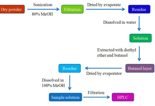
Extraction and determination of ginsenosides were carried out by modifying the method of Kwon et al. (2003). Ultrasound-assisted extraction was performed with an ultrasonic water bath (Branson ultrasonics, USA). The output power is 117 volts and the frequency is 50/60Hz. Sample powder 0.5 g was placed into a 100 mL conical flask, into which 30 mL of 80% (v/v) methanol–water were added. Then the flask was sonicated for 1 h in an ultrasonic water bath. The extract obtained was evaporated using a rotary evaporator under vacuum at 55℃. The evaporated residue (total extract yield) was dissolved in 20 mL of distilled water and washed twice with 20 mL of diethyl ether to remove the fat contents using a separatory funnel. The aqueous layer was extracted four times with 20 mL of water-saturated n-butanol. The butanol solution was washed twice with 30 mL of distilled water to remove the impurities, thereby obtaining crude saponins. The remaining butanolic solution was transferred to the tarred round bottom flask for the evaporation using a rotary evaporator under vacuum at 55℃ (Figure 4).
( )
( )
g
DW
Inoculated
g
DW
Harvested
ratio
Growth
=
Figure 4. Extraction procedures of crude saponin in Panax ginseng.
Determination of ginsenoside content by HPLC
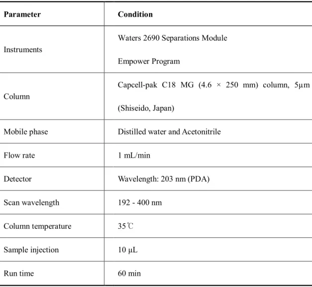
The HPLC conditions for ginsenoside assay was slightly modified the previous report (Park et al. 2007). Quantitative determinations were achieved by HPLC using a Capcell-pak C18 MG (4.6 × 250 mm) column (Shiseido, Japan), Waters 2998 Photodiode Array Detector, Waters 2690 Separations Module and Empower Program (Table 1).
Table 1. HPLC conditions for ginsenosides analysis.
Parameter Condition
Instruments
Waters 2690 Separations Module Empower Program
Column
Capcell-pak C18 MG (4.6 × 250 mm) column, 5µm (Shiseido, Japan)
Mobile phase Distilled water and Acetonitrile
Flow rate 1 mL/min
Detector Wavelength: 203 nm (PDA) Scan wavelength 192 - 400 nm
Column temperature 35℃ Sample injection 10 μL
Run time 60 min
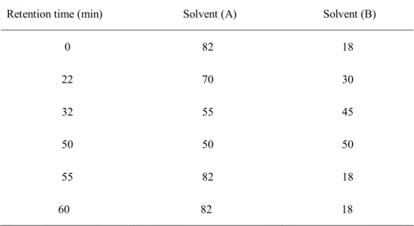
The solvents of HPLC grade were used. The water used in this study was treated with a Milli-Q water purification system (Millipore, USA). The mobile phase of HPLC gradient was conducted as follows (Table 2). Ginsenosides Rg1, Re, Rf, Rh1, Rb1, Rc, Rb2 and Rd standards were purchased from BTGin Co., Ltd (Daejeon, Korea). Digoxin was used as internal standard.
Table 2. Mobile phase of HPLC gradient condition for ginsenosides analysis. Retention time (min) Solvent (A) Solvent (B)
0 22 32 50 55 60 82 70 55 50 82 82 18 30 45 50 18 18 Solvent (A): Distilled water, Solvent (B): Acetonitrile.
Stock solutions for the 4 ginsenosides were prepared separately in 100% MeOH. Digoxin stock solution was prepared in 70% MeOH. Working solutions were prepared in methanol by mixing known amount of all the ginsenosides together. Five concentrations were made for standard curves, each concentration was 60, 120, 240, 320, 480 ppm. Ginsenosides were detected at a wavelength of 203 nm with the peak areas corresponding to ginsenosides from the samples matching retention times as authentic ginsenoside standards.
Analysis of ginsenosides contents was performed according to Son et al. (1999a) and Yu
et al. (2000). The total ginsenoside content was calculated as the sum of individual
The ginsenoside content of ginseng adventitious roots was calculated as:
(GC: ginsenoside content; SGC: sample ginsenoside concentration from HPLC; SV: sample volume; AR: adventitious root)
Method validation and statistical analysis
Stock solutions for the 8 ginsenosides were prepared separately in 100% methanol. Digoxin stock solution was prepared in 80% methanol. Working solutions were prepared in 100% methanol by mixing known amounts of all the compounds. The linear range, limit of detection (LOD) and limit of quantification (LOQ) were studied for the developed method. The linearity of calibration curve was tested by standard analysis. The calibration curves of individual ginsenosides were constructed using a range of five concentrations of the standard, and LOD and LOQ for each analyte were evaluated at signal-to-noise ratios (S/N) of 3:1 and 10:1, respectively.
Statistical analysis was performed according to the SPSS system. Mean and standard errors were used throughout and statistical significance between the mean values was assessed by applying a Duncan’s multiple range tests. A probability of P < 0.05 was considered significant.
) ( ) ( ) )( ( ) ( 1 1 g AR l SV X g mg fromHPLC SGC g mg GC -- =
RESULTS AND DISCUSSION
Effects of gamma irradiation on survival rate of adventitious roots
In this experiment, I first determined the survival rate of adventitious roots at different dosage of γ-irradiation. The survival rates of adventitious roots declined with increasing dosage of irradiation (Figure 5). No survived adventitious roots were observed at 125Gy. The growth of adventitious roots is inhibited over 50Gy treatment and the LD50 was established by 30Gy irradiation of adventitious root based on the survival rates of main root.
Panax ginseng was irradiated by 60CO gamma ray and the growth of hairy roots was
inhibited over 30Gy (Choi et al. 2002). LD50 was established by irradiation of shoot tip explants at 30Gy based on the survival of explants and shoot proliferation (Ali, 2006).
Effects of gamma irradiation on growth rate of cell lines
Five cell lines were selected at last according to the phenotype (Figure 6). After 30 days culture in flask, dry weight of control was 0.31g and the selected cell lines CL1, 3, 9, 13 and 14 were 0.46g, 0.45g, 0.29g, 0.34g and 0.48g respectively. The value of growth ratio of control was 4.42 and the selected cell lines CL1, 3, 9, 13, 14 were 6.57, 6.48, 4.1, 4.9 and 6.9, respectively (Figure 7). The values of growth ratio in cell lines CL1, CL3 and CL14 were increased 49%, 47% and 57%, respectively, compared to the control.
Panax ginseng was irradiated by gamma ray and cell lines were selected according to the
by gamma ray and the cell lines selected in terms of growth rate (similar with control) and appearance of shikonin (Chung et al. 2006). In this experiment, cell line CL1, 3 and 14 showed higher growth ratio.
Effects of gamma irradiation on ginsenoside production
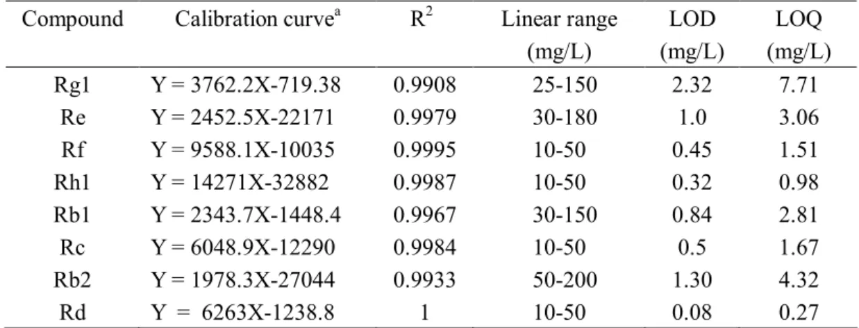
The spectrum of Rg1, Re, Rf, Rh1, Rb1, Rc, Rb2 and Rd were 203nm. The calibration curves and the LOD for the ginsenosides are shown in Table 3 and Figure 8. The correlation coefficients are all better than 0.99, which show good linearity.
The chromatograms for 8 major ginsenosides among each cell line and the internal standard shown in Figure 9, 10, 11, 12 and 13. The contents of the eight ginsenosides were significantly enriched, especially in the cell line CL1 (Figure 14). The total contents of ginsenosides (Rg1, Re, Rf, Rh1, Rb1, Rc, Rb2 and Rd) in cell line CL1 were 1.6-fold higher than that of control. In the cell line CL1, the level of ginsenoside Rg1, Re, Rh1and Rb2 contents were increased by 2-3 times than that of the control. In the cell line CL3, level of ginsenoside Re, Rh1, Rb2, Rc and Rd contents were increased by 1.6-2.3 times than that of the control. In the cell line CL9, level of ginsenoside Rg1, Re and Rh1 contents were increased by 1.7-2 times than that of the control. In the cell line CL14, level of ginsenoside contents were similar with that of the control. In case of the cell line CL13, the ginsenoside contents were affected negatively in ginseng adventitious root cultures. Overall, the highest ginsenoside contents were obtained in cell line CL1.
CONCLUSIONS
In the present study, I investigated the influence of gamma irradiation on the content of ginsenosides of Korean wild ginseng in order to search for better application of ginseng as a health food. As it is known, in root cultures, the ginsenoside yield depends not only on the accumulation of the commercially useful compounds but also on their biomass production capability. In this study, I selected cell line CL1 as the best cell line. Because of the ginsenoside content and the growth ratio of the CL1 were highest compared to control and other cell lines. So, the ginsenoside productivity of CL1 is better than other cell lines. In the next study, I will use these mutant cell lines to establish the best condition for regeneration system of Panax ginseng and this system also use for genetic transformation.
Figure 5. Survival rate among gamma-ray irradiated adventitious roots of Panax ginseng. After irradiation, the adventitious roots were cultured in the petridish for 5 weeks on MS medium with NAA, IAA and 3% sucrose at 23℃ in light condition. Bar shown are means ± standard deviations of three replicates.
Figure 6. Phenotypic characteristics of secondary roots in the gamma-irradiated cell lines of
Panax ginseng (scale bar = 1 cm). Control: non-irradiated adventitious roots, CL1-14: gamma-irradiated mutant cell lines.
Figure 7. Growth ratio of Panax ginseng adventitious root in the control and gamma-irradiated cell lines. The adventitious roots were cultured in the flask for 30 days with 50 mL MS medium. Bar shown are means ± standard errors of three replicates. Control: non-irradiated adventitious roots, CL1-14: selected mutant cell lines.
Table 3. Calibration curve, limit of detection (LOD), and limit of quantification (LOQ) for eight ginsenosides.
a)y is the peak area and x is the concentration of analyte.
Compound Calibration curvea R2 Linear range
(mg/L) LOD (mg/L) LOQ (mg/L) Rg1 Re Rf Rh1 Rb1 Rc Rb2 Rd Y = 3762.2X-719.38 Y = 2452.5X-22171 Y = 9588.1X-10035 Y = 14271X-32882 Y = 2343.7X-1448.4 Y = 6048.9X-12290 Y = 1978.3X-27044 Y = 6263X-1238.8 0.9908 0.9979 0.9995 0.9987 0.9967 0.9984 0.9933 1 25-150 30-180 10-50 10-50 30-150 10-50 50-200 10-50 2.32 1.0 0.45 0.32 0.84 0.5 1.30 0.08 7.71 3.06 1.51 0.98 2.81 1.67 4.32 0.27
Figure 8. Typical chromatograms obtained from the standard solution by UV detection at 203 nm. IS: internal standard (digoxin).
Figure 9. Typical chromatograms and ginsenoside content obtained from the standard, control and cell line CL1. The ginsenosides were extracted from adventitious roots cultured in the flask. Control: non-irradiated adventitious root, CL1: selected cell line 1 from gamma-irradiated adventitious roots, IS: internal standard (digoxin).
Figure 10. Typical chromatograms and ginsenoside content obtained from the standard, control and cell line CL3. The ginsenosides were extracted from adventitious roots cultured in the flask. Control: non-irradiated adventitious root, CL3: selected cell line 3 from gamma-irradiated adventitious roots, IS: internal standard (digoxin).
Figure 11. Typical chromatograms and ginsenoside content obtained from the standard, control and cell line CL9. The ginsenosides were extracted from adventitious roots cultured in the flask. Control: non-irradiated adventitious root, CL9: selected cell line 9 from gamma-irradiated adventitious roots, IS: internal standard (digoxin).
Figure 12. Typical chromatograms and ginsenoside content obtained from the standard, control and cell line CL13. The ginsenosides were extracted from adventitious roots cultured in the flask. Control: non-irradiated adventitious root, CL13: selected cell line 13 from gamma-irradiated adventitious roots, IS: internal standard (digoxin).
Figure 13. Typical chromatograms and ginsenoside content obtained from the standard, control and cell line CL14. The ginsenosides were extracted from adventitious roots cultured in the flask. Control: non-irradiated adventitious root, CL14: selected cell line 14 from gamma-irradiated adventitious roots, IS: internal standard (digoxin).
Figure 14. Total ginsenoside content of the control and mutant cell lines of Panax ginseng adventitious root. Control: non-irradiated adventitious root, CL1-14: selected cell lines from gamma-irradiated adventitious roots.
REFERENCE
Abe I, Rohmer M, Orestwich GD. 1993. Enzymatic cyclization of squalene and oxidosqualene to sterols and triterpene. Chemical Reviews 93: 2189-2206.
Atak M, Özcan S. 2006. Use of gamma rays to induce mutations in Four Pea (Pisum sativum L.) cultivars. Turk Journal of Biology 30: 29-37.
Ali MA. 2006. Optimal dose rate of gamma irradiation and EMS concentration for mutation induction on shoot tip of Banana cv. Grand Nain. The 38th Meeting of the NCHC,
Sudan 228-233.
Asaka I, Ii I, Hirotani M, Asada Y, Furuya T. 1993. Production of ginsenoside saponins by culturing ginseng (Panax ginseng) embryogenic tissues in bioreactors. Biotechnology
Letters 15: 1259-64.
Akao T, Kida H, Kanaoka M, Hattori M, Kobashi K. 1998b. Intestinal bacterial hydrolysis is required for the appearance of compound K in rat plasma after oral administration of ginsenoside Rb1 from Panax ginseng. Journal of Pharmacy and Pharmacology 50: 1155-1160.
Bae TW, Kim J, Song IJ, Song SY, Lim PO, et al. 2009. Production of unbolting lines through gamma-ray irradiation mutagenesis in genetically modified herbicide-tolerant
Zoysia japonica. Breeding Science 59: 103-5.
Bae EA, Choo MK, Park EK, Park SY, Shin HY, Kim DH. 2002a. Metabolism of ginsenoside Rc by human intestinal bacteria and its related antiallergic activity.
Biological & Pharmaceutical Bulletin 25: 743-747.
Basar-Eroglu C, Brand A, Hildebrandt H, Karolina Kedzior K, Mathes B, Schmiedt C. 2007. Working memory related gamma oscillations in schizophrenia patients. International
Journal of Psychophysiology 64: 39-45.
Senthamizh Selvi B, Ponnuswami V, Sumathi T. 2007. Identification of DNA polymorphism induced by gamma ray irradiation in alma Emblica Officinalis Gaertn. grafts of V1 M1 and V2 M1 generation. Journal of Applied Sciences Research, 3(12): 1933-1935. Bae EA, Shin JE, Kim DH. 2005. Metabolism of ginsenoside Re by human intestinal microflora
and its estrogenic effect. Biological & Pharmaceutical Bulletin 28: 1903-8.
Bae EA, Park SY, Kim DH. 2000. Constitutive beta-glucosidases hydrolyzing ginsenoside Rb1 and Rb2 from human intestinal bacteria. Biological & Pharmaceutical Bulletin 23: 1481-1485.
Bae EA, Han MJ, Choo MK, Park SY, Kim DH. 2002b. Metabolism of 20(S) - and 20(R)-ginsenoside Rg3 by human intestinal bacteria and its relation to in vitro biological activities. Biological & Pharmaceutical Bulletin 25: 58-63.
Bae EA, Han MJ, Kim EJ, Kim DH. 2004a. Transformation of ginseng saponins to ginsenoside Rh2 by acids and human intestinal bacteria and biological activities of their transformants. Pharmaceutical Society of Korea 27: 61-67.
Bae KH, Choi YE, Shin CG, Kim YY, Kim YS. 2006. Enhanced ginsenoside productivity by combination of ethephon and methyl jasmonate in ginseng (Panax ginseng C.A.
Briskin DP. 2000. Medicinal plants and phytomedicines. Linking plants biochemistry and physiology to human health. Plant physiology. 124: 507–514.
Chung BY, Lee YB, Baek MH, Kim JH, Wi SG, Kim JS. 2006. Effects of low-dose gamma-irradiation on production of shikonin derivatives in callus cultures of
Lithospermum erythrorhizon S. Radiation Physics and Chemistry 75: 1018-23.
Chang H, But P. ( Eds.). 1986. Pharmacology and applications of Chinese materia medica:
World Scientific Publishing Co., Inc.
Chatterjee C, MJ Corell, PJ Weathers, BE Wyslouzil, and DB Wacers. 1997. Simplified acoustic window mist bioreactor. Biotechnology Techniques 11:155–158.
Choi S. 2002. Epidermis proliferative effect of the Panax ginseng ginsenoside Rb2. Archives
of Pharmacal Research 25: 71-6.
Choi DW, Jung J, Ha YI, Park H-W, In DS, et al. 2005. Analysis of transcripts in methyl jasmonate-treated ginseng hairy roots to identify genes involved in the biosynthesis of ginsenosides and other secondary metabolites. Plant Cell Reports 23: 557-66. Choi KM, Kwon JH, SungHee Ban SH, Yang DC. 2002. Production of Ginsenoside in the
Hairy Roots Irradiated by 60Co γ-ray on Panax ginseng C.A Meyer. Journal of Ginseng Research 26 (4): 219-225.
Choi YE, Kim YS, Paek KY. 2006. Types and designs of bioreactors for hairy root culture. S.
Chan RY, Chen WF, Dong A, Guo D, Wong MS. 2002. Estrogen-like activity of ginsenoside Rg1 derived from Panax notoginseng. Journal of Clinical Endocrinology &
Metabolism. 87: 3691-5.
Chen JC, Chen LD, Tsauer W, Tsai CC, Chen BC, Chen YJ. 2001. Effects of Ginsenoside Rb2 and Rc on inferior human sperm motility in vitro. American Journal of Chinese
Medicine. 29: 155-60.
Charbaji T, Nabulsi I. 1999. Effect of low doses of gamma irradiation on in vitro growth of grapevine. Plant Cell, Tissue and Organ Culture 57: 129-32.
Chang TL, Ding HY, Kao YW. 2008. Role of Ginsenoside Rd in Inhibiting 26S Proteasome Activity. Journal of Agricultural and Food Chemistry 56: 12011-5.
Gyawali R, Seo HY, Lee HJ, Song HP, Kim DH, et al. 2006. Effect of γ-irradiation on volatile compounds of dried Welsh onion (Allium fistulosum L.). Radiation Physics
and Chemistry 75: 322-8.
Hobbs C. 1996. Ginseng: The Energy Herb, Botanica Press, Loveland, Connecticut, USA. Han J, Choi Y. 2009. Rapid induction of Agrobacterium tumefaciens-mediated transgenic
roots directly from adventitious roots in Panax ginseng. Plant Cell, Tissue and
Organ Culture 96: 143-9.
Hady MS Abdel, Ali ZA. 2006. Effect of gamma irradiation on wheat immature culture regenerated plants. Journal of Applied Sciences Research 2(6): 310-316.
synthesized 2-hydroxyethyl jasmonate. Process Biochemistry 43: 113-8.
Harrison RG. 1990. Hybrids and hybrid zones: historical perspective. Published by Oxford University Press. Inc.
Ingram B, Mavituna E. 2000. Effect of bioreactor configuration on the growth and maturation of Picea sitchensis somatic embryo cultures. Plant Cell, Tissue and
Organ Culture 61: 87–96.
Jeong C, Hyun J, Kim Y, Lee ES. 2003. Ginsenoside RB1 the anti-ulcer constituent from the head of Panax ginseng. Archives of Pharmacal Research 26: 906-11.
Jung JD, Park HW, Hahn Y, Hur CG, In DS, et al. 2003. Discovery of genes for ginsenoside biosynthesis by analysis of ginseng expressed sequence tags. Plant Cell Reports 22: 224-30. Joseph R, Yeoh HH, Loh CS. 2004. Induced mutations in cassava using somatic embryos and the identification of mutant plants with altered starch yield and composition.
Plant Cell Reports. 23: 91–98.
Kim O, Bang K, Kim Y, Hyun D, Kim M, Cha S. 2009. Upregulation of ginsenoside and gene expression related to triterpene biosynthesis in ginseng hairy root cultures elicited by methyl jasmonate. Plant Cell, Tissue and Organ Culture 98: 25-33. Kim Y, Hahn E, Murthy H, Paek K. 2005. Adventitious root growth and ginsenoside
accumulation in Panax ginseng cultures as affected by methyl jasmonate.
Biotechnology Letters 26: 1619-22.
Kwon JH, Bélanger JMR, Paré JRJ, Yaylayan VA. 2003. Application of the microwave-assisted process (MAP(TM)) to the fast extraction of ginseng saponins.
Food Research International 36: 491-8.
Kim O, Bang K, Kim Y, Hyun D, Kim M, Cha S. 2009. Upregulation of ginsenoside and gene expression related to triterpene biosynthesis in ginseng hairy root cultures elicited by methyl jasmonate. Plant Cell, Tissue and Organ Culture 98: 25-33. Kiong A, Lai A, Hussein S, Harun A. 2008. Physiological responses of Orthosiphon
stamineus plantles to gamma irradiation. American-Eurasian Journal of Sustainable
Agriculture 2: 135-49.
Kang KS, Kang BC, Lee BJ, Che JH, Li GX, et al. 2000. Preventive effect of epicatechin and ginsenoside Rb2 on the inhibition of gap junctional intercellular communication by TPA and H2O2. Cancer Letters 152: 97-106.
Ketchum REB, Gibson DM, Croteau RB, Shuler ML. 1999. The kinetics of taxoid accumulation in cell suspension cultures of Taxus following elicitation with methyl jasmonate. Biotechnology & Bioengineering 62: 97-105.
Lee MH, Jeong JH, Seo JW, Shin CG, Kim YS, et al. 2004. Enhanced triterpene and phytosterol biosynthesis in Panax ginseng overexpressing squalene synthase gene.
Plant Cell Physiology 45: 976-84.
Lee Y, Jin Y, Lim W, Ji S, Cho J, et al. 2003. Ginsenoside Rc and Re stimulate c-Fos expression in MCF-7 human breast carcinoma cells. Archives of Pharmacal
Research 26: 53-7.
Journal of Steroid Biochemistry and Molecular Biology 84: 463-8.
Ludwig-Müller J, Georgiev M, Bley T. 2008. Metabolite and hormonal status of hairy root cultures of Devil's claw (Harpagophytum procumbens) in flasks and in a bubble column bioreactor. Process Biochemistry 43: 15-23.
Lu MB, Wong HL, Teng WL. 2001. Effects of elicitation on the production of saponin in cell culture of Panax ginseng. Plant Cell Reports 20: 647–677.
Mabberley. DJ. 1987. The Plant-Book. Cambridge Univ. Press, Cambridge.
Mallol A, Cusidó RM, Palazón J, Bonfill M, Morales C, Piñol MT. 2001. Ginsenoside production in different phenotypes of Panax ginseng transformed roots.
Phytochemistry 57: 365-71.
Mokobia C, Okpakorese E-M, Analogbei C, Agbonwanegbe J. 2006. Effect of gamma irradiation on the grain yield of Nigerian Zea mays and Arachis hypogaea. Journal of
Radiological Protection 26: 423–427.
Ngan F, Shaw P, But P, Wang J. 1999. Molecular authentication of Panax species.
Phytochemistry 50: 787-91.
Nah SY, Kim DH, Rhim H. 2007. Ginsenosides: are any of them candidates for drugs Acting on the central nervous system? CNS Drug Reviews 13: 381-404.
Nam KY. 2002. Clinical applications and efficacy of Korean ginseng (Panax ginseng C.A. Meyer). Journal of Ginseng Research 26:111-131.
Nakata H, Kikuchi Y, Tode T, Hirata J, Kita T, et al. 1998. Inhibitory effects of ginsenoside Rh2 on tumor growth in nude mice bearing human ovarian cancer cells. Cancer