The IVC filter offers a safe and effective means for preventing pulmonary emboli and it further reduces the complications of DVT comparing with the classic tech- nique such as caval interruption (1). Even with the goal of preventing thrombus propagation, the thrombosis of the filter insertion site, DVT and vena caval occlusion have been the potential complications of the filters (1).
All the currently available filters have been considered to have similar caval thrombosis rates, but they have shown a wide range, from 1%-24%, in the reported lit- erature (2, 3). Treatments for caval thrombosis include early infusion of local thrombolytic agent, early thrombectomy and long-term anticoagulation, but if the
patients have no clinical symptoms or persisting con- traindication to anticoagulation, they can be followed up without any specific treatment. The recent advances in interventional radiology have given us another solution, i.e., aspiration thrombectomy with protection from propagation of thrombus via the insertion of another temporal filter. We report here on a case of a massively thrombosed filter-bearing IVC with pulmonary em- bolism, and this was successfully treated with another filter placement and aspiration thrombectomy.
Case Report
A 77-year-old man came to our hospital and presented with dyspnea. His medical history included cerebral in- farction, congestive heart failure and atrial fibrillation.
Three years prior to admission, he developed a swelling of the lower extremity. Doppler sonography and com- puted tomography (CT) confirmed the presence of DVT in the lower extremity (Fig. 1A, B).
1Department of Diagnostic Radiology, Keimyung University School of Medicine
2Department of Surgery, Keimyung University School of Medicine Received September 24, 2005 ; Accepted January 6, 2006
Address reprint requests to : Jin Soo Choi, M.D., Department of Diagnostic Radiology, Dongsan Medical Center, Keimyung University College of Medicine, 194, Dongsan-dong, Junggu, Daegu 700-712, Korea Tel. 82-53-250-7767 Fax. 82-53-250-7766 E-mail: [email protected]
of an inferior vena cava (IVC) filter in conjunction with anticoagulant therapy has been used to prevent pulmonary embolisms. However, for the patients who anticoagulant is contraindicated or if this is complicated, the use of an IVC filter without concurrent anticoagulation may become the sole treatment for pulmonary embolisms. In this situ- ation, the thrombi trapped in the IVC filter may cause significant clinical problems.
We report here on a case of IVC filter thrombosis that was successfully treated by aspi- ration thrombectomy after placing another filter proximal to the previous filter.
Index words :Venae cavae, filters Veins, thrombosis
Interventional procedures Thrombectomy
A
C B
Fig. 1. The contrast-enhanced CT scans in the lower abdomen show thrombotic filling defects (arrows) in the iliofemoral vein (A, B). A Greenfield IVC filter was deployed at the infrarenal level via a right internal jugular approach (C).
A B
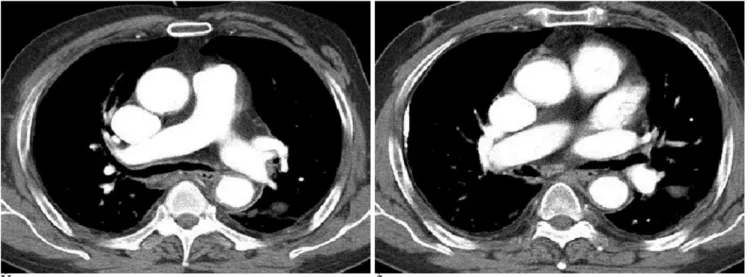
Fig. 2. The contrast-enhanced CT scans show thrombi in the main pulmonary artery and the interlobar artery (arrows).
tech, Watertown, MA, U.S.A.) and a 3-4 mm skin inci- sion is made along the guide wire. The long 12 Fr sheath was introduced under fluoroscopic guidance over a standard 0.035-inch guide wire to the intended implan- tation site in the IVC. After removing the guide wire, the
in the IVC (Fig. 1C). He was maintained on anticoagula- tion following filter placement. The dose of anticoagu- lant (Warfarin sodium, Daewha, Seoul) was 3.75 mg/day for 6 months.
After three months of anticoagulation therapy, he de-
A B C
Fig. 3. The findings of digital subtraction venography. It demonstrated an extensive thrombosis of the Greenfield filter-bearing IVC (arrows) with a cephalic extension to the level of the renal vein orifice (A). Before aspiration thrombectomy, another IVC filter (ar- row) was inserted at a suprarenal position via a right jugular approach to prevent pulmonary embolism (B). The thrombus was cleared after aspiration thrombectomy with using an 8 Fr Desilet-Hoffman sheath (C).
veloped nausea; we stopped the medication and the symptoms disappeared. However, he presented with dyspnea two months before the next scheduled visit to the hospital. Multiple thrombi were demonstrated in the main pulmonary artery and in both interlobar arteries on the contrast enhanced CT scan of the chest (Fig. 2A, B).
A venacavogram performed though the right femoral vein with a 5 Fr pigtail catheter placed below the filter revealed that a massive thrombus was captured in the filter with the cranial extension of the thrombus beyond the level of the filter (Fig. 3A). We planned an aspiration thrombectomy procedure with a Desilet-Hoffman sheath. However, the possibility of distal embolization during aspiration thrombectomy was one of our major concerns, as the shower of emboli might cause a fatal pulmonary embolus during the procedure. A temporary IVC filter was planed to prevent a thrombectomy-relat- ed pulmonary embolism, but the device was not avail- able and so we deployed an additional permanent IVC filter. By using a standard percutaneous procedure, the 6 Fr sheath was introduced under fluoroscopy though the right jugular vein over a standard 0.035-inch guide wire to the intended implantation site in the IVC. After removing the guide wire, the filter was introduced into the sheath and then advanced to the tip of the sheath by means of the pusher. A Trap Ease filter (Cordis, Miami, FL, U.S.A.) was successfully deployed at the suprarenal IVC (Fig. 3B). Aspiration thrombectomy was performed with an 8 Fr Desilet-Hoffman sheath (COOK, Bloomington, IN, U.S.A.) via the right common femoral vein. Successful removal of the thrombus in the original IVC filter was achieved along with the cranial extension
of the thrombus beyond the level of the filter. The ex- tracted specimen showed a red thrombus and the amount of thrombus was 10 gm. A venacavogram per- formed after the procedure showed restoration of IVC flow without residual thrombus (Fig. 3C).
A CT scan obtained 6 months after anticoagulation therapy revealed the complete resolution of thrombus in the pulmonary artery (Fig. 4A, B).
Discussion
Since the late 1960s, IVC filters have been available for the prevention of pulmonary embolism in patients with DVT (4). A recent clinical trial has documented the effec- tiveness of IVC filters for reducing the initial sympto- matic or asymptomatic pulmonary embolisms that are without major complications (5). However, repeated trapping of emboli by the IVC filter might cause throm- bosis around the filter and the IVC in about 5% of the pa- tients with implanted Greenfield filters (5, 6). The inci- dence of IVC thrombosis, though it is uncommon after IVC filter insertion, will increase up to 15.3% if concur- rent anticoagulation is not administered (6). The develop- ment of IVC filter thrombosis can markedly increase the incidence of pulmonary embolism to as high as 33% (7).
There are many treatment options available for IVC and IVC filter thrombosis, including systemic anticoagu- lation, systemic thrombolytic therapy, surgical thrombectomy, catheter directed thrombolysis and en- dovascular mechanical thrombectomy (8). Systemic an- ticoagulation and thrombolysis are contraindicated for the patients with recent intracranial hemorrhage or gas- trointestinal bleeding. Moreover, it is found that an ilio-
A B
Fig. 4. The CT scans of the chest show the complete resolution of thrombus in the pulmonary artery.
was performed. In case of contraindications to throm- bolytic therapy, MT was performed without any accom- panying pharmacologic thrombolysis for the primary treatment of an acutely thrombosed IVC. There is a po- tential risk that the MT device may catch on to one of the struts of the IVC filter during catheter manipulation.
This could create difficulties in withdrawing the devices or it could result in filter migration. There is a case re- port on the entanglement of the steel injection channel of a Hydrolyser catheter on a strut of a Gianturco Z-stent that was placed in the SVC, which could only be dissoci- ated with great technical difficulty (8).
Catheter-directed thrombolysis has been shown to be effective and safe for the management of IVC thrombo- sis (10). However, we are still concerned about the po- tential systemic effect of a regionally infused throm- bolytic agent, which may induce catastrophic intracra- nial hemorrhage or gastrointestinal bleeding.
Mechanical thrombectomy devices weren’t available in our country at that time and they are also very expen- sive, and for these reason our first line of treatment is generally aspiration thrombectomy. We didn’t use a thrombolytic agent.
In this case, we performed aspiration thrombectomy with a Desilet-Hoffman sheath. However, the possibility of distal embolization during aspiration thrombectomy was one of our major concerns, as the shower of emboli
References
1. Joels CS, Sing RF, Heniford BT. Complications of inferior vena ca- va filters. Am J Surg 2003;69:654-659
2. Millward SF, Peterson RA, Moher D, Lewandowski BJ, Burbridge BE, Aquino J, et al. LGM (Vena Tech) vena caval filter: experience at a single institution. J Vasc Interv Radiol 1994;5:351-356
3. Greenfield LJ, Proctor MC. Cho KJ, Cutler BS, Ferris EJ, McFarland D, et al. Extended evaluation of the titanium Greenfield vena cava filter. J Vasc Surg 1994;20:458-464
4. Greenfield LJ, Michna BA. Twelve-year clinical experience with the Greenfield vena cava filter. Surgery 1988;104:706-712
5. Decousus H, Leizorovicz A, Parent F, Page Y, Tardy B, Girard P, et al. A clinical trial of vena caval filters in the prevention of pul- monary embolism in patients with proximal deep-vein thrombo- sis. N Engl J Med 1998;338:409-415
6. Becker DM, Philbrick JT, Selby JB. Inferior vena cava filters: indi- cations, safety, and effectiveness. Arch Intern Med 1992;152:1985- 1994
7. Tardy B, Mismetti P, Page Y, Decousus H, Da Casta A, Zeni F, et al. Symptomatic inferior vena cava filter thrombosis: clinical study of 30 consecutive cases. Eur Respir J 1996;9:2012-2016
8. Poon WL, Luk SH, Yam KY, Lee AC. Mechanical thrombectomy in inferior vena cava thrombosis after caval filter placement: a re- port of three cases. Cardiovasc Intervent Radiol 2002;25:440-443 9. Vedantham S, Vesely TM, Parti N, Darcy MD, Pilgram TK, Sicard
GA, et al. Endovascular recanalization of the thrombosed filter- bearing inferior vena cava. J Vasc Interv Radiol 2003;14:893-903 10. Angle JF, Matsumoto AH, AI Shammari MA, Hagspiel KD,
Spinosa DJ, Humphries JE. Transcatheter regional urokinase ther- apy in the management of inferior vena cava thrombosis. J Vasc Interv Radiol 1998;9:917-925
대한영상의학회지 2006;55:123-128
다량의 혈전을 형성한 아래대정맥필터:
아래대정맥필터 추가 삽입과 흡인 혈전제거술을 이용한 치료11계명대학교 의과대학 진단방사선과학교실
2계명대학교 의과대학 외과학교실
최진수・김영환・조원현2・김형태2・구자현2・우성구
다량의 깊은 정맥혈전증(DVT)과 동반된 폐색전증의 치료에 아래대정맥필터 삽입과 항응고제를 사용해왔다. 그 러나 아래대정맥필터를 삽입한 환자에서 항응고제의 사용이 금기가 되거나 합병증이 발생하였으면, 아래대정맥필 터만으로 폐색전증의 치료가 이루어지며 이러한 경우 아래대정맥필터에 포획된 혈전이 심각한 임상적 문제를 야기 할 수 있다. 저자들은 아래대정맥필터 삽입 후 자발적으로 항응고제를 투여하지 않은 환자에서 아래대정맥필터에 다량의 혈전과 폐색전증이 발생하였고 아래대정맥필터 추가 삽입과 흡인 혈전제거술을 시행하여 성공적으로 혈전 제거를 하였기에 이를 보고하고자 한다.