INTRODUCTION
The primary purpose of root canal filling is to establish a hermetic seal of the root canal system and to entomb any residual micro-organisms. The apical seal is critical for preventing periapical tissue- derived fluid from nourishing the remaining micro- organisms and for protecting the root canal from bac-
terial contamination. Failure to establish a hermetic seal in endodontically treated teeth and the presence of bacteria at the apical portion of the filled root canal have been cited as the major causes of post- endodontic problems.1,2 Therefore, root canal filling materials must establish a fluid-tight barrier that prevents apical leakage. Although gutta-percha is the most commonly used canal filling material, it
The effects of total-etch, wet-bonding, and light-curing of adhesive on the apical seal of a resin-based root canal filling system
Won-Il Ryu1, Won-Jun Shon2, Seung-Ho Baek2, In-Han Lee2, Byeong-Hoon Cho2*
1Department of Dentistry, Seoul National University School of Dentistry
2Department of Conservative Dentistry, Seoul National University School of Dentistry and Dental Research Institute, Seoul, Korea
Objectives: This study evaluated the effects of adhesion variables such as the priming concepts of canal wall and the curing modes of adhesives on the sealing ability of a resin-based root canal filling system.
Materials and Methods:Apical microleakage of the Resilon-RealSeal systems filled with 3 different combina- tions of adhesion variables was compared with the conventional gutta-percha filling using a dye penetra- tion method. Experimental groups were SEDC, Resilon (Resilon Research LLC) filling with self-etch RealSeal (SybronEndo) primer and dual-cure RealSeal sealer; NELC, Resilon filling with no etching, Scotchbond Multi-Purpose (3M ESPE) primer application and light-curing adhesive; and TELC, Resilon filling with Scotchbond Multi-Purpose primer and adhesive used under total etch / wet bonding and light- cure protocols. GPCS, gutta-percha filling with conventional AH26 plus sealer, was the control group.
Results:The median longitudinal dye penetration length of TELC was significantly shorter than those of GPCS and SEDC (Kruskal-Wallis test, p < 0.05). In the cross-sectional microleakage scores, TELC showed significant differences from other groups at 2 to 5 mm from the apical foramen (Kruskal-Wallis test, p < 0.05).
Conclusions:When a resin-based root canal filling material was used, compared to the self-etching primer and the dual-cure sealer, the total etch/wet-bonding with primer and light-curing of adhesive showed improved apical sealing and was highly recommended. [J Kor Acad Cons Dent 2011;36(5):385-396.]
Key words:Adhesion variables; Light-curing; Microleakage; Resin-based root canal filling system;
Self-etching adhesives; Total-etching adhesives
-Received 23 June 2011; revised 31 July 2011; accepted 31 July 2011- ABSTRACT
1Ryu WI, BS, MS Student, Department of Dentistry, Seoul National University School of Dentistry,
2Shon WJ, DDS, PhD, Assistant Professor; Baek SH, DDS, PhD, Associate Professor; Lee IH, DMD, Assistant Clinical Professor; Cho BH, DDS, PhD, Professor, Department of Conservative Dentistry, Seoul National University School of Dentistry and Dental Research Institute, Seoul, Korea
*Correspondence to Byeong-Hoon Cho, DDS, PhD.
Professor, Department of Conservative Dentistry, Seoul National University School of Dentistry and Dental Research Institute, 28-2 Yeongeon- dong, Jongro-gu, Seoul, Korea 110-749
TEL, +82-2-2072-3514; FAX, +82-2-764-3514; E-mail, [email protected]
※This work was supported by the Korea Research Foundation (KRF) grant funded by the Korean government (MEST, No. 2009-0070771)
fails to prevent leakage effectively, and there is still no obturation material and method that provides a perfect gap-free seal.3,4
Resilon (Resilon Research LLC, Madison, CT, USA) is a thermoplastic polyester-based root canal- filling material used with self-etching Epiphany primer and dual curable Epiphany resin composite sealer (Pentron Clinical Technologies Ltd, Wallingford, CT, USA).5,6The Resilon-Epiphany sys- tem introduced a new adhesion concept to endodon- tics and offered improved sealing ability.5 Since the adhesion of the resin-based sealer to both Resilon filling material and root dentin creates a ‘mono- block’obturation, it was suggested that this adhe- sion provides better sealing ability and greater resis- tance to microbial leakage than conventional meth- ods, thereby improving the chances of root canal treatment success.5,7 Moreover, Resilon has user- friendly characteristics, such as handling properties similar to those of gutta-percha. It can be softened with heat or dissolved with solvents such as chloro- form for retreatment.5 Like Epiphany, RealSeal (SybronEndo, Glendora, CA, USA) is a resin-based dual-curable adhesive sealer system that is also used with Resilon and consists of a self-etching RealSeal primer and a dual-curable resin-based RealSeal seal- er. Like the adhesion procedures of composite resin restorations, this system adopts a ‘self-etching con- cept’for priming root canal dentin and a ‘dual-cure reaction’for the polymerization of the root canal adhesive sealer.
Adhesion efficiency can vary according to adhesion variables such as priming the root canal wall and adhesive curing. Priming of the root canal dentin surface can be accomplished by simultaneous self- etching/self-priming or traditional total-etching/wet- bonding protocols. Curing the adhesive, which corre- sponds to the RealSeal sealer in this system, can be performed with either dual-cure (usually starting with chemical-cure) or light-cure modes. For self- etching, Epiphany or RealSeal primers have acidic monomers, which hinder the self-cure portion of the polymerization reaction of the dual-cure root canal sealer.8Dentists have already experienced these con- ditions during core build-up procedures with self- etching adhesives and self-cure composite resin core
materials.9 The degree of conversion (DC) at the interface between the self-curing resin core material and the acidic self-etch adhesive was low, because the acidity of the oxygen-inhibited layer on the adhe- sive hindered the chemical cure of the core material at the interface.10,11 For adequate polymerization of the root canal sealer in the deepest apical part of the canal, dual curing of the sealer is required because of the limited penetration depth of the curing light through the opaque dentin and the Resilon filling material. However, the presence of a high percentage of hydrophilic acidic monomers and residual water in self-etching primers including the RealSeal primer can lead to sub-optimal DC, which results in poor mechanical properties and high permeability of the adhesive joint.12-15 Although the acidic monomers of self-etching adhesives need to be dissociated by water for curing, the dissociated acidic moieties hin- der the activity of the amine accelerators of the adhesive.13,16 Therefore, in order to obtain a more fluid-tight apical seal, it can be suggested that after the etchant is thoroughly washed, primer and/or adhesive with a neutral pH be used and light-cured for an extended period of time.
The objective of this study was to evaluate the effects of adhesion variables on the sealing ability of a resin-based root canal filling system. The null hypothesis assumed in the present study was that there would be no difference in the apical microleak- age between various combinations of the priming concepts of canal wall and the curing modes of adhe- sives used with a resin-based root canal filling sys- tem. For this purpose, the apical microleakages of the Resilon-RealSeal systems filled with 3 different combinations of adhesion variables were compared with the conventional gutta-percha filling as a con- trol, using a dye penetration method. The results were evaluated under a stereomicroscope and a scan- ning electron microscope (SEM, S-4700, HITACHI High Technologies Co., Tokyo, Japan).
MATERIALS AND METHODS Specimen preparation
A total of 40 straight and single-rooted (single
canal) human premolars were used for the experi- ment. After extraction, the teeth were stored in a 0.5% aqueous solution of chloramine T for 2 hours to prevent surface infection. After removing soft tissue, they were stored in isotonic saline solution until used. At 10 mm from the root apex, all the teeth were cut perpendicularly to the long axis of the tooth with a high-speed diamond bur. Canal preparation was done with a conventional step-back technique with K-files (25 mm, Mani, Inc., Tochigiken, Japan).
The working length was determined with bare eyes.
Size 10 K-file was inserted into the root canal until the file tip was just beyond the apical foramen, and the entire length of the root canal was measured.
The working length was established 0.5 mm short of the apical foramen.
Each canal was enlarged to a final apical size of 30 (ISO, International Organization for Standardization, 02 taper) to the working length and additionally to size 45 in a step-back mode by 1 mm with each increase in file size. After each filing, apical patency was confirmed with a size 10 K-file and the canal was irrigated with 1 mL of 5.25% sodium hypochlo- rite (NaOCl) solution.
After completing instrumentation, the canal was rinsed with 5 mL of 5.25% NaOCl followed by 5 mL of 17% ethylenediamine tetraacetic acid (EDTA) to remove dentin debris and smear layer and to elimi-
nate the peroxide which might have a retarding effect on the setting of the resin-based adhesives. The canals were then thoroughly flushed with 10 mL of distilled water to avoid the prolonged effects of NaOCl and EDTA solution. The root canals were dried with sterile paper points before obturation. The samples were always kept in wet condition, except the root canal treatment procedures.
Obturation procedures
The prepared roots were randomly assigned into one control group and three experimental groups con- taining 10 roots (10 canals) each as shown in Table 1.
Group 1. GPCS, gutta-percha and conventional sealer, gutta-percha filling with AH26 plus sealer
An ISO size 30, 02 taper, gutta-percha master cone was inserted to the working length and its adapta- tion was confirmed with tug-back sensation. The master cone was coated with AH26 plus sealer (Dentsply International Inc., York, PA, USA) and placed into the canal. Lateral condensation of the accessory gutta-percha cones were performed using a finger spreader until they could no longer be placed more than 3 mm into the canal. Excess cones were removed at the level of 6 mm from the apex with a warmed instrument (Duo-Alpha, B&L Biotech Co.
Table 1.Experimental groups and their assigned adhesion variables in root canal obturation protocols
Experimental groups Obturation protocols
Priming Sealer/Adhesive Filling material
Group 1, GPCSa - AH26 plus sealer (no cure) Gutta percha
Group 2, SEDC RealSeal primer (self-etch) RealSeal sealer as adhesive (dual-cure) Resilon Group 3, NELC SBMP primer (no etch)b SBMP adhesive as sealer (light-cure) Resilon Group 4, TELC phosphoric acid (total etch) SBMP adhesive as sealer (light-cure) Resilon
+ SBMP primer
GPCS, gutta-percha and conventional sealer; SEDC, self-etch with RealSeal primer and dual-cure of RealSeal sealer;
NELC, SBMP primer application with no etch of the root canal wall, complete dry, and light-cure of the SBMP adhe- sive; TELC, total-etch of the root canal wall with phosphoric acid, wet bonding with the SBMP primer, complete dry, and light-cure of the SBMP adhesive; SBMP, Scotchbond Multi-Purpose (3M ESPE, St. Paul, MN, USA).
a. Group 1 was used as the control group. b. For group 3, the SBMP primer was applied without additional etching after final rinsing with NaOCl, EDTA and distilled water, sequentially.
Ltd., Seoul, Korea) and the coronal 1 mm of the canal space was additionally filled with gutta-percha using a vertical condensation technique. The remain- ing root canal space was filled with Caviton (GC Corparation, Tokyo, Japan). The specimens were left under 100% relative humidity in a dark black jar containing distilled water for 24 hours at room tem- perature to allow the AH26 plus sealer to set.
Group 2. SEDC, self-etch and dual-cure, Resilon filling with RealSeal primer/RealSeal sealer
A size 30, 06 taper, Resilon master cone was inserted to the working length and its adaptation was confirmed with tug-back sensation. Self-etching RealSeal primer was applied onto the entire root canal wall using a microbrush. Excess primer was removed with paper points. The canal was dried with a gentle air stream from the coronal aspect. The Resilon master cone was coated with dual-curable RealSeal sealer and placed into the root canal.
Lateral condensation of the accessory Resilon cones was performed using a finger spreader until they could no longer be placed more than 3 mm into the canal. The specimen was light-cured with an LED light-curing unit (Free-Light2, 3M ESPE, St. Paul, MN, USA; Light intensity, 900 mW/cm2) for 30 sec- onds from coronal aspect. By gripping the specimen with a wet tissue, the dual-cure sealer at the apical area was prevented from curing additionally by later- ally-scattered light. Excess cones were removed at the level of 6 mm from the apex with a warmed instrument (Duo-Alpha) and the coronal 1 mm of the remaining root canal space was additionally filled with Resilon cones using a vertical condensation technique. The specimen was light-cured with the same light-curing unit for 30 seconds from the coro- nal aspect. The remaining root canal space was filled with Caviton. The specimens were left on the bench for appproximately 30 minutes to allow the RealSeal sealer to set in a self-cure mode5 and stored under 100% relative humidity in a dark black jar contain- ing distilled water for 24 hours at room temperature.
Group 3. NELC, no etch and light-cure, Resilon filling with SBMP primer/SBMP adhesive
Instead of using the RealSeal self-etching primer
and the dual-curable RealSeal sealer, Scotchbond Multi-Purpose (SBMP, 3M ESPE, St. Paul, MN, USA) primer and adhesive were used as the primer and as the sealer, respectively, in order to improve the integrity of the adhesive layer by increasing its DC with light-curing. However, except the removal of the smear layer by EDTA rinsing during canal irriga- tion, additional etching was not performed to omit surface priming, that is, we did not make any effort to create a relevant hybrid layer. The SBMP primer was applied twice onto the entire root canal wall using a microbrush. Excess primer was removed with paper points, and the canal was dried with a gentle air stream from the coronal aspect. The SBMP adhe- sive was then applied into the canal using a micro- brush. Excess adhesive was removed with paper points. The adhesive coated on the canal wall was cured for 20 seconds with the light passing from the coronal aspect through the empty canal using the same light-curing unit. By gripping the specimen firmly with a wet tissue, the adhesive at the apical area was prevented from curing additionally by later- ally-scattered light. After sufficient light-curing of SBMP adhesive, Resilon master cone was coated with the adhesive and placed into the root canal.
Lateral condensation of accessory Resilon cones, removal of excess cones, vertical condensation, light- curing, occlusal sealing, and storage procedures were done in the same manner as in group 2.
Group 4. TELC, total-etch and light-cure, Resilon filling with SBMP primer/SBMP adhesive using total etch/wet bonding and light-curing protocols
In this group, in order to maximize the effects of the priming root canal dentin and polymerization of the adhesive, the adhesion procedures were per- formed just like in composite resin restorations. To optimize priming, the root canal dentin surface was etched with 37% phosphoric acid (Phosphoric Acid Etchant Syringe, 3M ESPE) for 15 seconds and thor- oughly rinsed with distilled water. Excess water in the canal was roughly absorbed with paper points.
The hydrophilic and neutral SBMP primer was applied onto the wet decalcified dentin surface and dried completely. The SBMP adhesive was applied into the canal using a microbrush, and excess adhe-
sive was removed with paper points, so that only a thin coating of the adhesive was permitted to remain on the etched and primed dentinal wall within the root canal. With gripping the specimen as in group 2, the adhesive was then light-cured for 20 seconds with the light passing from the coronal aspect through the empty canal using the same light-curing unit. After sufficient light-curing of SBMP adhesive, Resilon master cone was coated with the adhesive and placed into the root canal. Lateral condensation of the accessory Resilon cones, and the rest of the canal filling procedures were done in the same man- ner as in group 2.
Evaluation of apical microleakage
Nail varnish was applied twice to all external sur- faces of the specimen except to the region 1 mm around the apical foramen. Specimens were immersed in 1% methylene blue dye solution for 12 hours at room temperature. Dye penetrated speci- mens were washed and mounted in an acrylic resin block with self-curing acrylic resin. Horizontal sec- tions perpendicular to the long axis of the root were obtained every 1 mm from the apex up to 6 mm along the root with a low-speed diamond saw (Isomet, Buehler Ltd, Lake Bluff, IL, USA). Cross- sectioned surfaces were inspected under ×40 magni- fication with a stereomicroscope (SZ40, Olympus Corp., Tokyo, Japan) to evaluate the penetration length and the mean number of segments stained with dye. In order to compare the microleakage quantitatively, the first set of data was obtained by reading the dye penetration lengths longitudinally at one millimeter interval. The second set of data was obtained cross-sectionally. For each section at one millimeter interval, the cross-section of the root canal filling was divided into 5 segments and the number of segments stained was counted. By drawing an imaginary circle at the center corresponding to the canal filling material and four lines radiating from the circle to the nearest line angle, 5 separate seg- ments were obtained from each cross-section (Figure 1). The stain at the interface between the dentinal wall and the canal filling material was regarded as belonging to the outer arc segment. If any trace of
dye was present in a segment, the segment was counted as a dye-penetrated one. Therefore, the microleakage score of each section was graded from 0 to 5. For each group, the cross-sectional microleak- age score was obtained at each level of the sections by dividing the total number of dye-penetrated seg- ments at the level by the sample number (n = 10, Table 2).
Scanning electron microscopic observation
The interfaces between the root canal filling mate- rials and the root canal dentin were also evaluated using a field-emission SEM operating at 15 kV. The sections randomly selected from the sections at 3 mm from the apex in each group were replicated with epoxy resin after treatment of the sectioned surface with 6N HCl for 3 seconds and 3.5% NaOCl for 5 minutes.
Statistical analysis
Statistical analysis of the data for longitudinal dye penetration lengths that were read at one millimeter intervals was performed with non-parametric Kruskal-Wallis test. The statistical differences in the cross-sectional microleakage scores, which were repeatedly measured along the distances from the apical foramen for the four groups, were evaluated Figure 1.Schematic illustration of the segments for scoring microleakage from each section.
Root surface Root dentin Root Canal Wall
Canal obturation (grey) divided into 5 regions
Two imaginary diagonal lines radiating from the circle at the center to the nearest line angle of the root
with Friedman 2-way Analysis of Variance (ANOVA) and Kruskal-Wallis tests. All statistical analyses were performed at a 5% level of significance.
RESULTS
Under a stereomicroscope, dye penetration was observed mostly at the interface between the root canal dentinal wall and the filling material. Filling materials such as gutta-percha and Resilon cones did not fill the entire canal space, but the remaining por- tion was filled with corresponding sealers.
Characteristic differences were not found in the leak- age patterns among sealers. However, in severe cases with dense bands of dye staining, the stain radiated outwards from the interface along the denti- nal tubules or spread inwards sparsely into the seal- er occupying the remaining canal space between the cones and the canal wall. In Group 4 (TELC), sec- tions having no stained segments were observed from 2 mm from the apical foramen.
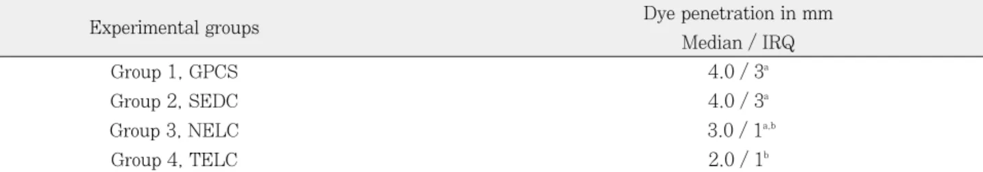
The median values of the longitudinal dye penetra- tion lengths read from each section at one millimeter intervals are summarized in Table 2. The median
longitudinal dye penetration length of Group 4 (TELC) was significantly shorter than Groups 1 (GPCS) and 2 (SEDC) (Kruskal-Wallis test, p <
0.05). The longitudinal dye penetration length in Group 2, where the self-etching RealSeal primer and dual-curing RealSeal sealer were used according to the instructions of the Resilon-RealSeal system, was statistically not different from the values of Group 1, the conventional gutta-percha filling with AH26 plus sealer. Group 3 was not different from any other groups.
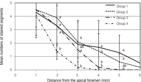
In Figure 2, The cross-sectional microleakage scores decreased significantly along the distance from the apical foramen (Friedman test, p < 0.05) and there was a significant interaction between the groups and the distances from the apical foramen (Friedman test, p = 0.042). The cross-sectional microleakage scores at each level of the sections also showed significant differences between groups. Group 4 (TELC) showed significant differences in the cross- sectional microleakage scores from all the other groups at 2 mm, from Groups 2 and 3 at 3 mm, and from Groups 1 and 2 at 4 and 5 mm from the apical foramen (Kruskal-Wallis test, p < 0.05).
Table 2.Longitudinal dye penetration length along the interface between the canal filling materials and the root canal dentin wall, which was read from each section obtained at one millimeter intervals
Experimental groups Dye penetration in mm
Median / IRQ
Group 1, GPCS 4.0 / 3a
Group 2, SEDC 4.0 / 3a
Group 3, NELC 3.0 / 1a,b
Group 4, TELC 2.0 / 1b
Statistical analysis was done with non-parametric Kruskal-Wallis test at a 5% level of significance. The same super- script meant that there was no significant difference.
IRQ, inter-quartile range; GPCS, gutta-percha and conventional sealer; SEDC, self-etch with RealSeal primer and dual-cure of RealSeal sealer; NELC, SBMP primer application with no etch of the root canal wall, complete dry, and light-cure of the SBMP adhesive; TELC, total-etch of the root canal wall with phosphoric acid, wet bonding with the SBMP primer, complete dry, and light-cure of the SBMP adhesive; SBMP, Scotchbond Multi-Purpose (3M ESPE, St.
Paul, MN, USA).
*Group 1 was used as the control group, For group 3, the SBMP primer was applied without additional etching after final rinsing with NaOCl, EDTA and distilled water, sequentially
In Group 1, there was a layer of conventional AH26 plus root canal sealer between the root canal dentin wall and gutta-percha canal filling material.
Continuous gaps were observed at the dentin side of the conventional root canal sealer (Figure 3a). In the other groups, both the self-etching RealSeal primer and the total-etch/wet bonding SBMP primer showed intimate contact with the root canal dentin wall, at which the junctions were not easily demarcated (Figures 3b, 3c and 3d). However, Group 2, where the dual-cure RealSeal sealer was used, showed a porous layer filled with a great number of small grains between the dentinal wall and the Resilon fill- ing material (Figure 3b). In Groups 3 and 4, instead of using the RealSeal sealer, the SBMP adhesive developed a thick adhesive layer between the dentin wall and the Resilon core material (Figures 3c and 3d). The quality of its continuity and thickness was reliable for both groups.
DISCUSSION
Even with a number of testing methods to evaluate the quality of seal in root canals, such as radioactive
isotope test, bacteria or bacterial metabolites leakage test, electrochemical technique, fluid filtration, and dye penetration, the conclusions of published reports on the apical sealing ability of canal filling materials have been still different or even conflicting.17The dye penetration test was the most popular, but the method was characterized by a great deal of variation among results and regarded to be unreliable in some dental materials, especially in calcium hydroxide and mineral trioxide aggregate.18 However, even in the study, Fuji II did not show decoloration over time.18 In our study, we used resinous filling materials and sealers and conventional gutta-percha and AH26 sealer (Group 1) showed more leakage than the resinous materials. If the leakage is evaluated from just one section of each specimen, the assessment of dye penetration cannot represent the spatial distrib- ution of the dye stain. The ordinal grades used in most leakage studies must also accept certain limita- tions in order to show the results quantitatively. In order to overcome the limitations of two-dimensional observations of dye penetration tests, in this study dye penetration was evaluated three-dimensionally along two axes, longitudinally and cross-sectionally.
Figure 2. Cross-sectional microleakage scores of the experimental groups at each level of section. The cross-sectional microleakage scores decreased significantly along the distance from the apical foramen. Group 4 (TELC) showed significant differences in the cross-sectional microleakage scores from the other groups at 2 mm, from Groups 2 and 3 at 3 mm, and from Groups 1 and 2 at 4 mm and 5 mm from the apical foramen.
Mean numbers of stained segments
5
4
3
2
1
0
0 1 2 3 4 5 6 Distance from the apical foramen (mm)
Group 1 Group 2 Group 3 Group 4 a
a a
b
a ab a
b
a a
b b
a a abb
Longitudinal dye penetration length was read from each section obtained at one millimeter intervals starting from the root apex to 6 mm from the apex and the cross-sectional microleakage score was calcu- lated as the mean number of the stained segments at each level of section in each group.
In this study, the longitudinal dye penetration length in Group 4 (TELC) was significantly less than those of Groups 1 (GPCS) and 2 (SEDC) (Kruskal- Wallis test, p < 0.05, Table 2). The values of the cross-sectional microleakage score of Group 4 (TELC) were significantly smaller than those of Figure 3. Scanning electron micrographs showing the interface between the root canal filling materials and the root canal dentin. a, Group 1 (GPCS), there were continuous gaps at the dentin side of the sealer; b, Group 2 (SEDC), the interface between the dentin wall and the sealer appeared tight, but the RealSeal sealer looked porous between the dentin wall and the Resilon filling material; c and d, Groups 3 (NELC) and 4 (TELC), the SMBP adhesive developed a thick adhesive layer between the dentin wall and the Resilon core filling material.
D, root canal dentin; G, gutta-percha root canal filling material; R, Resilon root canal filling material; A, AH26 plus sealer;
S, RealSeal sealer; M, Scotchbond Multi-Purpose adhesive; DAI, interface between the dentin and AH26 plus sealer; GAI, interface between the gutta-percha root canal filling material and AH26 plus sealer; DSI, interface between the dentin and the RealSeal sealer; RSI, interface between the Resilon root canal filling material and the RealSeal sealer; DMI, interface between the dentin and the Scotchbond Multi-Purpose adhesive; RMI, interface between the Resilon root canal filling material and the Scotchbond Multi-Purpose adhesive.
Groups 1 and 2 at each level of the sections from 2 mm to 5 mm from the apex (Friedman test, p <
0.05). Especially at the 2 mm sections, Group 4 showed significantly better sealing ability than all the other groups (Figure 2). According to the two sets of data from the longitudinal and the cross-sec- tional axes, the sealing ability of a resin-based Resilon-RealSeal system was not different from that of the conventional gutta-percha filling with AH26 sealer. However, when a total-etch 3-step SBMP adhesive system was used instead of the RealSeal primer / sealer (Group 4), the sealing ability of the canal filling was significantly improved (Kruskal- Wallis test and Friedman test, p < 0.05, Table 2 and Figure 2). Group 3, where SBMP adhesive was applied and light-cured without a pertinent priming procedure using total-etch and wet bonding proce- dures, exhibited intermediate dye penetration depth and microleakage scores. The group was statistically not different from both the groups showing worse sealing ability (Groups 1 and 2) and the best sealing ability (Group 4).
A number of studies have consistently criticized the sealing ability of conventional gutta-percha root canal filling material.3,4,19 Recently, there have been many reports on the improved sealing ability of the new resin-based Resilon root canal filling system.3,5,7,20 The improved sealing was attributed to ‘mono-block’
creation, which means an adhesion complex consist- ing of the dentin, closely adapted Resilon, and inter- posing Epiphany sealer.5 However, most of the reports focused on coronal leakage resulting from the light-curing step for coronal sealing5,7,20and inferior, or at most similar, results have also been reported.21-23 Within the coronal portion of the canals, the poly- merization shrinkage of the light-curing Resilon core material and inherent and highly unfavorable config- uration factor (C-factor) of the canal were attributed to the deterioration in the adhesion of the sealer to the dentin wall and core material.5,24
Our study focused on the apical microleakage from the apex. In the case of the apical portion of the canals, the extremely high C-factor resulted in the poor sealing of the resinous sealer and core material system because it deprived them of the chances to relieve shrinkage stress,25as did the less dense denti-
nal tubule configuration and dentinal sclerosis in the apical portion of adult teeth.22,26 In order to reduce the avenue for microleakage, ‘mono-block’creation through the removal of smear layer and close adapta- tion between the sealer and core material was sug- gested for direct chemical bonding of the sealers to the dentin wall and to the core material, respective- ly.5 In group 2, considering the penetration depth of the curing light through the opaque root dentin and the Resilon core material, the polymerization of the RealSeal sealer and the Resilon core material within the apical portion of the canal must have predomi- nantly occurred by a self-cure reaction. The Resilon cone coated with the dual-curable RealSeal sealer was delivered into the canal and condensed to the canal wall by lateral condensation. Therefore, the slow chemical reaction must have provided sufficient time to relieve the shrinkage stress through the flow of the curing resin network, and packing the core material with a spreader must have also reduced the detachment among the components of the ‘mono- block’adhesion complex. Nonetheless, in our study, there were no significant differences in the longitudi- nal dye penetration depth and the cross-sectional microleakage score between Groups 1 and 2 (Table 2 and Figure 2). The Resilon-RealSeal system was not superior to the conventional gutta-percha filling with AH26 sealer in terms of apical sealing ability.
However, in group 4, to make the adhesive layer strong enough to resist the shrinkage stress of the resinous canal filling material, the adhesive coated on the canal wall was cured sufficiently with the light passing through the empty canal.15When the SBMP adhesive system was used for strict wet bonding and light-cured instead of the RealSeal primer and sealer before packing core materials, the longitudinal dye penetration depth and the cross-sectional microleak- age score decreased significantly. From the observa- tion, the poor sealing ability of a resin-based dual- curing Resilon-RealSeal canal filling system might result from the adhesion quality of the ‘mono-block’
created by the self-etch RealSeal primer and the dual-cured RealSeal adhesive.27
Simplified adhesives are known to exhibit lower DC than multistep dentin adhesives and incomplete polymerization of the adhesives are also known to
result in poor mechanical properties and high perme- ability of the adhesive joint.12,14,15 In this study, we evaluated whether the sealing ability of the resin- based root canal filling system could be improved if it were used with the strict adhesion concept of multi- step adhesive systems instead of the simplified one of the RealSeal primer and sealer, a simplified self- etching dual-curing adhesive system. For the pur- pose, longitudinal dye penetration depth and cross- sectional microleakage scores were evaluated for the experimental groups that adopted different combina- tions of two adhesion variables; the priming concept of the canal wall and the curing mode of the adhe- sives. In Group 2, the self-etching RealSeal primer and the dual-curing RealSeal sealer was used accord- ing to the instructions of the manufacturer. The RealSeal adhesive near the apex must have been polymerized by chemical-cure mode due to the limi- tation in penetration depth of curing light. The adhe- sive layer was very porous (Figure 3b). In Group 4, instead of using the RealSeal primer and sealer, the SBMP total-etch 3-step adhesive system was used.
In the case of the SBMP adhesive system, the total- etch and wet bonding procedures could be performed strictly to obtain a reliable hybrid layer and maxi- mum DC and sufficient strength to resist possible polymerization shrinkage of the Resilon core material could be obtained by light-curing the adhesive before condensation of Resilon cones. Group 4, which exhib- ited a well-developed hybrid layer and a thick adhe- sive layer between the dentin wall and the Resilon core material, showed the best apical sealing ability among the experimental groups (Figure 3d). In Group 3, the adhesive was also light-cured for rele- vant DC before lateral condensation of Resilon cones, but the primer was applied without etching of the root canal dentinal wall in order to evaluate the effect of the hybrid layer. However, after completing instrumentation and NaOCl rinsing to remove dentin debris and smear layer, the canal was rinsed with 5 mL of 17% EDTA. As a result, the thick adhesive layer was intimately in contact with the root canal dentin wall and was observed around the Resilon core material. The quality of their continuity and thickness were comparable to that of Group 4 (Figure 3c).
In Group2, the acidic mononer of the RealSeal self- etching primer might have affected the bond integrity between the Resilon cones and the root canal dentin wall. The RealSeal primer is a solution of 2-acrylami- do-2-methylpropane sulfonic acid and hydrophilic monomer. The acidity of the self-etching primer caused reduction in the DC of RealSeal sealer and resulted in a decrease in mechanical strength and permeability. The difference in the polymer system between the SBMP adhesive and the Resilon cones might still have left intermittent gaps. In Group 2, the acidity of the RealSeal primer hindered the poly- merization of the sealer layer and might have left a large number of small grains (Figure 3b). The poor polymerization of the porous layer resulted in poor bond integrity. The quality and thickness of the hybrid and the adhesive layers were better in Group 4 than in the other groups. The observed bond integrity, which stands for the continuity and the thickness of the sealer or the adhesive, could be related with the sealing ability. Additionally, since the SBMP adhesive does not have the required prop- erties for root canal sealers such as radiopacity, strength for easy removal during retreatment, and biocompatibility, it must be considered when inter- preting this result and additional studies are needed.
CONCLUSIONS
From the results, the null hypothesis was rejected.
Adhesion variables such as the priming concepts of root canal dentin surface and the curing modes of adhesive or sealer had a significant effect on the sealing ability of the resin-based Resilon-RealSeal root canal filling system. When a resin-based root canal filling material was used, compared to the self- etching primer and the dual-cure sealer, the adhe- sive system using a separate etch and rinse step, strict wet-bonding concept, and light-curing of the adhesive before condensation of canal filling material, showed improved apical sealing ability and was high- ly recommended.
Conflict of Interest: No potential conflict of interest relevant to this article was reported.
REFERENCES
1. Baumgartner JC, Falkler WA Jr. Bacteria in the apical 5 mm of infected root canals. J Endod 1991;17:380- 383.
2. Ricucci D, Siqueira JF Jr, Bate AL, Pitt Ford TR.
Histologic investigation of root canal-treated teeth with apical periodontitis: a retrospective study from twenty- four patients. J Endod 2009;35:493-502.
3. Epley SR, Fleischman J, Hartwell G, Cicalese C.
Completeness of root canal obturations: Epiphany techniques versus gutta-percha techniques. J Endod 2006;32:541-544.
4. Hammad M, Qualtrough A, Silikas N. Evaluation of root canal obturation: a three-dimensional in vitro study. J Endod 2009;35:541-544.
5. Shipper G,
∅
rstavik D, Teixeira FB, Trope M. An evaluation of microbial leakage in roots filled with a thermoplastic synthetic polymer-based root canal fill- ing material (Resilon). J Endod 2004;30:342-347.6. Nagas E, Uyanik MO, Sahin C, Durmaz V, Cehreli ZC.
Effects of different light-curing units and obturation techniques on the seal of the Resilon/Epiphany system.
J Endod 2008;34:1230-1232.
7. Shipper G, Teixeira FB, Arnold RR, Trope M.
Periapical inflammation after coronal microbial inocu- lation of dog roots filled with gutta-percha or resilon. J Endod 2005;31:91-96.
8. Pawin′ska M, Kierklo A, Marczuk-Kolada G. New tech- nology in endodontics - the Resilon-Epiphany system for obturation of root canals. Adv Med Sci 2006;51 (Supplement 1):154-157.
9. Oook S, Miyazaki M, Rikut A, Moore BK. Influence of polymerization mode of dual-polymerized resin direct core foundation systems on bond strengths to bovine dentin. J Prosthet Dent 2004;92:239-244.
10. Cekic-Nagas I, Ergun G, Vallittu PK, Lassila LV.
Influence of polymerization mode on degree of conver- sion and micropush-out bond strength of resin core systems using different adhesive systems. Dent Mater J 2008;27:376-385.
11. Ariyoshi M, Nikaido T, Foxton RM, Tagami J.
Influence of filling technique and curing mode on the bond strengths of composite cores to pulpal floor dentin. Dent Mater J 2010;29:562-569.
12. Cadenaro M, Antoniolli F, Sauro S, Tay FR, Di Lenarda R, Prati C, Biasotto M, Contardo L, Breschi L. Degree of conversion and permeability of dental adhesives. Eur J Oral Sci 2005;113:525-530.
13. Navarra CO, Cadenaro M, Codan B, Mazzoni A, Sergo V, De Stefano Dorigo E, Breschi L. Degree of conver- sion and interfacial nanoleakage expression of three one-step self-etch adhesives. Eur J Oral Sci 2009;117:
463-469.
14. Ferracane JL, Greener EH. The effect of resin formula- tion on the degree of conversion and mechanical prop-
erties of dental restorative resins. J Biomed Mater Res 1986;20:121-131.
15. Dickens SH, Cho BH. Interpretation of bond failure through conversion and residual solvent measurements and Weibull analyses of flexural and microtensile bond strengths of bonding agents. Dent Mater 2005;21:354- 364.
16. Suh BI, Feng L, Pashley DH, Tay FR. Factors con- tributing to the incompatibility between simplified-step adhesives and chemically-cured or dual-cured compos- ites. Part III. Effect of acidic resin monomers. J Adhes Dent 2003;5:267-282.
17. Xu Q, Fan MW, Fan B, Cheung GS, Hu HL. A new quantitative method using glucose for analysis of endodontic leakage. Oral Surg Oral Med Oral Pathol Oral Radiol Endod 2005;99:107-111.
18. Wu MK, Wesselink PR. Endodontic leakage studies reconsidered. Part I. Methodology, application and rel- evance. Int Endod J 1993;26:37-43.
19. Chailertvanitkul P, Saunders WP, Mackenzie D. An assessment of microbial coronal leakage in teeth root filled with gutta-percha and three different sealers. Int Endod J 1996;29:387-392.
20. Wedding JR, Brown CE, Legan JJ, Moore BK, Vail MM. An in vitro comparison of microleakage between Resilon and gutta-percha with a fluid filtration model.
J Endod 2007;33:1447-1449.
21. Biggs SG, Knowles KI, Ibarrola JL, Pashley DH. An in vitro assessment of the sealing ability of Resilon/Epiphany using fluid filtration. J Endod 2006;
32:759-761.
22. Shemesh H, Wu MK, Wesselink PR. Leakage along apical root fillings with and without smear layer using two different leakage models: a two-month longitudinal ex vivo study. Int Endod J 2006;39:968-976.
23. Paque′ F, Sirtes G. Apical sealing ability of Resilon/Epiphany versus gutta-percha/AH Plus:
immediate and 16-months leakage. Int Endod J 2007;40:722-729.
24. Onay EO, Ungor M, Unver S, Ari H, Belli S. An in vitro evaluation of the apical sealing ability of new polymeric endodontic filling systems. Oral Surg Oral Med Oral Pathol Oral Radiol Endod 2009;108:e49-54.
25. Tay FR, Loushine RJ, Lambrechts P, Weller RN, Pashley DH. Geometric factors affecting dentin bonding in root canals: a theoretical modeling approach. J Endod 2005;31:584-589.
26. Paque′F, Luder HU, Sener B, Zehnder M. Tubular sclerosis rather than the smear layer impedes dye pen- etration into the dentine of endodontically instrument- ed root canals. Int Endod J 2006;39:18-25.
27. Hiraishi N, Papacchini F, Loushine RJ, Weller RN, Ferrari M, Pashley DH, Tay FR. Shear bond strength of Resilon to a methacrylate-based root canal sealer.
Int Endod J 2005;38:753-763.
국문초록
접착제의 접착변수가 레진계 근관충전제의 근단밀폐효과에 미치는 영향
류원일1∙손원준2∙백승호2∙이인한2∙조병훈2* 서울대학교 치의학대학원1치의학과, 2치과보존학교실
연구목적: 본 연구는 치근벽의 전처리 개념과 접착제의 중합방법이 레진계 근관충전제의 치근단 밀폐능에 미치는 영향을 평 가하였다.
연구 재료 및 방법: 3가지 다른 접착변수를 조합하여 충전한 Resilon-RealSeal 시스템의 근단미세누출을 색소침투법을 이 용하여 통상의 가타퍼쳐 충전과 비교하였다. 실험군은 자가부식형 RealSeal primer와 이중중합형 RealSeal sealer를 이용 하여 Resilon 충전한 SEDC군, 산부식없이 Scotchbond Multi-Purpose primer를 적용한 후 접착제는 광중합하고 Resilon 을 충전한 NELC군, 및 Scotchbond Multi-Purpose의 접착강화제와 접착제를 total etch/wet bonding과 광중합의 술식을 정확하게 지켜서 Resilon을 충전한 TELC군으로 구분하였다. 대조군(GPCS)에서는 통상의 AH26 plus sealer를 사용하여 가타퍼쳐를 충전 하였다.
결과: 치아장축방향의 색소침투는 TELC군에서 GPCS군과 SEDC군에 비해 유의하게 작았다(Kruskal-Wallis test, p <
0.05). 횡단면의 미세누출 점수도 TELC군이 근첨으로부터 2 - 5 mm의 범위에서 다른 군에 비해 유의하게 낮았다 (Kruskal-Wallis test, p < 0.05).
결론: 레진계 근관충전제를 사용 시에는, 자가부식형 전처리제와 이중중합형 sealer보다는, total etch/wet bonding 개념과 적절한 광중합을 도모하는 전처리제 및 접착제의 사용이 치근단 미세누출을 감소시키므로 추천된다.
주요단어: 광중합; 레진계 근관충전제; 미세누출; 자가부식형 접착제; 접착변수; Total-etching 접착제