ISSN 2234-3806 • eISSN 2234-3814
https://doi.org/10.3343/alm.2020.40.5.361
Diagnostic Approach for Double-Hit and Triple-Hit Lymphoma Based on Immunophenotypic and
Cytogenetic Characteristics of Bone Marrow Specimens
Heyjin Kim , M.D.1, Hee-Jin Kim , M.D., Ph.D.2, and Sun-Hee Kim , M.D., Ph.D.2
1Department of Laboratory Medicine, Korea Cancer Center Hospital, Seoul, Korea; 2Department of Laboratory Medicine and Genetics, Samsung Medical Center, Sungkyunkwan University School of Medicine, Seoul, Korea
Background: High-grade B-cell lymphoma with rearrangements of MYC and BCL2 and/or BCL6 (BCL2/BCL6), also known as double-hit lymphoma (DHL) and/or triple-hit lym- phoma (THL), is a new entity of B-cell lymphoma in the 2017 WHO Classification. We ret- rospectively investigated D/THL and their clinico-laboratory features among cases of large B-cell lymphoma involving the bone marrow (BM), including diffuse large B-cell lym- phoma, Burkitt lymphoma, and B-cell lymphomas with medium to large lymphoid cells, by additional FISH analysis of BM aspirates.
Methods: A total of 111 patients diagnosed with aggressive B-cell lymphomas or B-cell lymphoma involving the BM with medium to large-sized malignant lymphocytes were re- viewed from January 2000 to January 2018. Patients with available BM aspirates were evaluated by immunophenotyping by flow cytometry, chromosome, and FISH analysis for MYC and/or BCL2/BCL6 rearrangements.
Results: In total, 23/111 (20.7%) showed MYC rearrangement, and eight (7.2%) were re- classified as D/THL on BM after FISH analysis for MYC and BCL2/BCL6. The detection of CD5(-)/CD10(+) based on flow cytometry was strongly associated with D/THL. A complex karyotype with aberrations related to regions in MYC and BCL2/BCL6 was significantly as- sociated with D/THL. When the MYC FISH results of 28 BM aspirates and formalin-fixed paraffin-embedded tissue specimens were compared, 14% were discrepant.
Conclusions: Immunophenotypic and cytogenetic characteristics facilitate the diagnosis of D/THL in the cases with BM-involving aggressive B-cell lymphomas.
Key Words: Double-hit lymphoma, Triple-hit lymphoma, Diffuse large B-cell lymphoma, Burkitt lymphoma, Aggressive B-cell lymphoma, MYC, BCL2, BCL6
Received: October 21, 2019
Revision received: December 16, 2019 Accepted: March 13, 2020
Corresponding author:
Sun-Hee Kim, M.D., Ph.D.
Department of Laboratory Medicine and Genetics, Samsung Medical Center, Sungkyunkwan University School of Medicine, 81 Irwon-ro, Gangnam-gu, Seoul 06351, Korea
Tel.: +82-2-3410-2704 Fax: +82-2-3410-2719 E-mail: [email protected]
© Korean Society for Laboratory Medicine This is an Open Access article distributed under the terms of the Creative Commons Attribution Non-Commercial License (https://creativecom- mons.org/licenses/by-nc/4.0) which permits unrestricted non-commercial use, distribution, and reproduction in any medium, provided the original work is properly cited.
INTRODUCTION
Identification of concurrent rearrangements of MYC (MYC-R), BCL2 (BCL2-R), and/or BCL6 (BCL6-R) is a key factor in diag- nosing double-hit lymphoma (DHL) and triple-hit lymphoma (THL). These lymphomas are mainly described in the 2017 WHO Classification as high-grade B-cell lymphomas (HGBL)
with MYC-R, BCL2-R, and/or BCL6-R [1]. DHL and THL (D/
THL) exhibit a very low prevalence and are characterized by the presence of clinical, cytomorphological, and genetic ambiguity, especially in diffuse large B-cell lymphoma (DLBCL) and Burkitt lymphoma (BL), representing a biological gray zone [1–4]. D/
THL can also occur due to the transformation of follicular lym- phoma (FL) or other low-grade B-cell lymphomas [1, 5]. The
2017-03-16 https://crossmark-cdn.crossref.org/widget/v2.0/logos/CROSSMARK_Color_square.svg
lack of an optimal treatment and the presence of aggressive clinical features, including advanced stages frequently involving the central nervous system (CNS) and poor prognosis, necessi- tate strategies for highly accurate diagnosis [4–9]. D/THL is di- agnosed based on interphase FISH for MYC, BCL2, and BCL6 (MYC/BCL2/BCL6) [1]. However, no consensus has been reached regarding the patient groups requiring additional FISH- based D/THL diagnosis, and it remains unclear whether all pa- tients with DLBCL or aggressive B-cell lymphomas require fur- ther analysis or whether the decision should be based on im- munohistochemistry (IHC) [9–15].
Excisional biopsies collected from lymph nodes (LN) and/or extranodal tissues are primarily used to evaluate suspected non- Hodgkin lymphoma (NHL). Bone marrow (BM) aspirates and biopsies are commonly used to stage NHL using simple antigen markers [16–19]. The diagnostic workup of lymphomas was performed at two different clinical laboratories, the departments of Pathology and Laboratory Medicine, at Samsung Medical Center in Korea. Ancillary tools, such as molecular and cytoge- netic analyses, are used as necessary in each laboratory investi- gating different types of specimens. The usefulness of these an- cillary tools, including flow cytometry (FCM) and cytogenetic studies, for the initial staging of NHL has been demonstrated in several studies [18–22]. However, many clinical laboratories do not actively conduct FCM and chromosome analysis for lym- phoma diagnosis, especially with tissue specimens, because of technical and specimen limitations.
Aggressive B-cell lymphomas, particularly D/THL, show a high prevalence of BM involvement [1, 4]. Although several studies have suggested that a BM aspirate with malignant lym- phoid cells is appropriate for FCM and cytogenetic analyses, there is insufficient data regarding aggressive B-cell lympho- mas, particularly D/THL [1, 4]. We retrospectively investigated D/THL and their clinico-laboratory features after additional FISH analysis in BM aspirates from cases with DLBCL, BL, and B-cell lymphomas with medium to large-sized malignant lymphocytes.
In addition, we present immunophenotypic and cytogenetic characteristics of D/THL, which have never been reported, es- pecially with a focus on BM specimen.
MATERIALS AND METHODS
Patients and specimensWe retrospectively reviewed electronic medical records, includ- ing BM reports, of 111 patients diagnosed as having BM in- volvement in aggressive B-cell lymphomas such as DLBCL, BL,
DHL, B-cell lymphoma, unclassifiable lymphomas with features intermediate between DLBCL and BL (BCLU), and FL with large-sized and/or blastoid malignant lymphocytes from January 2000 to January 2018 at Samsung Medical Center, Seoul, Ko- rea. The initial diagnosis revealed 86 cases of de novo lympho- mas and 25 cases of relapsed aggressive B-cell lymphoma.
Fresh BM aspirates were subjected to further workup for immu- nophenotyping via FCM and cytogenetic analyses including chromosome and FISH at the time of diagnosis. In 22 cases without FISH results, the BM aspirates stored as cell pellets at -70°C with median storage duration of 72 months (18–181 months) were used for FISH to confirm MYC-R, BCL2-R, and/or BCL6-R. At least two experts in hematopathology confirmed the agreement of the reclassification of previous pathologic diagno- sis based on cytogenetic and/or FCM data. This study was ap- proved by the Institutional Review Board of Samsung Medical Center (IRB#-2018-01-133-001), which waived the need for in- formed consent.
Immunophenotypic methods FCM
Sixty-seven of 111 cases were evaluated by FCM at the time of diagnosis to detect blastoid cells or atypical lymphoid cells ex- pressing several surface markers, including CD3, CD5, CD10, CD19, CD20, and immunoglobulin (IG) kappa and lambda, us- ing a fluorescence-activated cell sorter (FACS) Canto II (Becton- Dickinson, San Jose, CA, USA). Some of the cases, particularly those expressing lymphoblastic features, were also evaluated for immature cell markers such as CD34 and nuclear terminal de- oxynucleotidyl transferase (TdT). The data were analyzed using BD FACSDiva software (Becton-Dickinson) and Kaluza software version 1.3 (Beckman Coulter, Brea, CA, USA).
IHC
IHC was performed using nodal or extranodal formalin-fixed paraffin-embedded (FFPE) tissue biopsies prepared at the time of diagnosis. Based on the diagnosis of initial non-BM speci- mens, the BM biopsies were evaluated using hematoxylin and eosin staining and IHC of CD3 and CD20 antigen markers to es- tablish malignant lymphomas. In 24 cases with cytopenia, fever of unknown origin, or suspicion of acute leukemia, the BM bi- opsy was used as the initial tissue specimen for a workup in- volving various IHC stains. The molecular subtypes were strati- fied according to the cell of origin (COO) based on IHC results using the Hans’ algorithm [23]. The FFPE tissue specimens were analyzed by IHC for CD10, BCL6, and MUM1/IRF4. Ac-
cording to the Hans’ algorithm, the DLBCL phenotype based on COO was divided into germinal center B-cell (GCB)-like (CD10+/
CD10-, BCL6+, MUM1-) and non-GCB-like (CD10-, BCL-6-, or CD10-BCL6+ MUM1+) cases by semi-quantitatively scoring the fraction of tumor cells stained using a 30% threshold.
Cytogenetic studies
Chromosome analysis was performed using heparinized, fresh BM aspirate specimens at the time of diagnosis. The specimens were cultured and harvested using standard cytogenetic meth- ods for cancer detection. Twenty cells in metaphase were sub- jected to routine chromosome analysis. A complex karyotype was defined as more than three numerical and/or structural ab- errations. Interphase FISH was performed on BM aspirate spec- imens using commercially available probes, which were previ- ously validated to detect MYC-R/BCL2-R/BCL6-R. MYC-R was evaluated with the Locus-Specific Identifier (LSI)-MYC dual color break-apart (B-A) probe (Cytocell, Cambridge, UK) targeting 8q24 and/or cytocell LSI immunoglobulin heavy (IGH)/MYC dual color FISH probe detecting t(8;14)(q24;32) in all cases. In most cases, BCL2/BCL6 FISH analyses were conducted de- pending on positive MYC-R results. BCL2-R was determined using FISH probes for the Cytocell BCL2 dual color B-A probe to identify rearrangements in 18q21. BCL6-R was identified us- ing FISH probes for Cytocell BCL6 dual color, a B-A probe that detects rearrangements in 3q27. A total of 200 nuclei were in- vestigated and the threshold for positivity was 2.5% for each probe. Interphase FISH was also performed using FFPE tissues with dual color B-A probes for Vysis LSI MYC and/or BCL2 and/
or BCL6 (Abbott Diagnostics, Maidenhead, UK). Fifty non-over- lapping nuclei were counted. A cutoff value with 3% positivity was used for each probe in the FFPE tissues.
Statistical analysis
Chi-squared and Fisher’s exact tests were used to correlate the frequencies of categorical variables between D/THL and non-D/
THL groups. The Mann-Whitney test or a two-sample t-test was used to analyze continuous variables. Cohen’s kappa (κ) coeffi- cient was used to estimate the agreement between the results of chromosome and FISH analyses. Overall survival (OS) was determined from the time of initial diagnosis to death from any cause or last follow-up. The Kaplan-Meier method was used to estimate OS, and the results were compared using the log-rank test. Data were analyzed using SPSS software version 19 (IBM Corp., Armonk, NY, USA). P <0.05 indicated statistically signifi- cant differences.
RESULTS
Patient characteristics
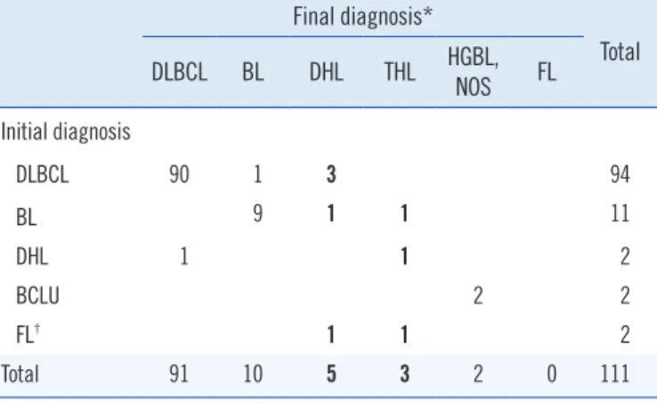
Of the 111 patients diagnosed with aggressive B-cell lymphoma, 10 were reclassified after additional FISH analysis (Tables 1 and 2). One of the cases previously diagnosed as having DHL based solely on MYC and BCL2 FISH was reclassified as THL after BCL6 FISH analysis. Patient characteristics are shown in Table 3. Serum lactate dehydrogenase (LD) levels (reference range, 4.01–8.02 µkat/L) were significantly higher in D/THL than in non-D/THL (P =0.016). With a median follow-up of 22 months (0–267 months) for all patients, the median OS was significantly shorter in D/THL than in non-D/THL (P =0.003, log-rank P = 0.017). Other baseline characteristics, including sex, age, com- plete blood count parameters (Hb, white blood cell, and platelet counts), hemophagocytic lymphohistiocytosis of BM, and CNS involvement were not significantly associated with D/THL status.
Immunophenotypic characteristics
CD5(-)/CD10(+) was significantly associated with D/THL com- pared with non-D/THL (P =0.002; Table 3). Other CD5(-)/
CD10(+) phenotypes were expressed in eight BL, five DLBCL, and one HBCL-NOS in the non-D/THL specimens. When the COO was classified using the Hans’ algorithm in 69 of our co- hort cases, the GCB-like origin was not associated with D/THL
Table 1. Re-classification based on additional FISH analysis of BM aspirates for the diagnosis of double-hit or triple-hit lymphomas
Final diagnosis*
Total DLBCL BL DHL THL HGBL,
NOS FL Initial diagnosis
DLBCL 90 1 3 94
BL 9 1 1 11
DHL 1 1 2
BCLU 2 2
FL† 1 1 2
Total 91 10 5 3 2 0 111
Bold indicates cases reclassified as DHL or THL.
*Final diagnosis was assessed with BM specimens after additional FISH analysis for the diagnosis of D/THL; †Two cases were diagnosed as having FL characterized by large-sized or blastoid malignant lymphocytes (grades 1–2 and grade 3, respectively).
Abbreviations: BM, bone marrow; DLBCL, diffuse large B-cell lymphoma;
BL, Burkitt lymphoma; HGBL NOS, high-grade B-cell lymphoma, not other- wise specified; FL, follicular lymphoma; DHL, double-hit lymphoma; THL, triple-hit lymphoma; BCLU, B-cell lymphomas, unclassifiable, with features intermediate between DLBCL and BL.
(P =0.079). IHC staining for BCL2 (N=61), BCL6 (N=69), and Ki-67 (%, N=77) did not show significant results in distinguish- ing D/THL (P =0.559, P =0.355, and P =0.524, respectively).
Cytogenetic characteristics Chromosome and FISH analyses
Cytogenetic characteristics are shown in Tables 3 and 4. Of the
MYC-R (+) cases, eight tested positive for BCL2-R, and three (13%, THL) tested positive for BCL6-R in addition to BCL2-R (+). A good agreement was observed between chromosomal al- terations in 8q24 and MYC FISH (κ=0.865, P <0.001). Three cases, which showed add(8)(q24.1) or add(8)(q24.2)t(8;14) (q24.1;q32.1) in chromosome analysis, were within the normal range of B-A signals or dual fusion signals in FISH (data not Table 2. Clinical and laboratory features of D/THL patients
Case Sex Age (yr)
LD (4.01–
8.02, µkat/L)
CD5
(FCM/IHC) CD10
(FCM/IHC) Karyotype (BM-ASP)
FISH
(BM-ASP/FFPE tissue) COO (Hans’)
Initial diagnosis
(tissue type)
BM final diagnosis
Clinical course
(OS, months) MYC-R BCL2-R BCL6-R
1 M 62 227.12 −/ND +/N.D 46,XY,t(1;9)(q25;p24),dup(2)
(q31q33),del(3)(q25),add(8)(q24.1)×
2,?del(14)(q32.1),der(16)t(16;17) (p13.3;q11.2),del(18)(q21.3) [3]/46,idem,?del(14)(q32.1)x2 [31]
+/ND +/ND +/ND ND BL (BM) THL 4.5
2 M 70 124.70 −/ND +/+ 44,X,−Y,add(1)(q42),−2, add(4) (p12),der(5)t(1;5)(q21;q35), add(8) (q24.1),der(9)t(1;9)
(q25;p21),−10,−13,add(14)(q22), add(17)(p12),add(18)(q23),+2mar [7]/46,XY [10]
+/ND +/− −/ND GCB BL
(calf, soft tissue)
DHL 6.8
3 M 62 19.51 −/ND +/+ 48,XY,der(3)t(2;3)(q31;p25)add(3) (q26.2),del(6)(q23),+7,t(8;14) (q24.1;q32),+12,t(14;18)(q32;q21.3) [7]/50,sl,+del(X)(q24),+10,-12,+13 [13]
+/+ +/ND +/ND GCB DHL (LN) THL 9.5
4* M 44 7.93 −/− +/+ 49,XY,+del(1)(q21),der(1)del(1)(p21) add(1)(q32),del(2)(q24),der(4)t(4;18) (p16;q21.1),+7,add(8)(q24),der(8)t(8;9) (p21;q21),−9,+11, t(14;18)
(q32;q21.3),+mar [5]/49,sl,add(17)(p13) [3]/49,sl,add(1)(q42),del(3)(q24),add(6) (p22) [6]/46,XY [6]
+/− +/− −/ND non-
GCB
DLBCL (LN)
DHL 1.1
5 M 47 33.78 −/ND +/+ 46,XY,add(1)(p36.1),t(8;14) (q24.1;q32),t(14;18)(q32;q21.3) [8]/47,sl,+12 [12]
+/ND +/ND −/ND ND DLBCL
(LN)
DHL 0.9
6 F 71 57.03 ND/ND ND/+ 51,X,−X,+7,+8,+8,der(8)t(8;14) (q24.1;q32)t(14;18)(q32;q21.3)× 2, +12,t(14;18),+19,+mar
[1]/51,idem,del(12)(q13q22) [19]
+/ND +/ND −/ND GCB FL Grade 1-2 (LN)
DHL Alive
7 M 50 245.62 ND/− ND/+ 47,XY,t(3;4)(q27;p13),del(6)(q13),t(8;14) (q24.1;q32),t(14;18)(q32;q21.3),+21 [10]/48,idem,+20 [10]
+/− +/+ +/+ GCB FL Grade
2-3 (BM) THL Alive 8 M 77 44.97 −/ND +/+ 50,XY,+X,t(1;14)(q42;q32),t(2;10)
(q33;q24),del(4)(q21q25),+7,der(8)t(1;8) (q21;p23),+der(10)t(2;10),+12,t(14;18) [18]/46,XY [2]
+/+ +/+ −/− GCB DLBCL
(thigh, soft tissue)
DHL Alive
*The case was previously diagnosed as DLBL derived from FL following treatment, and the other D/THL cases were diagnosed de novo.
Abbreviations: BM, bone marrow; ASP, aspirate; DLBCL, diffuse large B-cell lymphoma; F, female; M, male; LD, lactate dehydrogenase; LN, lymph node;
FCM, flow cytometry; IHC, immunohistochemistry; FFPE, formalin-fixed paraffin-embedded; COO, cell of origin; GCB, germinal center B-cell, FL, follicular lymphoma; D/THL, double-hit lymphoma and triple-hit lymphoma; R, rearrangement; ND, not determined; OS, overall survival; BL, Burkitt lymphoma.
shown). In contrast, two of the 23 cases presented split signals only in MYC FISH analysis, without any aberration involving the chromosomal MYC region. The partner genes for MYC-R were inferred from the combined chromosome analysis and MYC FISH results (Table 4).
Nineteen of the 23 cases showed juxtaposition to IG loci: 15 to IGH (14q32), three to immunoglobulin kappa (IGK) (2p12), and one to immunoglobulin lambda (IGL) (22q11.2). Nine of the 19 cases were confirmed using the IGH/MYC FISH probe.
Four of the nine cases were analyzed with both the MYC B-A probe and MYC/IGH dual fusion probe. MYC-R (+) was de- tected in only four cases using the MYC/IGH dual fusion probe (Table 4). A good agreement was observed between chromo-
some and FISH analyses for BCL2-R and BCL6-R, although only a small number of patients were compared (BCL2, N=34, κ=0.795, P <0.001; BCL6, N=24, κ=0.833, P =0.002). Of the 34 BCL2 FISH results, four showed aberrations involving the 18q21 region of the chromosome despite testing negative for BCL2 FISH (data not shown).
Discrepant results between different types of specimens
Evaluation of 28 of the 111 MYC FISH cases analyzed using FFPE tissue sections showed discrepancies across different spe- cimens. Although a good agreement was observed (κ=0.673, P =0.001) when the MYC FISH results of the BM aspirates and FFPE tissues were compared, 14% (4/28) demonstrated dis- Table 3. Patient characteristics by double-hit and/or triple-hit status
Characteristics Total (N=111) Non-D/THL (N=103) D/THL (N=8) P*
Age (range, yr), median 55 (1–86) 55 (1–86) 60 (44–77) 0.436
>60 yr 48/63 43/60 5/3 0.289
Sex (male/female) 70/41 63/40 7/1 0.254
CBC
Hb (range, g/L), median 107 (60–163) 107 (60–163) 107 (85–136) 0.801
WBC (range, ×109/L), median 6.73 (1.22–79.49) 6.60 (1.22–79.49) 7.46 (1.96–17.15) 0.873
PLT (range, ×109/L), median 140 (9–690) 152 (9–578) 72 (23–690) 0.576
LD, median (range, µkat/L) 17.05 (3.96–285.04) 15.98 (3.96–285.04) 51.00 (7.93–245.12) 0.016
HLH on BM 15/96 14/89 1/7 1
CNS involvement (N=109) 30/79 28/73 2/6 1
Chromosome
Complex karyotype 68/43 60/43 8/0 0.022
8q24 aberration† 24 17 7 <0.001
18q21 aberration† 14 6 8 <0.001
3q27 aberration† 9 3 6 0.017
FCM with BM aspirates (N=67)
CD5 (-) CD10 (-) 38 38 0 0.002
CD5 (-) CD10 (+) 20 14 6
CD5 (+) CD10 (-) 8 8 0
CD5 (+) CD10 (+) 1 1 0
COO
GCB/non-GCB (N=69) 30/39 25/38 5/1 0.079
Ki-67 (%, N=77), Median 80 (5–99) 80 (5–99) 90 (60–96) 0.524
OS (range, months), Median 22 (0–267) 20 (0–267) 7 (0–18) 0.003
Bold indicates statistically significant results.
*Chi-square test or Fisher’s exact test when appropriate; †The criteria for chromosomal aberration at appropriate cutoff points followed the “two-band rule,”
which only recognizes the cut points within the two-band or two-sub-bands from the target cut points at >400-band levels.
Abbreviations: D/THL, double-hit lymphoma and triple-hit lymphoma; CBC, complete blood count; WBC, white blood cell; PLT, platelet; LD, lactate dehydro- genase; HLH, hemophagocytic lymphohistiocytosis; CNS, central nervous system; FCM, flow cytometry; BM, bone marrow; GCB, germinal center B-cell; OS, overall survival; COO, cell of origin.
crepant MYC FISH results; cases 4 and 7 were MYC-R (+), with BM aspirates showing aberrations in the MYC region in the chromosome analysis (Table 5). In contrast, cases 55 and 67 were MYC-R (-), with the BM aspirates showing a normal karyo- type (Table 5). Among these 28 cases, comparable FISH results between BM aspirate and FFPE tissue were only observed in four and two cases for BCL2 and BCL6 FISH, respectively. A single case (1/4, case 4) with BCL2-R (-) in the FFPE LN tissue, but BCL2-R (+) in the BM aspirate, had aberrations in BCL2 re- gion in the chromosome analysis (Table 5).
DISCUSSION
Diagnosing D/THL is challenging without further FISH analysis.
However, further FISH analysis in all patients with DLBCL or other aggressive B-cell lymphomas is hindered due to practical limitations [10, 11]. In this study, most patients were diagnosed histopathologically using LN or extranodal specimens before BM examination. Together with morphological findings, we con- firmed two diagnostic parameters for D/THL during BM workup.
First, immunophenotypic characteristics, such as CD5(-) CD10(+) combined with B-cell antigen markers, are indicators that allow for rapid screening for further FISH analysis of MYC/
BCL2/BCL6. Second, complex karyotypes comprising chromo- somal aberrations located near specific regions, such as MYC/
BCL2/BCL6 on malignant lymphocytes of the BM aspirate, strongly reflect the FISH analysis results for MYC/BCL2/BCL6.
CD5(-) CD10(+) phenotypes in lymphoproliferative disorders Table 4. Site-specific comparison of chromosome and FISH analyses results for diagnosis of D/THL in 23 MYC-R (+) cases with BM aspi- rates
MYC-R (+) cases 8q24 aberration (CHR) MYC partner (CHR/FISH) 18q21 aberration (CHR) BCL2-R FISH 3q27 aberration (CHR) BCL6-R FISH
1 add(8)(q24.1) ND/ND del(18)(q21.3) P del(3)(q25)* P
2 add(8)(q24.1) ND/IGH add(18)(q23)* P N N
3 t(8;14)(q24.1;q32) IGH/IGH t(14;18)(q32;q21.3) P add(3)(q26.2)* P
4 add(8)(q24.1) ND/ND t(14;18)(q32;q21.3) P N N
5 t(8;14)(q24.1;q32) IGH/ND t(14;18)(q32;q21.3) P N N
6 der(8)t(8;14)(q24.1;q32) IGH/ND t(14;18)(q32;q21.3) P N ND
7 t(8;14)(q24.1;q32) IGH/IGH t(14;18)(q32;q21.3) P t(3;4)(q27;p13) P
8 N ND/ND t(14;18)(q32;q21.3) P N N
9 t(8;14)(q24.1;q32) IGH/ND inv(18)(p11.3q21) N N N
10 t(8;14)(q24.1;q32) IGH/IGH N N N N
11 t(2;8)(p12;q24.1) 2p12 (IGK)/ND N N N N
12 t(8;22)(q24.1;q11.2) 22q11.2 (IGL)/ND N N N N
13 N ND/ND N N N N
14 t(2;8)(p12;q24.1) 2p12 (IGK)/ND N N N ND
15 t(2;8)(p12;q24.1) 2p12 (IGK)/ND N N N N
16 t(8;14)(q24.1;q32) IGH/ND N N N N
17 t(8;14)(q24.1;q32) IGH/IGH† t(16;18)(p13.1;q21.1) N N N
18 t(8;14)(q24.1;q32) IGH/ND N N N N
19 t(8;14)(q24.1;q32) IGH/ND N N N ND
20 t(8;14)(q24.1;q32) IGH/IGH† N N N N
21 t(8;14)(q24.1;q11.2) IGH/IGH† N N N N
22 t(8;14)(q24.1;q32) IGH/IGH† N N N N
23 t(8;14)(q24.1;q32) IGH/IGH N N N N
*The criteria for chromosomal aberration at appropriate cut points followed the “two-band rule,” which only recognizes the cut points within the two bands or two sub-bands from the target cut points at >400-band levels; †Four cases were analyzed with both the MYC break-apart FISH probe and MYC/IGH probe; however, MYC rearrangement was detected only with the MYC/IGH probe.
Abbreviations: D/THL, double-hit lymphoma and triple-hit lymphoma; R, rearrangement; BM, bone marrow; CHR, chromosome; IGH, immunoglobulin heavy chain; IGK, immunoglobulin kappa; IGL, immunoglobulin lambda; N, negative; P, positive; ND, not determined.
have been observed in DLBCL, BL, FL, and hairy cell leukemia [24]. The immunophenotypic characteristics of DHL have been defined by the expression of CD10, high expression of CD38, and frequent under-expression of CD19, CD20, and the light chain [24–28]. Additional diagnostic clues based on the results of chromosome analysis suggested the need for further FISH analysis in approximately 22% (24/111) of our cohort, particu- larly those carrying aberrations involving the 8q24 region. Chro- mosome analysis can facilitate the detection of D/THL in the fi- nal step of diagnosis, although cryptic translocations cannot be detected. Based on our results, the combined results of FCM and chromosome analysis can improve D/THL diagnosis and may reduce the number of cases wherein further FISH analysis is recommended. Although concurrent evaluation of MYC/
BCL2/BCL6 can rapidly reveal D/THL, many clinical laboratories use a two-step approach for their diagnostic workflow, including initial FISH for MYC-R followed by BCL2/BCL6-R FISH if re- quired [7]. The diagnostic workflow can be determined based on a consensus between laboratory professionals and clinicians at the hospital.
Fourteen percent of our cohort showed discrepant MYC-R re- sults between different types of specimens (BM aspirates and FFPE tissues; Table 5). Different types of specimens containing different types or numbers of malignant lymphoid cells or probe type may yield false-positive or false-negative results because of the quality of the specimens and analysis or inter-observer varia- tion. The BM aspirate is considered as a more practical speci- men than tissue specimens for FCM and chromosomal analy- ses, especially when suspected malignant cells are morphologi- cally identified. Although the primary diagnosis is performed with LN or extranodal tissues (non-BM), the possibility of D/THL should also be closely investigated with BM specimens in the fi-
nal diagnostic process.
A selection bias was observed in this retrospective analysis. In cases that were not fully evaluated at diagnosis, additional FISH analysis was used only in cases with available BM specimens.
FCM analysis was conducted only in 67 patients at diagnosis.
We did not analyze other surface markers specifically, except for CD5 and CD10, owing to the limitations of retrospective studies.
Additionally, fewer specimens were analyzed compared with re- cent studies evaluating large cohorts for D/THL [6, 29]. The prevalence of MYC-R (+) cases was higher than in other studies, as our cohort included cases of BM involving aggressive B-cell lymphoma, which was related to poor prognosis in MYC-R (+) cases [29–31]. Second, MYC partner genes were not fully ex- plored, although they have been associated with prognosis in a recent study [29]. The FISH probe type for identifying rearrange- ments was selected according to the laboratories’ preference based on technical or clinical issues [32]. Many MYC-R cases were evaluated with the MYC B-A probe, while the IGH/MYC dual fusion probe was used in only a few cases. Interestingly, al- though four cases from our cohort were analyzed with both the MYC B-A probe and MYC/IGH probe, MYC-R was detected only with the MYC/IGH probe (Table 4). False-negative results from the MYC B-A probe could be generated by cryptic insertion trans- location of IGH promoter/enhancer sequences into the MYC gene region [33, 34]. Therefore, some studies have suggested that both MYC B-A and MYC/IGH probes should be used to identify a wide range of 8q24 breakpoints that occur very close to the MYC gene region and that they could help detect MYC/non-IGH or non-IG rearrangement [33, 34]. FISH analyses for IGK (2p12) and IGL (22q11) genes were not conducted. However, the part- ner genes were predicted based on chromosome analysis of 19 of the 23 MYC-R (+) cases (Table 4). Many studies revealed a high prevalence of non-IG partners in DHL, while none of the cases showed non-IG partners in our five D/THL and the other 14 MYC-R (+) cases [35, 36]. These results might be attributed to the small cohort size.
In conclusion, a rapid and rational approach for diagnosing D/
THL can be established based on CD5(-)/CD10(+) results using FCM and complex karyotypes with aberrations on MYC/BCL2/
BCL6 regions, particularly in cases of BM-involving aggressive B-cell lymphomas. Based on these results, further FISH analy- sis can be used effectively to establish a definitive diagnosis of D/THL among aggressive B-cell lymphomas during the BM workup.
Table 5. Discrepant MYC and BCL2 FISH results between BM as- pirate and FFPE tissues
Case FISH*
FISH results BM aspirate
(diagnosis) FFPE (tissue, diagnosis)
4 MYC-R/BCL2-R P/P (DHL) N/N (LN, DLBCL)
7 MYC-R P (THL) N (BM biopsy, FL)
55 MYC-R N (DLBCL) P (Breast, DLBCL)
67 MYC-R N (DLBCL) P (Stomach, DHL)
*Dual color break-apart probes were used for MYC-R and BCL2-R.
Abbreviations: BM, bone marrow; LN, lymph node; DLBCL, diffuse large B- cell lymphoma; FFPE, formalin-fixed paraffin-embedded; FL, follicular lym- phoma; DHL, double-hit lymphoma; THL, triple-hit lymphoma; N, negative;
P, positive; R, rearrangement.
ACKNOWLEDGEMENTS
None.
AUTHOR CONTRIBUTIONS
H.K. analyzed data and wrote the manuscript; H.-J.K. provided expertise about laboratory data; S.-H.K. supervised the research study and edited the manuscript; and all authors were approved the manuscript.
CONFLICTS OF INTEREST
No potential conflicts of interest relevant to this article are re- ported.
RESEARCH FUNDING
None declared.
ORCID
Heyjin Kim https://orcid.org/0000-0002-2213-2579 Hee-Jin Kim https://orcid.org/0000-0003-3741-4613 Sun-Hee Kim https://orcid.org/0000-0002-7542-5551
REFERENCES
1. Swerdlow SH, Campo E, et al. WHO classification of tumours of haema- topoietic and lymphoid tissues. revised 4th ed. Lyon: International Agency for Research on Cancer (IARC), 2017:335-41.
2. Campo E, Swerdlow SH, Harris NL, Pileri S, Stein H, Jaffe ES. The 2008 WHO classification of lymphoid neoplasms and beyond: evolving con- cepts and practical applications. Blood 2011;117:5019-32.
3. Said JW. Aggressive B-cell lymphomas: how many categories do we need? Mod Pathol 2013;26:S42-56.
4. Lindsley RC and LaCasce AS. Biology of double-hit B-cell lymphomas.
Curr Opin Hematol 2012;19:299-304.
5. Snuderl M, Kolman OK, Chen YB, Hsu JJ, Ackerman AM, Dal Cin P, et al. B-cell lymphomas with concurrent IGH-BCL2 and MYC rearrange- ments are aggressive neoplasms with clinical and pathologic features distinct from Burkitt lymphoma and diffuse large B-cell lymphoma. Am J Surg Pathol 2010;34:327-40.
6. Oki Y, Noorani M, Lin P, Davis RE, Neelapu SS, Ma L, et al. Double hit lymphoma: the MD Anderson Cancer Center clinical experience. Br J Haematol 2014;166:891-901.
7. Li S, Lin P, Fayad LE, Lennon PA, Miranda RN, Yin CC, et al. B-cell lym- phomas with MYC/8q24 rearrangements and IGH@BCL2/t(14;18) (q32;q21): an aggressive disease with heterogeneous histology, germi- nal center B-cell immunophenotype and poor outcome. Mod Pathol 2012;25:145-56.
8. Merron B and Davies A. Double hit lymphoma: how do we define it and how do we treat it? Best Pract Res Clin Haematol 2018;31:233-40.
9. Carbone A, Gloghini A, Kwong YL, Younes A. Diffuse large B cell lym- phoma: using pathologic and molecular biomarkers to define sub- groups for novel therapy. Ann Hematol 2014;93:1263-77.
10. Friedberg JW. How I treat double-hit lymphoma. Blood 2017;130:590- 6.
11. Nabhan C and Mato AR. Emerging strategies in treating double hit lym- phomas. Clin Lymphoma Myeloma Leuk 2017;17:563-8.
12. Sakr H and Cook JR. Identification of “Double Hit” lymphomas using updated WHO criteria: insights from routine MYC immunohistochemis- try in 272 consecutive cases of aggressive B-cell lymphomas. Appl Im- munohistochem Mol Morphol 2019;27:410-5.
13. Sesques P and Johnson NA. Approach to the diagnosis and treatment of high-grade B-cell lymphomas with MYC and BCL2 and/or BCL6 rear- rangements. Blood 2017;129:280-8.
14. Aukema SM, Siebert R, Schuuring E, van Imhoff GW, Kluin-Nelemans HC, Boerma EJ, et al. Double-hit B-cell lymphomas. Blood 2011;117:
2319-31.
15. Scott DW, King RL, Staiger AM, Ben-Neriah S, Jiang A, Horn H, et al.
High-grade B-cell lymphoma with MYC and BCL2 and/or BCL6 rear- rangements with diffuse large B-cell lymphoma morphology. Blood 2018;131:2060-4.
16. Zelenetz AD, Gordon LI, Wierda WG, Abramson JS, Advani RH, An- dreadis CB, et al. Diffuse large B-Cell lymphoma version 1.2016. J Natl Compr Canc Netw 2016;14:196-231.
17. Bain BJ. Bone marrow trephine biopsy. J Clin Pathol 2001;54:737-42.
18. Talaulikar D and Dahlstrom JE. Staging bone marrow in diffuse large B- cell lymphoma: the role of ancillary investigations. Pathology 2009;41:
214-22.
19. Lee SH, Erber WN, Porwit A, Tomonaga M, Peterson LC; International Council for Standardization in Hematology. ICSH guidelines for the stan- dardization of bone marrow specimens and reports. Int J Lab Hematol 2008;30:349-64.
20. Mazur G, Hałoń A, Wróbel T, Jeleń M, Kuliczkowski K. Contribution of flow cytometric immunophenotyping and bone marrow trephine biopsy in the detection of lymphoid bone marrow infiltration in non-Hodgkin’s lymphomas. Neoplasma 2004;51:159-63.
21. Kim B, Lee ST, Kim HJ, Kim SH. Bone marrow flow cytometry in staging of patients with B-cell non-Hodgkin lymphoma. Ann Lab Med 2015;35:
187-93.
22. Morice WG, Kurtin PJ, Hodnefield JM, Shanafelt TD, Hoyer JD, Rem- stein ED, et al. Predictive value of blood and bone marrow flow cytometry in B-cell lymphoma classification: comparative analysis of flow cytometry and tissue biopsy in 252 patients. Mayo Clin Proc 2008;83:776-85.
23. Hans CP, Weisenburger DD, Greiner TC, Gascoyne RD, Delabie J, Ott G, et al. Confirmation of the molecular classification of diffuse large B-cell lymphoma by immunohistochemistry using a tissue microarray. Blood 2004;103:275-82.
24. Jaffe ES, Campo E, Harris NL, Pileri SA, Stein H, Swerdlow SH. Intro- duction and overview of the classification of lymphoid neoplasms. In:
Swerdlow SH, Campo E, et al. WHO classification of tumours of haema- topoietic and lymphoid tissues. revised 4th ed. Lyon: International Agency for Research on Cancer (IARC), 2017:190-8.
25. Dorfman DM. Clinical flow cytometry: state-of-the-art and new ap- proaches. Clin Lab Med 2017;37:xiii-xiv.
26. Wu D, Wood BL, Dorer R, Fromm JR. ‘Double-Hit’ mature B-cell lym- phomas show a common immunophenotype by flow cytometry that in- cludes decreased CD20 expression. Am J Clin Pathol 2010;134:258- 65.
27. Roth CG, Gillespie-Twardy A, Marks S, Agha M, Raptis A, Hou JZ, et al.
Flow cytometric evaluation of double/triple hit lymphoma. Oncol Res 2016;23:137-46.
28. Schniederjan SD, Li S, Saxe DF, Lechowicz MJ, Lee KL, Terry PD, et al.
A novel flow cytometric antibody panel for distinguishing Burkitt lym- phoma from CD10+ diffuse large B-cell lymphoma. Am J Clin Pathol 2010;133:718-26.
29. Copie-Bergman C, Cuillière-Dartigues P, Baia M, Briere J, Delarue R, Canioni D, et al. MYC-IG rearrangements are negative predictors of sur- vival in DLBCL patients treated with immunochemotherapy: a GELA/
LYSA study. Blood 2015;126:2466-74.
30. de Jonge AV, Roosma TJ, Houtenbos I, Vasmel WL, van de Hem K, de Boer JP, et al. Diffuse large B-cell lymphoma with MYC gene rearrange- ments: current perspective on treatment of diffuse large B-cell lympho- ma with MYC gene rearrangements; case series and review of the litera- ture. Eur J Cancer 2016;55:140-6.
31. Landsburg DJ, Falkiewicz MK, Petrich AM, Chu BA, Behdad A, Li S, et al. Sole rearrangement but not amplification of MYC is associated with a poor prognosis in patients with diffuse large B cell lymphoma and B cell
lymphoma unclassifiable. Br J Haematol 2016;175:631-40.
32. Ventura RA, Martin-Subero JI, Jones M, McParland J, Gesk S, Mason DY, et al. FISH analysis for the detection of lymphoma-associated chro- mosomal abnormalities in routine paraffin-embedded tissue. J Mol Di- agn 2006;8:141-51.
33. May PC, Foot N, Dunn R, Geoghegan H, Neat MJ. Detection of cryptic and variant IGH-MYC rearrangements in high-grade non-Hodgkin’s lymphoma by fluorescence in situ hybridization: implications for cytoge- netic testing. Cancer Genet Cytogenet 2010;198:71-5.
34. Muñoz-Mármol AM, Sanz C, Tapia G, Marginet R, Ariza A, Mate JL.
MYC status determination in aggressive B-cell lymphoma: the impact of FISH probe selection. Histopathology 2013;63:418-24.
35. Chong LC, Ben-Neriah S, Slack GW, Freeman C, Ennishi D, Mottok A, et al. High-resolution architecture and partner genes of MYC rearrange- ments in lymphoma with DLBCL morphology. Blood Adv 2018;2:2755- 65.
36. Sarkozy C, Traverse-Glehen A, Coiffier B. Double-hit and double-protein- expression lymphomas: aggressive and refractory lymphomas. Lancet Oncol 2015;16:e555-67.