A Rare Manifestation of Solitary
Primary Bone Lymphoma of the Finger:
a Case Report
INTRODUCTION
Primary bone lymphoma is a rare disease, accounting for less than 5% of primary bone tumors and 3% of malignant bone tumors (1, 2). Most of the primary bone lymphomas represent non-Hodgkin lymphoma, especially diffuse large B-cell lymphoma (DLBCL) (3). Clinically, bone lymphomas present as palpable masses with intermittent bone pain (4). According to a previous study (5), the metadiaphysis of the femur is the most common site involved. Other sites include pelvis, tibia, head and neck, and vertebra. Lymphoma localized to peripheral sites such as the feet and the hands, or the toes and the fingers, is rarely reported. Here, we report a very rare case of solitary primary bone lymphoma located in the middle phalanx of the third finger.
CASE REPORT
A 71-year-old male presented to our institution complaining of swelling in the right third finger, with a six-month history of symptoms. He had no specific medical history.
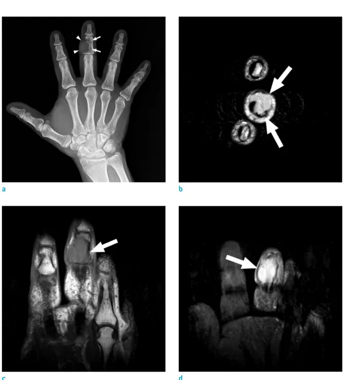
Laboratory testing revealed elevated erythrocyte sedimentation rate and C-reactive protein (51 mm/h and 5.8 mg/dL, respectively). Plain radiograph revealed evidence of bony mass causing cortical disruption at the radial aspect of the third middle phalanx (Fig. 1a). The patient underwent magnetic resonance imaging (MRI) for further evaluation. T2-weighted MRI showed a well-defined mass of high signal intensity, involving the adjacent 3rd middle phalangeal bone with large soft tissue component.
The mass was thought to have originated in the bone rather than soft tissue, because the center of the mass was near the cortex of the phalangeal bone and normal cortical
This is an Open Access article distributed under the terms of the Creative Commons Attribution Non-Commercial License (http://creativecommons.org/licenses/
by-nc/3.0/) which permits unrestricted non-commercial use, distribution, and reproduction in any medium, provided the original work is properly cited.
Received: November 19, 2018 Revised: December 11, 2018 Accepted: December 12, 2018 Correspondence to:
You Seon Song, M.D.
Department of Radiology, Pusan National University Hospital, 179 Gudeok-ro, Seo-gu, Busan 49241, Korea.
Tel. +82-51-240-7354 Fax. +82-51-244-7534 E-mail: [email protected]
Copyright © 2018 Korean Society of Magnetic Resonance in Medicine (KSMRM)
Case Report
Primary extranodal bone lymphoma involving the peripheral extremities is extremely rare. Here, we report a definitive case of diffuse large B-cell lymphoma involving the phalangeal bone of the 3rd finger. Systemic evaluation revealed the lesion as the only site of lymphoma involvement.Keywords: Primary lymphoma; Bone; Finger
Jeong A Yeom2, You Seon Song1*, In Sook Lee1, Kyung Un Choi3, Jeung Il Kim4
1Department of Radiology, Pusan National University Hospital, Busan, Korea
2Department of Radiology, Pusan National University Yangsan Hospital, Busan, Korea
3Department of Pathology, Pusan National University Hospital, Busan, Korea
4Department of Orthopedic Surgery, Pusan National University Hospital, Busan, Korea Magnetic resonance imaging
bone was noted within the boundary of the mass. The mass abutted with the flexor tendon of the third finger (Fig. 1b).
T1-weighted imaging revealed a mass of intermediate signal intensity (Fig. 1c). The contrast-enhanced MRI showed strong and homogeneous enhancement (Fig. 1d). Based on
the imaging findings, the differential diagnosis included mimics of primary bone tumor such as glomus tumor or intraosseous epidermal inclusion cyst and metastatic bone tumor, despite its rarity. Total excision was performed for symptom relief.
a b
Fig. 1. A 71-year-old-man with DLBCL involving 3rd finger. (a) Plain radiograph shows osteolytic bony mass (arrows) with soft-tissue component (arrowheads) involving right third middle phalanx. (b) Axial T2-weighted image reveals a high- signal intensity mass (arrows) associated with the right third middle phalanx. The mass originated in the intramedullary cavity constituting the large soft-tissue component, abutting the flexor tendon of the third finger. (c) Coronal T1-weighted image indicates a mass of intermediate signal intensity (arrow). (d) Coronal T1-weighted image with fat suppression after gadolinium administration shows homogenous enhancement of the mass (arrow).
c d
A yellowish-white soft tissue mass causing lysis of the middle phalangeal bone was visible in the operative field.
Staining with Hematoxylin and Eosin (H&E) revealed diffuse infiltration of atypical cells (Fig. 2a). Immunohistochemical analysis showed positive test results for leukocyte common antigen (LCA) (Fig. 2b) and CD20, and negative findings for CD45R0, pan cytokeratin, synaptophysin, CD56, and CD99.
Therefore, the final pathologic diagnosis was DLBCL. Chest
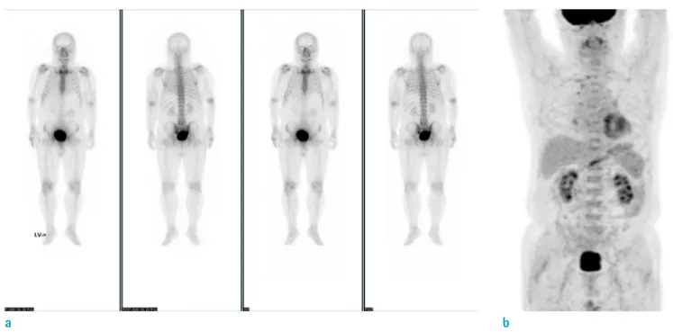
and abdominal computed tomography (CT) scan, bone scan (Fig. 3a), and positron emission tomography (PET)-CT (Fig.
3b) were performed to determine the involvement of other organs. However, no evidence of lymphoma involvement was detected at any other site, nor was any evidence of disease spread to other organs detected during the 6 months of follow-up.
Fig. 2. Microscopic findings of the finger mass. (a) Microscopic finding (Hematoxylin & Eosin stain, × 200) shows diffuse infiltration of atypical cells. (b) Immunohistochemical staining was positive for leukocyte common antigen.
a b
Fig. 3. Systemic evaluation of other organ involvement. (a) Bone scan reveals no evidence of other bone involvement. (b) PET-CT reveals no evidence of other organ involvement of lymphoma.
a b
DISCUSSION
Lymphoma is the most common hematologic disorder of the lymphoreticular tissue. However, lymphomas can also involve organs outside of the lymphatic system, referred to as extranodal lymphoma. Extranodal lymphoma may occur in the gonads, gastrointestinal tract, lung, central nervous system and bone. In primary bone lymphoma, axial rather than appendicular skeleton is more commonly affected (6).
Moreover, the small bones of the peripheral extremities are very rarely involved in bone lymphoma. To our knowledge, only few cases (7), which were confirmed as primary bone lymphoma, involved the hand.
The diagnostic criteria include the presence of lymphoma involving an osseous site, without evidence of any other organ involvement for at least 6 months after the initial diagnosis (8). The regional lymph node involvement cannot exclude a diagnosis of primary bone lymphoma. In our case, the palpable mass on the third finger was the initial manifestation of the disease, without any masses involving other organs on systemic evaluation. The patient visited our hospital in May, following initial plain radiograph at another hospital in January of the same year, showing the finger mass. Therefore, no evidence of metastatic spread was found about 6 months after the initial presentation.
Thus, it is reasonable to define our case as primary bone lymphoma.
According to a previous report (6), the majority of primary bone lymphoma occurs in older adults, and more than 90% of patients are over the age of 30 years. In a large series, patients diagnosed with primary bone DLBCL were younger (median age, 56 years) than patients with nonosseous DLBCL and secondary bone lymphoma (9). In our case, the patient was 71 years old. Additionally, men are more frequently affected than women, with a ratio ranging from 1.2 to 1.8 (6). Femur is the most common site of involvement in nearly 50% of the cases. The pelvis is affected in about 20%, and other sites including the vertebra, ribs and tibia account for the remainder (5).
DLBCL is the most common histological subtype of primary bone lymphoma, as in our case (6). Rarely, follicular lymphoma, small lymphocytic lymphoma and marginal zone lymphoma may manifest as primary bone lymphoma.
Imaging findings of primary bone lymphoma vary, and may be non-specific in some patients (4). Although, the imaging findings of bone lymphoma exhibit a wide spectrum, a few features are characteristic of the disease.
Typically, lymphomatous bone involvement is defined by a solitary lytic lesion near the end of a long bone with permeative or moth-eaten appearance, and aggressive periosteal reaction (10). In addition, extensive surrounding soft-tissue masses without significant cortical destruction has been reported exclusively in round cell tumors including bone lymphoma, multiple myeloma and Ewing sarcoma (3).
However, in our case, none of these typical imaging findings of bone lymphoma was detected. Therefore, the initial radiologic diagnosis was not focused on lymphoma, but rather on the more commonly encountered masses associated with the finger.
Here, we reported a very rare case of solitary primary bone lymphoma involving the third finger. Although rare with unusual location and imaging findings, the possibility of this disease should be considered to avoid misdiagnosis.
REFERENCES
1. Krishnan A, Shirkhoda A, Tehranzadeh J, Armin AR, Irwin R, Les K. Primary bone lymphoma: radiographic-MR imaging correlation. Radiographics 2003;23:1371-1383; discussion 1384-1387
2. Pobirci O, Rosca E, Pobirci DD, Nichita A. Primary diffuse large B-cell lymphoma of the humerus. Case report and review of literature. Med Con 2014;9:69-72
3. Mulligan ME, McRae GA, Murphey MD. Imaging features of primary lymphoma of bone. AJR Am J Roentgenol 1999;173:1691-1697
4. de Camargo OP, dos Santos Machado TM, Croci AT, et al.
Primary bone lymphoma in 24 patients treated between 1955 and 1999. Clin Orthop Relat Res 2002:271-280 5. Vincent JM, Ng YY, Norton AJ, Armstrong P. Case report:
primary lymphoma of bone--MRI appearances with pathological correlation. Clin Radiol 1992;45:407-409 6. Jawad MU, Schneiderbauer MM, Min ES, Cheung MC,
Koniaris LG, Scully SP. Primary lymphoma of bone in adult patients. Cancer 2010;116:871-879
7. Galati V, Wortmann F, Stang FH, Thorns C, Mailander P, Kisch T. A rare manifestation of primary bone lymphoma:
solitary diffuse large B-cell lymphoma of the little finger. J Hand Surg Am 2018;43:779 e771-779 e774
8. Coley BL, Higinbotham NL, Groesbeck HP. Primary reticulum-cell sarcoma of bone; summary of 37 cases.
Radiology 1950;55:641-658
9. Li X, Xu-Monette ZY, Yi S, et al. Primary bone lymphoma exhibits a favorable prognosis and distinct gene expression
signatures resembling diffuse large B-cell lymphoma derived from centrocytes in the germinal center. Am J Surg Pathol 2017;41:1309-1321
10. Phillips WC, Kattapuram SV, Doseretz DE, et al. Primary lymphoma of bone: relationship of radiographic appearance and prognosis. Radiology 1982;144:285-290