Partial ALPPS with a longer wait between procedures is safe and yields adequate future liver remnant hypertrophy
Nagappan Kumar1, Trish Duncan1, David O’Reilly1, Zsolt Káposztás2, Craig Parry3, John Rees3, and Sameer Junnarkar4
1Cardiff Liver Unit, University Hospital of Wales, Cardiff, UK, 2Moritz Kaposi Teaching General Hospital, Kaposvár, Hungary, 3Department of Radiology, University Hospital of Wales, Cardiff, UK, 4Department of Surgery, Tan Tock
Seng Hospital, Singapore
Backgrounds/Aims: Associating Liver Partition and Portal Vein Ligation for Staged Hepatectomy (ALPPS) has gen- erated controversy due to high morbidity and mortality. We present our series of patients with 30-40% parenchymal transection and minimal hilar dissection. Methods: Patients who had partial ALPPS between April 2015 and April 2016 were included. Patients with colorectal liver metastases (CRLM) had their future liver remnants (FLR) cleared with metastasectomies. The liver was divided along the future line of transection to 30-40%, right portal vein was stapled and divided without extensive hilar dissection, with minimal handling of right liver, which was not mobilised. We pre- served the middle hepatic vein. Data were collected prospectively for hypertrophy of the FLR, morbidity and mortality.
Results: Among the 8 patients (age 25-68) investigated, one patient with cholangiocarcinoma had portal vein emboliza- tion prior to partial ALPPS. All patients completed two stages with adequate FLR hypertrophy at a median of 28 days.
No mortality was found. The median length of stay after stages 1 and 2 was 9 and 9.6 days, respectively. The median increase in FLR was 38%. Conclusions: A limited transection of 30-40%, minimal hilar dissection and longer wait be- tween stages yielded adequate FLR hypertrophy with low morbidity and no mortality. (Ann Hepatobiliary Pancreat Surg 2019;23:13-19)
Key Words: Liver resection; Future liver remnant; ALPPS; Hypertrophy; Colorectal liver metastases
Received: June 14, 2018; Revised: July 23, 2018; Accepted: July 26, 2018 Corresponding author: Nagappan Kumar
Cardiff Liver Unit, University Hospital of Wales, Cardiff, CF144XW, UK
Tel: +44-29-2074-4793, Fax: +44-29-2074-3348, E-mail: [email protected]
Copyright Ⓒ 2019 by The Korean Association of Hepato-Biliary-Pancreatic Surgery
This is an Open Access article distributed under the terms of the Creative Commons Attribution Non-Commercial License (http://creativecommons.org/
licenses/by-nc/4.0) which permits unrestricted non-commercial use, distribution, and reproduction in any medium, provided the original work is properly cited.
Annals of Hepato-Biliary-Pancreatic Surgery ∙ pISSN: 2508-5778ㆍeISSN: 2508-5859
INTRODUCTION
Liver resection offers the best chance for long-term sur- vival of patients with colorectal liver metastases (CRLM) and hilar cholangiocarcinoma. A combination of better chemotherapeutic regimens and surgical innovations has increased the resection rates in patients with CRLM.
Several technical limiting factors such as location of the tumor and distribution preclude liver resection. A future liver remnant (FLR) of 20-30% is deemed safe in patients with normal background liver and at least 40% FLR in patients with compromised liver (steatohepatitis, fibrosis, cirrhosis).1 The techniques that have proved successful in increasing resection rates include neoadjuvant chemotherapy in patients with CRLM,2-4 portal vein embolization (PVE),5 portal vein ligation (PVL),6 two-stage liver resections,7
Associating Liver Partition and Portal vein ligation in Staged hepatectomy (ALPPS)8 and Associating Portal Em- bolization and Artery Ligation (APEAL).9
The ALPPS procedure created tremendous interest among liver surgeons because of the pace of liver regeneration that it allowed, enabling completion of the two stages of liver resection within 9 to 11 days. The procedure has yet to be widely adopted mainly due to concerns with increas- ing morbidity, mortality10 and potentially higher recurrence rates.11,12
We performed the first ALPPS procedure in April 2015 using only partial transection of liver and other technical modifications involving minimal dissection. This approach produced significant hypertrophy of the FLR and we have performed 8 procedures with no mortality and acceptable morbidity. We proceeded to the second stage in all pa-
Fig. 2. Intraoperative photograph showing metastasectomies in segment 4.
Fig. 3. Right portal vein stapling (arrow) with minimal hilar dissection.
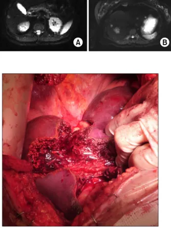
Fig. 1. Metastases detected in the hepatic segment 6 (A), seg- ments 2, and 7 (B), segments 3, 4, and 8 (C).
tients after a longer interval than the classical ALPPS.
The aim of this report is to show the feasibility of safe two-stage liver resection via transection of only 30-40%
of liver (partial ALPPS). A longer interval between the two procedures may lead to less liver failure.
PATIENTS AND METHODS
In order to describe the modified version of the ALPPS procedure, the first operation is briefly explained below.
The patient was a 68-year-old lady with obstructing carcinoma of descending colon and synchronous liver metastases. She underwent left hemicolectomy with end stoma in January 2014 for a T4bN0M1 tumor. The liver metastases involved all segments except segment 1 and 5 (Fig. 1). The patient had palliative chemotherapy with 4 cycles of XELOX (capecitabine plus oxaliplatin) follow- ing progression with FOLFIRI (leucovorin calcium, 5-flu- orouracil, and irinotecan) regimen as second-line treatment.
Aflibercept was added to augment the response, and was
completed in February 2015. An Fludeoxyglucose 18F (FDG)-positron emission tomography (PET) scan revealed liver only metastases and a pair of foci in the left lateral abdominal wall suggestive of local recurrence. However, review of cross-sectional images in this area revealed no masses and after discussion with the hepatobiliary multi- disciplinary team (MDT) meeting it was decided to ex- plore the possibility of two-stage liver resection.
Using laparotomy, the tumors in the segments 2, 3 and 4b were removed as metastasectomies (Fig. 2) with mac- roscopically clear margins. The FLR was inadequate.
Cholecystectomy was carried out and the right portal vein (RPV) was ligated with minimal dissection by incising the peritoneum on the right and posterior aspects of the hilum.
The RPV was stapled and divided (Fig. 3). Metastasectomy of the tumor in the segment 4b led to a large defect in the liver with only a small sliver of tissue between this defect and the gall bladder fossa. This was divided down to the hilum resulting in transection of nearly a third of liver. The right liver was not mobilized. A silastic drain
was placed along the cut surface of the liver. The patient recovered adequately and was discharged on postoperative day 6 with the drain in situ. Histological examination re- vealed complete resection with a margin of at least 1 mm.
A computed tomography (CT) scan was performed to measure the volume of the FLR 12 days after the first stage procedure and volumes were assessed using standard procedure described below.13
The CT assessment showed an increase in FLR from 408 ml to 630 ml, or an increase of 54%. She then under- went a completely extended right hemihepatectomy 21 days after the first stage procedure, using and completing the previous transection plane. Histologically, the tumor in the segment 7 close to the inferior vena cava extended to within 1 mm of the resection margin. The patient recov- ered without any complication and was discharged 7 days after the operation.
The above experience indicated that FLR hypertrophy was feasible with partial transection of the liver. We per- formed similar procedures in seven other patients reported here. We defined 30-40% transection for an extended right hepatectomy as resection of the liver along the falci- form ligament and removing the segment 4b pedicle. The cut was extended up to the segment 4a pedicle which was left in situ. The middle hepatic vein was not divided in the first operation.
Liver volume assessment
Liver volume was assessed using commercial volume- try software, Volume Viewer advanced imaging platform (GE Healthcare Version 2.0, AW Server) and calculated by manually defining the contours of the liver paren- chyma on representative sequential axial 0.625 mm slices of the CT acquisition on a liquid crystal display (1920×1200 resolution, 24-inch wide screen display, LA2405, HP Compaq).
The volume rendering tool automatically generates a 3D volume and calculates the total liver volume (TLV) from the selected slices. This process was repeated for all definable liver lesions to yield the estimated total lesion volume and the two volumes subtracted to determine the estimated Total Functional Liver Volume (TFLV).
The selected liver volume was used to define the pre- dicted Functional Residual Liver Volume (FRLV) by seg- menting and excluding the hepatic parenchyma planned
for resection as directed by the liver surgeon. The ratio of predicted functional residual liver volume (%FRLV) was then calculated.
The actual FRLV was subsequently measured on the post-ALPPS axial CT performed on day 14 to determine the extent of hypertrophy and feasibility of complete hepatectomy.
Hepatobiliary (HIDA) assessment
We performed a HIDA scan as part of the evaluation following the first stage for differential evaluation of the function of FLR. Hepatobiliary scintigraphy was per- formed by injecting 70 MBq of 99mTc-mebrofenin intra- venously after a 4-h fast. Dynamic anterior and posterior images were acquired for 60 min, with the patient lying supine on a large field-of-view dual-headed gamma cam- era (Millennium Hawkeye, GE) equipped with low-energy high-resolution collimators. Images were grouped into twelve 5-minute frames. The images were assessed, both to calculate the function of the future liver remnant, and also to exclude biliary leak.
Quantitation of the FLR was performed by drawing re- gions of interest around the left and right liver on both the anterior and posterior 0-5-min grouped image, as de- fined by the prior hepatic cut. The % contribution of each half of the liver was calculated by geometric mean.
In summary, all patients with CRLM had a CT chest, abdomen and pelvis, magnetic resonance imaging of the liver and a FDG PET scan before surgery. The patients were discharged after the first stage and exposed to a CT scan of abdomen and a HIDA scan 2 weeks from the procedure. The second stage procedure was scheduled 3 weeks from first stage. Chemotherapy was completed at least 6 weeks before the first stage procedure in all pa- tients with CRLM.
We recorded the complications and classified them ac- cording to the Clavien-Dindo system.14 Post-hepatectomy liver failure (PHLF) was classified according to the grades proposed by the International Study Group of Liver Surgery.15
We followed up patients at 4 weeks after surgery and at 3 monthly intervals. The CT scan of chest abdomen and pelvis was carried out at 3 months, 6 months and a year from the second stage procedure. Recurrence data and mortality were censored at December 2016.
The procedure was approved by the Clinical Effectiveness Committee at the University Hospital of Wales (UK).
RESULTS
A total of 8 patients, with a median age of 61 years (25-68), 6 with CRLM and 2 with hilar cholangiocarcinoma were studied. All patients proceeded to a second stage op- eration with adequate FLR hypertrophy at a median inter- val of 32 (21-36) days between the procedures. No mor- tality was detected. The pre and post mini-ALPPS FLR, morbidity and mortality data are presented in Table 1.
The last patient had a pre-operative PVE and a trial dis- section for a hilar cholangiocarcinoma. The liver volume was inadequate at the time of surgery despite a reasonable response to PVE and a partial parenchymal transection was performed. This patient developed post-operative bile leak requiring surgical washout. Following recovery, the second-stage resection was performed on day 36. Otherwise, following first-stage resection, 1 patient developed a bile leak and another patient presented with hospital-acquired pneumonia. Following second-stage resection, 1 patient developed transient liver failure and 2 patients developed bile leaks (1 requiring endoscopic retrograde cholangio- pancreatography). All of the patients underwent CT scan at a median of 14 days (11-35) of first-stage surgery to assess the volume of the FLR. A hypertrophy of 10% to 78% was achieved. Second-stage operations included right trisectionectomies. Patients were followed up for a median of 390 days (233-472).
DISCUSSION
We have shown that FLR can hypertrophy enough to allow second-stage resection by only dividing the liver partially. Besides, other techniques listed below were used to reduce morbidity and mortality. The right liver which was scheduled for resection in the second stage was not mobilized at all. The hilar dissection only involved an in- cision of the peritoneal layer on the right and posterior aspects of the free edge of the lesser omentum to allow access to the right portal vein. This was divided using a stapler. No bag was used to isolate the liver for sub- sequent resection, thus reducing the risk of infection. A
silastic drain was left where the liver was divided to en- Tabl
e 1. Demographics, pre and post partial ALPPS FLR, morbidity and blood transfusion in patients undergoing partial ALPPS AgePathologyPreop FLR (ml) Post 1st stage FLR (ml)
SLV (ml)
Percent increase in FLR Differential function in FLR on HIDA scan post stage 1 Time to second stage (days) Morbidity stage 1S1 blood lossS1 blood transfusionMorbidity stage 2
S2 blood loss
S2 blood transfusion 68CRLM408630116754n/a21None3500None3000 25CRLM26544010266652%21None1000None2500 62CRLM55074915483628%28None3500Bile leak25004 55Hilar cholangio carcinoma41958615634042%35Wound infection1000None23003 43CRLM34261013017869%28Bile leak6000None6002 68CRLM35147614703646%35None6600Liver failure (ISGLS Grade B)36502 63CRLM750860160515n/a21HAP8000Bile leak12002 59Hilar cholangio Carcinoma682 (371 before PVE)
75014211031%36Sepsis Laparotomy and wash out 4000Intra abdominal sepsis needing drainage 15002 CRLM, colorectal liver metastases; FLR, future liver remnant; SLV, standard liver volume; S1, stage 1; S2, stage 2
able easy access to the second procedure. We left a long polydioxanone (PDS) suture around the hilum as a loop for easy identification of the hilum to facilitate Pringle’s maneuver during the second operation.
de Santibañes et al.16 have described a similar procedure in four patients. However, they did not perform any hilar dissection during the first stage. Instead, they performed intraoperative PVE using the inferior mesenteric vein and designated the procedure as Mini-ALPPS. They claim that the lack of hilar dissection is beneficial for the second stage. We perform minimal hilar dissection and only the right and posterior peritoneum on the hilum is dissected to allow access to the right portal vein, which is stapled.
We have not encountered any difficulties related to this during the second stage. We started to perform the proce- dure before the Santibanes paper was published, and hence our approach of minimal hilar dissection. This ap- proach also avoids any potential logistic difficulties in performing PVE in the theatre. Li et al.17 described sim- ilar operation of PVE 2 days after the first procedure in patients with tumor involving the hilum.
The advantages of ALPPS include the higher completion rates of the two stages compared with two-stage hepatec- tomies (TSH). However, critics have shown a poorer long-term outcome in patients undergoing ALPPS com- pared with TSH.18 Our study is an observational study and hence prone to many biases. A randomized controlled trial is the only way to compare the long-term outcomes of ALPPS vs. TSH. Although no randomized controlled trial compared ALPPS and TSH, a systematic review and meta-analysis of all the comparative studies showed that the overall survival was not different between the two approaches. ALPPS was associated with higher morbidity and mortality.19 In addition, higher rates of liver failure followed ALPPS despite volumetric increase in the rem- nant liver. Matsuo et al. have shown that the hepatocytes that regenerate after ALPPS were morphologically im- mature compared with PVE.20 Only one patient developed ISGLS grade B liver failure in our series, with a 35-day interval between the procedures. The median interval be- tween the two procedures was 21 days and 5 out of the 8 patients underwent the second procedure within 30 days of the first intervention. This longer interval compared with the 9-11 days described in most series may have re- duced the liver failure, which requires confirmation in a
larger series or a randomized controlled trial. The last two patients in the series did not manifest significant hyper- trophy (15% and 10% respectively), which is most likely due to portoportal collaterals found on the post-partial ALPPS CT scan. The existence or development of porto- portal collaterals is a significant factor impeding hyper- trophy following PVE.21
The median increase in hypertrophy in our patients was 38% (10-78%), which was lower than in classical ALPPS, where the range varied from 58% to 110%.22 and attrib- uted to unidentified portoportal collaterals. Indeed the mechanism for significant hypertrophy following ALPPS is not fully understood. Our technique yielded enough hy- pertrophy such that all patients proceeded to the second stage operation.
It could be argued that the initial liver volumes were adequate to perform a single-stage procedure. However, we preferred two stages based on the quality of the liver at the time of the first stage along with the volume. Røsok et al.23 reported the Scandinavian experience involving a small number of patients, who were converted to ALPPS during a planned single-stage operation. The factors un- derlying the decision included detection of additional le- sions or suspected poor quality of liver, which was cri- tiqued by Belghiti et al.24 in their editorial in the same issue of the journal, suggesting that many of these patients could have been operated with a single-stage procedure.
Liver failure after resection carries high mortality. Currently, except for the volume, no predictors are available for the development of liver failure. However, Cieslak et al.25 showed that 99mTc-mebrofenin scintigraphy with a cut-off value of 2.7/min/m2 may be used to assess adequacy of FLR function. Further, no strict correlation with increase in volume was observed with CT volumetry. The HIDA scan was not used in the same way. The differential vol- ume of the liver was calculated and in the presence of adequate FLR (i.e.>25%) we proceeded to the second stage of ALPPS.
We have shown that partial ALPPS is a safe alternative to the ALPPS originally described. It facilitates safe and rapid two-stage procedure for tumor clearance in patients with CRLM. Many previous studies report a near 100%
success in proceeding to the second stage. However, sub- sequent to this report we found a 79-year-old patient who failed to proceed to second-stage ALPPS due to a heart
block and was thus contraindicated for the procedure. The patient lacked adequate hypertrophy of the FLR and un- derwent PVE due to abnormal anatomy that allowed per- fusion of the right liver. The long-term outcomes in pa- tients undergoing ALPPS for CRLM are questionable as well.
There are several limitations to our study. This is a small study of only 8 patients, and the technique used to measure volumes by HIDA scan was not validated. It is possible that the recorded volumes may have been ad- equate for a single-stage procedure. We feel that the safe- ty of the procedure facilitates liver surgeons to consider this option that is intermediate between traditional ALPPS and TSH. A longer interval between the two procedures along with the use of 99mTc-mebrofenin scintigraphy to as- sess FLR function may prevent postoperative liver failure.
This preliminary experience may allow prospective consideration of interventions in patients requiring FLR clearance that is inadequate without an additional proce- dure such as PVE or PVL. Modifications to the original ALPPS described facilitate safe two-stage hepatectomy, mainly in patients with CRLM.
REFERENCES
1. Adams RB, Aloia TA, Loyer E, Pawlik TM, Taouli B, Vauthey JN. Selection for hepatic resection of colorectal liver metastases:
expert consensus statement. HPB (Oxford) 2013;15:91-103.
2. Bismuth H, Adam R, Lévi F, Farabos C, Waechter F, Castaing D, et al. Resection of nonresectable liver metastases from color- ectal cancer after neoadjuvant chemotherapy. Ann Surg 1996;
224:509-520; discussion 520-522.
3. Folprecht G, Gruenberger T, Bechstein WO, Raab HR, Lordick F, Hartmann JT, et al. Tumour response and secondary resect- ability of colorectal liver metastases following neoadjuvant che- motherapy with cetuximab: the CELIM randomised phase 2 trial.
Lancet Oncol 2010;11:38-47.
4. Lam VW, Spiro C, Laurence JM, Johnston E, Hollands MJ, Pleass HC, et al. A systematic review of clinical response and survival outcomes of downsizing systemic chemotherapy and rescue liver surgery in patients with initially unresectable color- ectal liver metastases. Ann Surg Oncol 2012;19:1292-1301.
5. van Lienden KP, van den Esschert JW, de Graaf W, Bipat S, Lameris JS, van Gulik TM, et al. Portal vein embolization before liver resection: a systematic review. Cardiovasc Intervent Radiol 2013;36:25-34.
6. Capussotti L, Muratore A, Baracchi F, Lelong B, Ferrero A, Regge D, et al. Portal vein ligation as an efficient method of increasing the future liver remnant volume in the surgical treat- ment of colorectal metastases. Arch Surg 2008;143:978-982; dis- cussion 982.
7. Adam R, Laurent A, Azoulay D, Castaing D, Bismuth H.
Two-stage hepatectomy: a planned strategy to treat irresectable
liver tumors. Ann Surg 2000;232:777-785.
8. Schnitzbauer AA, Lang SA, Goessmann H, Nadalin S, Baumgart J, Farkas SA, et al. Right portal vein ligation combined with in situ splitting induces rapid left lateral liver lobe hypertrophy en- abling 2-staged extended right hepatic resection in small-for-size settings. Ann Surg 2012;255:405-414.
9. Dupré A, Hitier M, Peyrat P, Chen Y, Meeus P, Rivoire M.
Associating portal embolization and artery ligation to induce rap- id liver regeneration in staged hepatectomy. Br J Surg 2015;102:
1541-1550.
10. Schadde E, Raptis DA, Schnitzbauer AA, Ardiles V, Tschuor C, Lesurtel M, et al. Prediction of mortality after ALPPS stage-1:
an analysis of 320 patients from the International ALPPS Registry.
Ann Surg 2015;262:780-785; discussion 785-786.
11. Figueras J, Belghiti J. The ALPPS approach: should we sacrifice basic therapeutic rules in the name of innovation? World J Surg 2014;38:1520-1521.
12. Oldhafer KJ, Donati M, Jenner RM, Stang A, Stavrou GA.
ALPPS for patients with colorectal liver metastases: effective liver hypertrophy, but early tumor recurrence. World J Surg 2014;
38:1504-1509.
13. Urata K, Kawasaki S, Matsunami H, Hashikura Y, Ikegami T, Ishizone S, et al. Calculation of child and adult standard liver volume for liver transplantation. Hepatology 1995;21:1317-1321.
14. Dindo D, Demartines N, Clavien PA. Classification of surgical complications: a new proposal with evaluation in a cohort of 6336 patients and results of a survey. Ann Surg 2004;240:205-213.
15. Rahbari NN, Garden OJ, Padbury R, Brooke-Smith M, Crawford M, Adam R, et al. Posthepatectomy liver failure: a definition and grading by the International Study Group of Liver Surgery (ISGLS).
Surgery 2011;149:713-724.
16. de Santibañes E, Alvarez FA, Ardiles V, Pekolj J, de Santibañes M. Inverting the ALPPS paradigm by minimizing first stage im- pact: the Mini-ALPPS technique. Langenbecks Arch Surg 2016;401:557-563.
17. Li J, Kantas A, Ittrich H, Koops A, Achilles EG, Fischer L, et al. Avoid “all-touch” by hybrid ALPPS to achieve oncological efficacy. Ann Surg 2016;263:e6-e7.
18. Adam R, Imai K, Castro Benitez C, Allard MA, Vibert E, Sa Cunha A, et al. Outcome after associating liver partition and por- tal vein ligation for staged hepatectomy and conventional two- stage hepatectomy for colorectal liver metastases. Br J Surg 2016;103:1521-1529.
19. Moris D, Ronnekleiv-Kelly S, Kostakis ID, Tsilimigras DI, Beal EW, Papalampros A, et al. Operative results and oncologic out- comes of Associating Liver Partition and Portal Vein Ligation for Staged Hepatectomy (ALPPS) versus Two-Stage Hepatectomy (TSH) in patients with unresectable colorectal liver metastases:
a systematic review and meta-analysis. World J Surg 2018;42:
806-815.
20. Matsuo K, Murakami T, Kawaguchi D, Hiroshima Y, Koda K, Yamazaki K, et al. Histologic features after surgery associating liver partition and portal vein ligation for staged hepatectomy versus those after hepatectomy with portal vein embolization.
Surgery 2016;159:1289-1298.
21. Zeile M, Bakal A, Volkmer JE, Stavrou GA, Dautel P, Hoeltje J, et al. Identification of cofactors influencing hypertrophy of the future liver remnant after portal vein embolization-the effect of collaterals on embolized liver volume. Br J Radiol 2016;89:
20160306.
22. Schadde E, Schnitzbauer AA, Tschuor C, Raptis DA, Bechstein WO, Clavien PA. Systematic review and meta-analysis of feasi- bility, safety, and efficacy of a novel procedure: associating liver partition and portal vein ligation for staged hepatectomy. Ann
Surg Oncol 2015;22:3109-3120.
23. Røsok BI, Björnsson B, Sparrelid E, Hasselgren K, Pomianowska E, Gasslander T, et al. Scandinavian multicenter study on the safety and feasibility of the associating liver partition and portal vein ligation for staged hepatectomy procedure. Surgery 2016;
159:1279-1286.
24. Belghiti J, Dokmak S, Schadde E. ALPPS: innovation for in-
novation's sake. Surgery 2016;159:1287-1288.
25. Cieslak KP, Olthof PB, van Lienden KP, Besselink MG, Busch OR, van Gulik TM, et al. Assessment of liver function using (99m)Tc-mebrofenin hepatobiliary scintigraphy in ALPPS (Asso- ciating Liver Partition and Portal Vein Ligation for Staged Hepatectomy). Case Rep Gastroenterol 2015;9:353-360.