Sclerosing encapsulating peritonitis after living-donor liver transplantation: A case series, Kyoto experience
Vusal Aliyev1, Shintaro Yagi1, Ahmed Hammad1,2, Amr Badawy1,3, Yudai Sasaki1, Yuki Masano1, Gen Yamamoto1, Naoko Kamo1, Kojiro Taura1, Hideaki Okajima1, Toshimi Kaido1, and Shinji Uemoto1
1Division of Hepato-Biliary-Pancreatic and Transplant Surgery, Department of Surgery, Graduate School of Medicine, Kyoto University, Kyoto, Japan, 2Department of General Surgery, Mansoura University, Mansoura,
3Department of Surgery, Alexandria University, Alexandria, Egypt
Sclerosing encapsulating peritonitis (SEP), or abdominal cocoon is a rare cause of intestinal obstruction, and still etiol- ogy remains unknown. We report a series of 4 patients with abdominal cocoon, and all the 4 patients had previously undergone living-donor liver transplantation (LDLT). There was no evidence of SEP before and during LDLT. At the time of diagnosis of SEP, 3 out of 4 patients had ascites. First and fourth patients had multiple episodes or attacks of cholangitis, which were managed by percutaneous transhepatic biliary drainage and hepaticojejunostomy, respectively. All 4 patients presented with intestinal obstruction and 3 of them underwent a successful operation. The fourth patient died due to liver failure and complications of the SEP. The first 3 patients are doing well without SEP recurrence. Our experience suggest that the prognosis of SEP is poor in patients with poor graft liver functions after LDLT. (Ann Hepatobiliary Pancreat Surg 2018;22:144-149)
Key Words: Sclerosing encapsulating peritonitis; Abdominal cocoon; Liver transplantation; Intestinal obstruction
Received: December 5, 2017; Revised: January 29, 2018; Accepted: January 30, 2018 Corresponding author: Shintaro Yagi
Division of Hepato-Biliary-Pancreatic and Transplant Surgery, Department of Surgery, Graduate School of Medicine, Kyoto University, 54 Kawahara-Cho, Shogoin, Sakyo-Ku, Kyoto 606-8507, Japan
Tel: +81-75-751-4323, Fax: +81-75-751-4348, E-mail: [email protected]
Copyright Ⓒ 2018 by The Korean Association of Hepato-Biliary-Pancreatic Surgery
This is an Open Access article distributed under the terms of the Creative Commons Attribution Non-Commercial License (http://creativecommons.org/
licenses/by-nc/4.0) which permits unrestricted non-commercial use, distribution, and reproduction in any medium, provided the original work is properly cited.
Annals of Hepato-Biliary-Pancreatic Surgery ∙ pISSN: 2508-5778ㆍeISSN: 2508-5859
INTRODUCTION
Sclerosing encapsulating peritonitis (SEP), or abdomi- nal cocoon is a rare clinical condition which can lead to bowel obstruction, and its etiology is still unclear. The dense fibrocollagenous membrane, which forms several cocoons, frequently encases small bowel either partially or totally.1 Reported cases of SEP were mostly noted in patients undergoing peritoneal dialysis (PD),2,3 end-stage liver disease (ESLD) with ascites, liver transplantation (LT),4-9 and in cirrhotic patients after peritoneal-venous shunting (PVS).10 SEP has also been reported in adoles- cent females, the occurrence of which is potentially linked to retrograde menstruation and in patients on -blockers treatment.11,12 Usually, SEP is diagnosed at the time of laparotomy. Early diagnosis can be made with pre- operative abdominal computed tomography (CT) scan. We herein report 4 patients with SEP, who developed SEP af-
ter living-donor LT (LDLT). Three patients were success- fully treated with adhesiolysis and partial small bowel resection. Unfortunately, the fourth patient developed SEP due to chronic liver disease secondary to hepatitis C virus (HCV) recurrence while he was waiting for retransplantation. His liver function was very poor, and condition was too risky to perform any surgical intervention. The patient died from liver failure and SEP complication.
The patients provided written informed consent before the initiation of the study, which was approved by the Ethics Committee of Kyoto University in accordance with the Declaration of Helsinki of 1996.
CASE
Case 1
A 62-years-old man had hepatocellular carcinoma
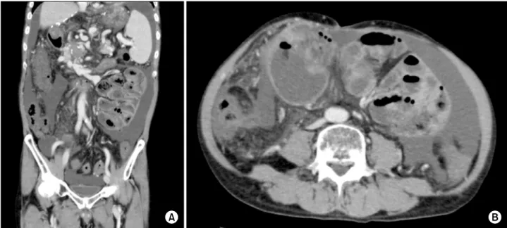
Fig. 1. Abdominal computed tomography (CT) scan reveals remarkable ascites, thickening of the peritoneum and fibrous sheaths surrounding the small intestine, and presence of a me- chanical intestinal obstruction.
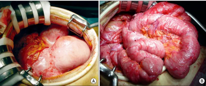
Fig. 2. On laparotomy, the small intestine appears to be encapsulated with a fibrous peel (A). We removed the fibrous membrane, and a total enteroclysis was performed (B).
(HCC) on top of chronic HCV. The patient previously un- derwent esophageal variceal ligation. The patient was on warfarin treatment for portal and superior mesenteric vein thrombosis. At the time of admission, he had massive as- cites and the Child-Pugh classification was B. The patient underwent LDLT and splenectomy in December 2010. A left lobe was used as a graft harvested from his brother.
After LT, the patient was readmitted several times to our clinic due to cholangitis secondary to anastomotic biliary strictures and was managed by percutaneous transhepatic biliary drainage (PTBD) and balloon dilatation. Eight months after transplant, he returned with a complaint of nausea, vomiting, and abdominal distension. The labo- ratory values were: white blood cells (WBCs) 6.7×10⁹/L, hemoglobin (Hb) 10 g/dl, platelet count (PLT) 348,000/l, aspartate transferase (AST) 39 IU/L, alanine transferase (ALT) 32 IU/L, gama-glutamyl-transferase (GGT) 295 IU/L, alkaline phosphatase (ALP) 1,094 IU/L, total bilir- ubin (T.bil) 0.7 mg/dl, creatinine (Cr) 1.5 mg/dl, and C-reactive protein (CRP) 2.6 mg/dl. An abdominal com- puted tomography (CT) scan revealed remarkable ascites, thickening of the peritoneum, fibrous membranes sur- rounding the small intestine leading to paralytic ileus, and eventually mechanical intestinal obstruction (Fig. 1). The CT scan findings were consistent with SEP. On lapa- rotomy, we found the small intestine encapsulated by a fibrous membrane. We removed the fibrous membrane, and subsequently, total adhesiolysis and enteroclysis were
performed (Fig. 2A, B). The postoperative course was un- eventful, and the patient was discharged.
Case 2
A 54-years-old man suffered from chronic HCV in- fection and was a chronic carrier of Human T-Cell Lymphotropic Virus (HTLV). At the time of admission, a huge HCC was detected in the right liver lobe that was complicated with portal vein thrombosis (PVT), and lung metastases during his surveillance in 2007. After receiving two cycles of interferon/5-fluorouracil (IFN/5FU) therapy by hepatic artery infusion, the tumor shrank and follow-up
Fig. 3. Abdominal CT scan reveals small intestinal loops en- cased by a fibrous sheath with secondary ileus and fluid collection.
Fig. 4. Small intestine wrapped around in a fibrous mem- brane, consistent with intra-abdominal cocoon. The fibrous peel was removed and adhesiolysis was performed.
chest CT images had been negative since then. Right hep- atectomy was performed. In 2008, the patient underwent splenectomy to increase platelets count. In August 2014, the patient underwent LDLT for end-stage of liver disease secondary to liver cirrhosis and the Child-Pugh classi- fication was B. Right lobe liver graft was used. The post- operative course was uneventful and the patient was discharged. On the 59th postoperative day, the patient de- veloped fever and abdominal pain. Further examination revealed hepatic artery pseudoaneurysm and intra- abdominal fluid collection. Coiling embolization and fluid collection drainage were performed. Enterococcus was de- tected in the sampled drained fluid. In July 2015, the pa- tient had repeated ileus episodes. Abdominal CT scan re- vealed small intestinal loops encased by a fibrous sheath with secondary ileus (Fig. 3). The laboratory values were:
WBC 7.7×10⁹/L, Hb 11.5 g/dl, PLT 337,000/l, AST 21 IU/L, ALT 16 IU/L, ALP 397 IU/L, GGT 56 IU/L, T.Bil 0.4 mg/dl, D.Bil 0.1 mg/dl, albumin 2.7 g/dl, Cr 0.9 mg/dl, and CRP 0.2 mg/dl. The patient did not respond to conservative treatment, and could not tolerate oral feed- ing, so we decided to perform surgery. An exploratory laparotomy revealed extensive membranous adhesions in the abdominal cavity. The entire small intestine was wrap- ped around with the fibrous membrane, consistent with in- tra-abdominal cocoon (Fig. 4). Subsequently, the fibrous peel was removed and adhesiolysis was performed. The patient's postoperative course was uneventful and he was discharged thereafter.
Case 3
A 63-years-old man suffered from ESLD secondary to HCV cirrhosis. The patient underwent LDLT in July 2002. Previously, the patient had received interferon and ribavirin treatment for HCV recurrence. Five years after transplant, the patient developed refractory ascites due to PVT, and warfarin administration was started. The patient had suffered from hepatic encephalopathy (HE) in November 2015 and spontaneous bacterial peritonitis (SBP) in February 2016, respectively. The patient devel- oped portal hypertension and chronic liver disease. The liver function was poor, and he was on the waiting list for cadaveric liver transplantation. The Child-Pugh classi- fication was C. Due to deterioration in condition; he was admitted to the intensive care unit (ICU) in October 2016.
He had HE, and his laboratory values were: WBC 4.3×10⁹/L, Hb 9 g/dl, PLT 63,000/l, AST 19 IU/L, ALT 16 IU/L, GGT 26 IU/L, ALP 360 IU/L, T.bil 1.5 mg/dl, CRP 0.7 mg/dl, Cr 1.5 mg/dl, and PT/INR 1.9. Blood am- monia level was 193 g/dl. An abdominal CT revealed ascites, splenomegaly, and PVT with collateral veins. The CT scan also showed the presence of a fibrous sheath around the intestine with intestinal dilatation (Fig. 5).
According to these findings, a diagnosis of the abdominal cocoon was made. We considered doing a surgical inter- vention but the liver function and clinical condition of the patient worsened and unfortunately, we lost the patient.
Fig. 5. Abdominal CT reveals ascites, splenomegaly, and portal vein thrombosis with collateral veins (A). The CT scan also shows the presence of a fibrous sheath around the intestine with intestinal dilatation (B).
Fig. 6. Abdominal CT reveals ascites, small bowel dilatation, and wall inflammation consistent with sclerosing peritonitis (A).
Repeated abdominal CT shows that contrast material did not pass through the colon, and the ileus continued (B).
Case 4
A 60-year-old man had ESLD due to liver cirrhosis secondary to chronic HCV. He underwent LDLT in July 2003. Ten years after LT, a relapsing HCV episode was treated with interferon administration. Recurrent episodes of cholangitis due to biliary stricture, with hep- aticojejunostomy, carried out. In 2014, the patient was di- agnosed with diabetes mellitus, coronary artery disease, and chronic kidney disease. Subsequently, he underwent coronary artery stenting and peritoneal dialysis. In
December 2016, the patient experienced abdominal pain, nausea, and vomiting. Abdominal CT revealed ascites, small bowel dilatation, and peritoneal wall inflammation consistent with sclerosing peritonitis (Fig. 6A). The pa- tient was admitted to our clinic, and conservative therapy was started (oral intake was stopped, a nasogastric tube was placed for decompression, and intravenous fluid was given). At the time of admission, his laboratory values were: WBC 4.5×10⁹/L, Hb 8.6 g/dl, PLT 86,000 /l, AST 45 IU/L, ALT 39 IU/L, GGT 210 IU/L, T.bil 1.0 mg/dl,
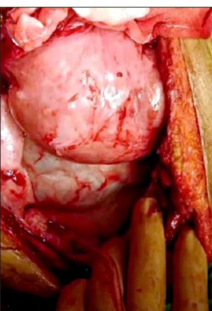
Fig. 7. Thick fibrotic peel wrapping loops of small bowel.
D.bil 0.7 mg/dl, albumin 2.7 g/dl, PT/INR 1.05, Cr 5.95 mg/dl, and CRP 0.5 mg/dl. Despite conservative treat- ment, no improvement in the patients’ conditions was observed. Repeated abdominal CT showed that contrast material did not pass through the colon, and the ileus con- tinued (Fig. 6B). Surgery was decided and laparotomy was performed. At laparotomy, we found dense, thick fi- brotic peel wrapping loops of small bowel (Fig. 7). After the excision of the peel, an edematous appearance of the affected ileal loops was found and multiple serosal in- juries occurred during dissection. Around 30 cm of the affected ileal segment was resected and end-to-end anasto- mosis was performed. The patient developed fever and ab- dominal pain on the 8th postoperative day, and on further examination, an anastomotic leakage was detected. The patient was reoperated, and ileostomy and abdominal washing were performed. The postoperative course was uneventful, and the patient was discharged. After 3 months, the patient was readmitted and the stoma was closed.
DISCUSSION
To the best of our knowledge, this is the first case ser- ies describing four SEP cases all following LDLT. All pa- tients had no evidence of SEP before or during LT. A wide variation for post-LDLT intervals till the develop- ment of SEP findings was seen in all the 4 cases; 8 months, 12 months, 13 years, and 14 years, respectively.
Homogeneous immunosuppressive therapy of Tacrolimus and Mycophenolate sodium was adopted in the 4 cases.
All 4 patients had a history of potential sepsis on SEP diagnosis. Three out of 4 patients had ascites. The first patient had an anastomotic biliary stricture and possible biliary leak which might have contributed to SEP development. The first and the fourth patients experienced multiple episodes of cholangitis, which were managed by PTBD or hepaticojejunostomy, respectively. The third pa- tient had a history of SBP, while the fourth patient had peritoneal dialysis.
All the patients presented with obstructive gastro- intestinal symptoms. Initially, contrast studies were per- formed in all the four cases and delayed transit time for the contrast material to pass through the small intestine was observed for all patients. On abdominal CT, abdomi- nal cocoon findings were detected. The third patient ex- hibited extremely poor liver function at the time of SEP diagnosis to perform surgery. Unfortunately, the patient was lost due to deteriorated liver failure and SEP complications. The other three patients were successfully operated upon (mortality rate was 25%). Until now, all the 3 patients are doing well, without SEP recurrence.
Only 2 case series and 3 case reports regarding SEP after LT were previously reported.4-8 Maguire et al.4 de- scribed the development of SEP in 5 patients within 2 months after LT. As per the report, 3 patients underwent deceased donor LT and 2 patients received right lobe grafts from living donors. The contrast studies and ab- dominal CT scans findings were consistent with the re- sults of our study. All patients underwent adhesiolysis.
One patient died due to intracerebral infarction and pul- monary aspiration. Mortality rate was 20%. However, a specific pre-LT history with regards to ascites or SBP was not illustrated. It is hypothesized that such conditions could play a substantial role in the pathogenesis of SEP.
The second case series published by Mekeel et al de- scribed 3 SEP cases, of which one case was a retransplantation.5 One patient was diagnosed with SEP during LT, and the other 2 cases were diagnosed 4 and 5 months after LT, respectively. However, a pre-LT his- tory of refractory ascites, multiple paracentesis, and SBP was reported in this series. Two patients had encasement of their grafts, which subsequently resulted in biliary and outflow obstruction. Adhesiolysis was performed in all the
cases but unfortunately, the mortality rate was 100%.
Causes of mortality were sepsis and multiorgan failure.
Few reports have demonstrated the development of an interesting gender distribution of SEP cases after LT.
Until date, a total of 16 cases including ours have been reported in the literature, and only one of them was a female. However, a causal association between SEP and gender is still unproven.
The pathogenesis of SEP remains unclear; however, it is believed that both chronic irritation and inflammation triggers this condition. The SEP can be fatal, as evident from published literature. Immunosuppression has been proven to exacerbate the symptoms of the disease result- ing in delayed diagnosis. Therefore, correct and early di- agnosis is important. Pre- and post-transplant sepsis-sug- gestive history (SBP, PD, ascites or paracentesis) should be borne in mind. Generally, surgical intervention (adhesiolysis and small bowel resection) is necessary for SEP treatment. However, Takeuchi et al. reported success- ful treatment of SEP in LDLT patient with tamoxifen.8 Tamoxifen was occasionally used as a therapeutic option in 4 SEP cases.13 Moustafellos et al.,14 reported that ta- moxifen treatment led to improvement in SEP in 2 cases, diagnosed after kidney transplantation. Tamoxifen prob- ably interferes with transforming growth factor beta-1 (TGF-beta-1) and is thus regarded as potentially useful in the treatment of SEP.15
Reaching to preoperative diagnosis of SEP could be puzzling. High threshold of suspicion is required.
Abdominal CT scans are pivotal. The prognosis for SEP is poor in patients with poor graft liver functions which need to be optimized prior to prompt surgical adhesiolysis. Endoscopic drainage might be better than PTBD for management of biliary stricture with respect to SEP prevention.
REFERENCES
1. Yip FW, Lee SH. The abdominal cocoon. Aust N Z J Surg 1992;62:638-642.
2. Godlewski G, Kadi B, Branger B, Vécina F, Deschodt G, Joujoux JM, et al. Encapsulating peritonitis after chronic peri- toneal dialysis. Apropos of 3 cases. Chirurgie 1994;119:686-689;
discussion 690.
3. Holland P. Sclerosing encapsulating peritonitis in chronic ambu- latory peritoneal dialysis. Clin Radiol 1990;41:19-23.
4. Maguire D, Srinivasan P, O’Grady J, Rela M, Heaton ND.
Sclerosing encapsulating peritonitis after orthotopic liver transplantation. Am J Surg 2001;182:151-154.
5. Mekeel K, Moss A, Reddy KS, Douglas D, Mulligan D.
Sclerosing peritonitis and mortality after liver transplantation.
Liver Transpl 2009;15:435-439.
6. Abul S, Al-Oazweni H, Zalat S, Al-Sumait B, Asfar S. Cocoon abdomen in a liver transplant patient. J R Coll Surg Edinb 2002;47:579-581.
7. Lin CH, Yu JC, Chen TW, Chan DC, Chen CJ, Hsieh CB.
Sclerosing encapsulating peritonitis in a liver transplant patient:
a case report. World J Gastroenterol 2005;11:5412-5413.
8. Takeichi T, Narita Y, Lee KJ, Yamamoto H, Asonuma K, Inomata Y. Sclerosing encapsulating peritonitis after living donor liver transplantation: a case successfully treated with tamoxifen:
report of a case. Surg Today 2013;43:1326-1329.
9. Yamamoto S, Sato Y, Takeuchi T, Kobayashi T, Hatakeyama K. Sclerosing encapsulating peritonitis in two patients with liver cirrhosis. J Gastroenterol 2004;39:172-175.
10. Stanley MM, Reyes CV, Greenlee HB, Nemchausky B, Reinhardt GF. Peritoneal fibrosis in cirrhotics treated with peri- toneovenous shunting for ascites. an autopsy study with clinical correlations. Dig Dis Sci 1996;41: 571-577.
11. Wig JD, Goenka MK, Nagi B, Vaiphei K. Abdominal cocoon in a male: rare cause of intestinal obstruction. Trop Gastroenterol 1995;16:31-33.
12. Narayanan R, Bhargava BN, Kabra SG, Sangal BC. Idiopathic sclerosing encapsulating peritonitis. Lancet 1989;2:127-129.
13. Eltoum MA, Wright S, Atchley J, Mason JC. Four consecutive cases of peritoneal dialysis-related encapsulating peritoneal scle- rosis treated successfully with tamoxifen. Perit Dial Int 2006;26:203-206.
14. Moustafellos P, Hadjianastassiou V, Roy D, Velzeboer NE, Maniakyn N, Vaidya A, et al. Tamoxifen therapy in encapsulat- ing sclerosing peritonitis in patients after kidney transplantation.
Transplant Proc 2006;38:2913-2914.
15. Allaria PM, Giangrande A, Gandini E, Pisoni IB. Continuous ambulatory peritoneal dialysis and sclerosing encapsulating peri- tonitis: tamoxifen as a new therapeutic agent? J Nephrol 1999;
12:395-397.