Letter to the Editor
Vol. 28, No. 1, 2016 115
Received October 24, 2014, Revised March 2, 2015, Accepted for publication March 21, 2015
Corresponding author: Ken Igawa, Department of Dermatology, Tokyo Medical and Dental University, Graduate School of Medicine, Yushima 1-5-45, Bunkyo-ku, Tokyo 1138519, Japan. Tel: 81-3-5803-5286, Fax: 81-3-5803-0143, E-mail: k.igawa.derm@tmd.ac.jp
This is an Open Access article distributed under the terms of the Creative Commons Attribution Non-Commercial License (http://
creativecommons.org/licenses/by-nc/4.0) which permits unrestricted non-commercial use, distribution, and reproduction in any medium, pro- vided the original work is properly cited.
http://dx.doi.org/10.5021/ad.2016.28.1.115
Increasing Numbers of Mast Cells in Skin Lesions of Hyperpigmented Mycosis Fungoides with Large-Cell Transformation
Mayo Kondo, Ken Igawa, Takichi Munetsugu, Sayaka Shibama, Aya Nishizawa, Shown Tokoro, Hiroo Yokozeki
Department of Dermatology, Tokyo Medical and Dental University, Graduate School of Medicine, Tokyo, Japan
Dear Editor:
Mycosis fungoides (MF) is a common type of cutaneous T-cell lymphoma. In recent years, several reports have been published on an uncommon and rare form of MF:
hyperpigmented MF1-4. Herein, we report a case of hyper- pigmented MF and discuss the mechanisms of “hyper- pigmentation.”
A 55-year-old Japanese man presented at the dermato- logical outpatient clinic of our hospital for widespread, pruritic, hyperpigmented plaques. The lesions, which first appeared nearly 15 years ago, gradually developed; they were diagnosed as MF, and were subsequently treated with topical corticosteroids and/or radiation with nar- row-band ultraviolet B (UVB). However, no therapeutic re- sponse was observed and small nodular lesions developed.
On physical examination of the patient, we observed hy- perpigmented plaques with no atrophy on the face, trunk, and extremities (Fig. 1A). We found small nodules on the back. Blood cell count, urinalysis, and serum chemistry were within normal limits. Anti-human T-cell leukemia vi- rus type I antibody was negative. Computed tomography scan from the neck to the pelvic area was normal.
Histopathological analysis of skin specimens collected from a hyperpigmented plaque on the buttocks revealed slight acanthosis with atypical lymphocytic infiltration within the epidermis and upper dermis, containing some inflammatory cells and abundant melanophages (Fig. 1B).
Rearrangement of the T-cell receptor (β chain) was de- tected using reverse transcriptase polymerase chain re-
action method, but not Southern blot analysis. Immuno- histochemically, most of the atypical lymphoid cells were CD4-positive (Fig. 1C and D). We diagnosed the lesions as MF, particularly, hyperpigmented MF. After X-ray irradi- ation to the nodules on the back, oral retinoid plus long-wave UVA therapy was started. Although CD30 (+) large-cell transformation was found during the therapeutic course, the patient’s general health and skin improved over time.
So far, the etiology of hyperpigmentation in certain MF cases is not clear. Pavlovsky et al.4 reported that hyper- pigmented MF was characterized by a predominantly CD8-positive phenotype. Those authors speculated that CD8-positive tumor cells could have stronger cytotoxicity and affect neighboring cells such as melanocytes or basal keratinocytes, thereby causing interface changes and marked melanin incontinence, leading to the clinical ap- pearance of hyperpigmentation4. However, in the present case, CD4-positive cells were dominant; therefore, hyper- pigmentation cannot be attributed to this hypothesis.
Yamamoto et al.1 reported the association of mast cell number with hyperpigmented MF. Melanocytes are strongly stimulated by stem cell factor (SCF), which also activates the mast cells. Increasing numbers of mast cells in the skin with lesions and an upregulation of SCF in sera of hyperpigmented MF cases were found1. Thus, we com- pared the numbers of mast cells in the skin with lesion with normal skin controls and non-pigmented MF cases.
Mast cell counts were averaged from three random fields
Letter to the Editor
116 Ann Dermatol
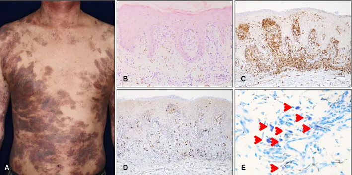
Fig. 1. (A) Clinical manifestation. Hyperpigmented plaques on the trunk. (B) Histology of the lesional skin of a hyperpigmented plaque.
Slight acanthosis with atypical lymphocytic infiltration within the epidermis and upper dermis, containing abundant melanophages (H&E, ×100). (C, D) Immunohistochemical analysis (×100). (C) CD4 staining and (D) CD8 staining. (E) Mast cells in lesional skin.
Red arrowheads indicate mast cells (toluidine blue, ×400).
Table 1. Mast cell counts in skin
Variable Cells/HPF
Present case 43.67
Mycosis fungoides
1 19.33
2 19.67
Normal human skin
1 8.67
2 6.67
3 11.67
Values are presented as number. HPF: high-power field.
(cells/high-power field) in toluidine blue-stained skin spe- cimens (Fig. 1E). As shown in Table 1, compared with nor- mal and non-pigmented MF skin, the numbers of mast cells clearly increased in skin lesions of hyperpigmented MF. Unfortunately, we did not examine the serum levels of SCF. However, the increasing numbers of mast cells
suggest the existence of similar underlying mechanisms as reported by Yamamoto et al.1
REFERENCES
1. Yamamoto T, Katayama I, Nishioka K. Role of mast cell and stem cell factor in hyperpigmented mycosis fungoides. Blood 1997;90:1338-1340.
2. Lee JS, Yun SJ, Lee JB, Kim SJ, Won YH, Lee SC. A case of hyperpigmented mycosis fungoides: a rare variant. J Eur Acad Dermatol Venereol 2007;21:983-985.
3. Erbil H, Sezer E, Koseoglu D, Filiz N, Kurumlu Z, Bülent Taştan H, et al. Hyperpigmented mycosis fungoides: a case report. J Eur Acad Dermatol Venereol 2007;21:982-983.
4. Pavlovsky L, Mimouni D, Amitay-Laish I, Feinmesser M, David M, Hodak E. Hyperpigmented mycosis fungoides: an unusual variant of cutaneous T-cell lymphoma with a frequent CD8+ phenotype. J Am Acad Dermatol 2012;
67:69-75.