J Korean Surg Soc 2012;83:83-87
http://dx.doi.org/10.4174/jkss.2012.83.2.83
ORIGINAL ARTICLE
Journal of the Korean Surgical Society
JKSS
pISSN 2233-7903ㆍeISSN 2093-0488
Received March 27, 2012, Revised June 4, 2012, Accepted June 19, 2012 Correspondence to: Dong Yi Kim
Division of Gastroenterologic Surgery, Department of Surgery, Chonnam National University Medical School, 42 Jebong-ro, Dong-gu, Gwangju 501-746, Korea
Tel: +82-62-220-6450, Fax: +82-62-227-1635, E-mail: dockim@chonnam.ac.kr
cc Journal of the Korean Surgical Society is an Open Access Journal. All articles are distributed under the terms of the Creative Commons Attribution Non-Commercial License (http://creativecommons.org/licenses/by-nc/3.0/) which permits unrestricted non-commercial use, distribution, and reproduction in any medium, provided the original work is properly cited.
Preoperative predictors of malignant gastric submucosal tumor
Ho Goon Kim, Seong Yeob Ryu, Sang Kwon Yun, Jae Kyoon Joo, Jae Hyuk Lee
1, Dong Yi Kim
Division of Gastroenterologic Surgery, Department of Surgery, 1Department of Pathology, Chonnam National University Medical School, Gwangju, Korea
Purpose: The preoperative prediction of malignant potential in patients with gastric submucosal tumors (SMTs) plays an im- portant role in decisions regarding their surgical management. Methods: We evaluated the predictors of malignant gastric SMTs in 314 patients with gastric SMTs who underwent surgery in Chonnam National University Hospital. Results: The ma- lignant SMTs were significantly associated with age (odds ratio [OR], 1.067; 95% confidence interval [CI], 1.042 to 1.091; P < 0.0001), presence of central ulceration (OR, 2.690; 95% CI, 1.224 to 5.909; P = 0.014), and tumor size (OR, 1.791; 95% CI, 1.483 to 2.164; P < 0.0001). Receiver operating characteristic curve analysis showed that tumor size was a good predictor of malig- nant potential. The most relevant predictor of malignant gastric SMT was tumor size with cut-offs of 4.05 and 6.40 cm.
Conclusion: Our findings indicated that age, central ulceration, and tumor size were significant preoperative predictors of malignant SMTs. We suggest that 4 cm be selected as a threshold value for malignant gastric SMTs. In patients with a gastric SMT larger than 4 cm with ulceration, wide resection of the full thickness of the gastric wall or gastrectomy with adequate margins should be performed because of its malignant potential.
Key Words: Stomach neoplasms, Submucosal tumor, Malignant factor, Preoperative predictor
INTRODUCTION
Gastric submucosal tumors (SMTs) account for less than 2% of all neoplasms of the stomach and stromal tumors are the most common tumors of the gastric submucosa [1,2].
Most SMTs are asymptomatic and benign, but 15 to 30%
are malignant [3,4]. Endoscopic ultrasonography (EUS) is widely used following endoscopy for the evaluation of SMTs, but EUS is not yet reliable enough to differentiate between benign and malignant SMTs. Brand et al. [5] re-
ported a sensitivity of 95% and a specificity of 72% for di- agnosing gastrointestinal stromal tumors, while Oguz et al. [6] reported a sensitivity of 50% and specificity of 72%
for diagnosing gastrointestinal SMTs. More than 30% of the patients with malignant tumors develop local re- currence and distant metastases [7]. Therefore, the pre- operative prediction of malignant potential in patients with gastric SMTs plays an important role in the decision regarding surgical management. This study examined the preoperative predictors of malignant gastric SMTs.
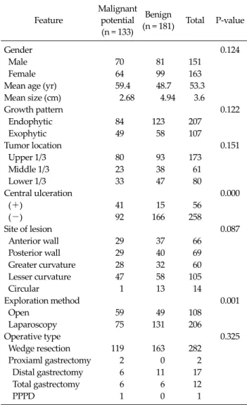
Table 1.Clinicopathological features of the malignant potential and benign groups
Feature
Malignant potential (n = 133)
Benign
(n = 181) Total P-value
Gender 0.124
Male 70 81 151
Female 64 99 163
Mean age (yr) 59.4 48.7 53.3 Mean size (cm) 2.68 4.94 3.6
Growth pattern 0.122
Endophytic 84 123 207
Exophytic 49 58 107
Tumor location 0.151
Upper 1/3 80 93 173
Middle 1/3 23 38 61
Lower 1/3 33 47 80
Central ulceration 0.000
(+) 41 15 56
(-) 92 166 258
Site of lesion 0.087
Anterior wall 29 37 66
Posterior wall 29 40 69
Greater curvature 28 32 60
Lesser curvature 47 58 105
Circular 1 13 14
Exploration method 0.001
Open 59 49 108
Laparoscopy 75 131 206
Operative type 0.325
Wedge resection 119 163 282
Proxiaml gastrectomy 2 0 2 Distal gastrectomy 6 11 17 Total gastrectomy 6 6 12
PPPD 1 0 1
PPPD, pylorus-preserving pancreaticoduodenectomy.
METHODS
Patients and specimens
From January 2004 to December 2009, 314 patients (151 males, 163 females) with suspected SMT of the stomach underwent surgery in Chonnam National University Hospital, Gwangju, Korea. Patient information was gath- ered from the hospital records retrospectively. Eleven var- iables were evaluated for each patient: patient age and gender, the use of EUS, pre- and postoperative diagnosis, tumor growth pattern, exploration method, type of oper- ation, presence of central ulceration, and tumor location and size. To establish the diagnosis and determine the ex- tent of the disease, all patients underwent a preoperative work-up including esophagogastroduodenoscopy, EUS, and computed tomography (CT). We classified the pa- tients into malignant potential and benign groups. The ag- gressive risk was defined according to the size and mitotic rate of the tumors, as proposed by Fletcher et al. [8]. We evaluated the accuracy of the preoperative diagnosis and the sensitivity and specificity of the preoperative diag- nosis in all cases. Furthermore, we compared the sensi- tivity and specificity of diagnosis with and without per- forming EUS.
Operation
The operative indications included a SMT > 20 mm in size and definitely visible by endoscopy, irrespective of symptoms. Tumors < 20 mm in size measured on EUS or CT were observed or a gastrectomy was performed when the patient requested surgery because of concern. Laparo- scopic resection and local excision were performed using three methods: extragastric wedge resection was perfor- med for SMTs with an exophytic growth pattern; trans- gastric resection was performed for endophytic SMTs; and intragastric resection was performed for SMTs located at the esophagogastric junction. Intraoperative gastroscopy was used to identify and mark small tumors and to ensure that the tumor was excised with an adequate margin.
Conventional open surgery was performed via an upper midline laparotomy.
Statistical analysis
To identify significant independent correlates of overall and malignant risk, a stepwise procedure was applied for selected factors with P < 0.05 in order to identify in- dependent potent risk factors. A multiple logistic model was applied to evaluate the odds ratios of the major risk factors. Statistical analyses were conducted using PASW ver. 18.0 (IBM Co., Armonk, NY, USA).
RESULTS
Table 1 shows the clinicopathological features of the pa-
Table 3. Multivariate analyses of factors related to malignant gastric submucosal tumors
Predictive factor for
malignancy P-value Odds ratio (95% CI)
Age 0.000 1.067 (1.042-1.091)
Gender 0.356
Presentation of central ulcer 0.000 2.690 (1.224-5.909)
Growth patter 0.171
Location of tumor
Upper 1/3 0.082
Middle 1/3 0.035
Lower 1/3 0.268
Anterior wall 0.100
Posterior wall 0.910
Greater curvature 0.246 Lesser curvature 0.184
Circular 0.774
Size 0.000 1.791 (1.483-2.164)
CI, confidence interval.
Table 2. Sensitivity, specificity, PPV, and NPV of the diagnosis of malignant submucosal tumor (exclusion criterion: preoperative diagnosis not described, n = 63)
Sensitivity (%) Specificity (%) PPV (%) NPV (%)
Diagnosis for malignant SMTs without EUS 93.8 35.1 71.7 76.4
Diagnosis for malignant SMTs with EUS 88.0 46.7 39.3 90.9
PPV, positive predictive value; NPV, negative predictive value; SMTs, submucosal tumors; EUS, endoscopic ultrasonography.
Fig. 1. The receiver operating characteristic curve of tumor size for predicting malignant gastric submucosal tumors.
tients with gastric SMTs. In total, 314 patients with a SMT in the stomach underwent surgery. Of the patients, 151 (48.1%) were male and 163 (51.9%) were female. The mean age was 53.3 years and the mean tumor size was 3.6 ± 2.5 cm in maximum diameter (range, 0.6 to 20.0 cm). The growth pattern was endophytic in 207 cases (65.9%) and exophytic in 107 cases (34.1%). The lesions were located in the upper one third in 173 (55.1%) patients including the esophagogastric junction, in the middle third in 61 (19.4%) patients, and in the lower one third in 80 (25.5%) patients.
Fifty-six tumors (17.8%) were ulcerated. Wedge resection was the procedure carried out most frequently in patients with gastric SMTs. The preferred procedure was laparo- scopic surgery.
The sensitivity, specificity, and negative and positive predictive values of diagnosis without EUS for malignant SMT were 93.8, 35.1, 71.7, and 76.4%, respectively. The cas- es diagnosed with EUS had respective values of 88.0, 46.7,
39.3, and 90.9% for potential malignancy (Table 2).
Multivariate analysis of the predictors of malignant SMTs is summarized in Table 3. The malignant SMTs were significantly associated with age (odds ratio [OR], 1.067;
95% confidence interval [CI], 1.042 to 1.091; P < 0.0001), the presence of central ulceration (OR, 2.690; 95% CI, 1.224 to 5.909; P = 0.014), and tumor size (OR, 1.791; 95% CI, 1.483 to 2.164 for a 1-cm increase; P < 0.0001). No relationship between the other factors (gender, growth pattern, tumor location) and malignant potential was found.
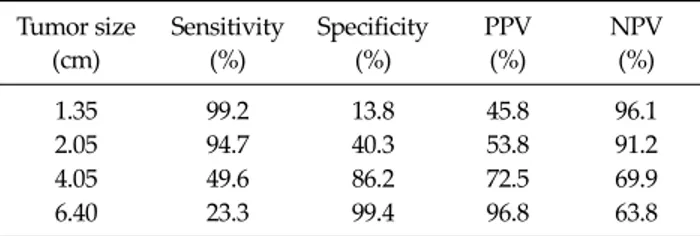
The receiver operating characteristic (ROC) curve anal- ysis showed that tumor size was a good predictor of malig- nant potential (Fig. 1). The area under the ROC curve was 0.772 ± 0.026 (95% CI, 0.721 to 0.823). The diagnostic per- formance of malignant potential was further analyzed us- ing two cut-off values: a lower value to improve sensitivity and a higher value to improve specificity. Table 4 shows the different threshold values, and the most relevant ones for malignant SMT prediction were 4.05 and 6.40 cm. A cut-off value of 4.05 cm gave a sensitivity of 49.6%, specif- icity of 86.2%, positive predictive value of 72.5%, and neg- ative predictive value of 69.9%. When the cut-off value was
Table 4. Sensitivity, specificity, PPV, and NPV of the diagnosis of malignant submucosal tumor using size to identify the malignant potential
Tumor size (cm)
Sensitivity (%)
Specificity (%)
PPV (%)
NPV (%)
1.35 99.2 13.8 45.8 96.1
2.05 94.7 40.3 53.8 91.2
4.05 49.6 86.2 72.5 69.9
6.40 23.3 99.4 96.8 63.8
PPV, positive predictive value; NPV, negative predictive value.
increased to 6.40 cm, the specificity reached 99.4% and the positive predictive value 96.8%.
DISCUSSION
The surgical principles for managing SMTs include lap- aroscopic resection for small tumors and avoidance of tu- mor rupture, wedge resection, and complete resection of gross disease and adherent organs [9-12]. A wide resection of the full thickness of the gastric wall with adequate mar- gins is a satisfactory procedure for malignant gastric SMTs, while mass enucleation may be sufficient for benign gastric SMTs [12,13]. The preoperative prediction of ma- lignant potential in patients with gastric SMTs plays an important role in deciding on the operative method.
However, it is difficult to diagnose whether a tumor is ma- lignant preoperatively; the definitive diagnosis of gastric SMTs is possible only with pathological confirmation.
EUS is used widely in the evaluation of gastric SMTs.
Unfortunately, EUS is not yet reliable enough for differ- entiating between benign and malignant SMTs. Palazzo et al. [14] reported that the three most predictive EUS fea- tures for malignancy were irregular margins, cystic spaces, and lymph nodes with a malignant pattern. They found that the presence of at least one of these criteria had a sensi- tivity of 91%, specificity of 88%, positive predictive value of 83%, and negative predictive value of 94% for potential malignancy. We found that EUS has a high diagnostic sen- sitivity for the malignant potential of SMT, in agreement with previous studies [8,9,14]. However, EUS had a low specificity and positive predictive value for the diagnosis of malignant SMTs in our study. Therefore, we need better
preoperative predictors for malignant SMTs to establish the surgical plan.
In this study, three factors predicted malignant SMTs in the multivariate analysis. The first was age. Rabin et al.
[15] found a significant correlation between age and ma- lignant potential and reported that the younger the pa- tient, the higher the incidence of malignancy. Contrary to their results, the mean age was higher in the malignant group in our series. No similar data regarding age as a pre- dictor of malignant SMTs were found when reviewing other reports.
Second, mucosal ulceration is a common feature of gas- tric SMTs. Miettinen et al. [16] reported that the presence of ulceration has no predictive value or prognostic sig- nificance, although it may be related to tumor size. In an- other study, Miettinen et al. [17] demonstrated that ulcer- ation was common in all histologic subtypes but, never- theless, was an adverse prognostic factor, probably be- cause of its consistent presence in malignant SMTs. In con- trast to their result, we found that tumor ulceration pre- dicted malignant SMTs.
Third, tumor size was another predictor of malignant SMTs in our series. Tumor size is an easily applicable mor- phologic criterion for predicting tumor behavior. In some studies, tumor size > 6 cm has been suggested as a thresh- old value for malignancy [18,19]. Miettinen et al. [17] sug- gested that 5 cm was a threshold value for malignant SMTs, despite an unpredictable, but low, frequency of un- expected progressive disease among patients with rela- tively small tumors and low mitotic activity, although this frequency does not exceed 2 to 3%. Hsu et al. [20] also re- ported that tumor size ≥ 10 cm carried both a higher risk of recurrence and worse survival in SMTs. We used ROC curves to determine the optimal cut-off value for size to distinguish malignant and benign tumors. Examining dif- ferent threshold values, the most relevant for malignant SMTs prediction were 4.05 and 6.40 cm. A cut-off value of 4.05 cm had a sensitivity of 49.6%, specificity of 86.2%, pos- itive predictive value of 72.5%, and negative predictive value of 69.9%. When the cut-off value was increased to 6.40 cm, the specificity reached 99.4% and the positive pre- dictive value was 96.8%. We propose 4 cm as the threshold value.
In conclusion, malignant SMTs were significantly asso- ciated with age, the presence of central ulceration, and tu- mor size. We suggest that a tumor size of 4 cm be selected as the threshold value for malignant SMTs. If an ulcerated SMT is bigger than 4 cm, we recommend a wide wedge re- section of the full thickness of the gastric wall, or gas- trectomy with adequate margins, because it has high po- tential for malignancy.
CONFLICTS OF INTEREST
No potential conflict of interest relevant to this article was reported.
REFERENCES
1. Campbell F, Bogomoletz WV, Williams GT. Tumours of the oesophagus and stomach. In: Fletcher CD, editor. Diagnos- tic histopathology of tumours. London: Churchill Living- stone; 1995. p.193-242.
2. Kempson RL, Hendrickson MR. Gastrointestinal stromal (smooth muscle) tumours. In: Whitehead R, editor. Gastro- intestinal and oesophageal pathology. 2nd ed. Edinburgh:
Churchill Livingstone; 1995. p.727-39.
3. Chak A. EUS in submucosal tumors. Gastrointest Endosc 2002;56(4 Suppl):S43-8.
4. Connolly EM, Gaffney E, Reynolds JV. Gastrointestinal stromal tumours. Br J Surg 2003;90:1178-86.
5. Brand B, Oesterhelweg L, Binmoeller KF, Sriram PV, Bohnacker S, Seewald S, et al. Impact of endoscopic ultra- sound for evaluation of submucosal lesions in gastro- intestinal tract. Dig Liver Dis 2002;34:290-7.
6. Oguz D, Filik L, Parlak E, Disibeyaz S, Cicek B, Kacar S, et al. Accuracy of endoscopic ultrasonography in upper gas- trointestinal submucosal lesions. Turk J Gastroenterol 2004;15:82-5.
7. Krajinovic K, Germer CT, Agaimy A, Wunsch PH, Isbert C.
Outcome after resection of one hundred gastrointestinal stromal tumors. Dig Surg 2010;27:313-9.
8. Fletcher CD, Berman JJ, Corless C, Gorstein F, Lasota J, Longley BJ, et al. Diagnosis of gastrointestinal stromal tu- mors: a consensus approach. Hum Pathol 2002;33:459-65.
9. Samelis GF, Ekmektzoglou KA, Zografos GC. Gastrointes- tinal stromal tumours: clinical overview, surgery and re- cent advances in imatinib mesylate therapy. Eur J Surg Oncol 2007;33:942-50.
10. Sasaki A, Koeda K, Nakajima J, Obuchi T, Baba S, Waka- bayashi G. Single-incision laparoscopic gastric resection for submucosal tumors: report of three cases. Surg Today 2011;41:133-6.
11. Eisenberg BL, Judson I. Surgery and imatinib in the man- agement of GIST: emerging approaches to adjuvant and neoadjuvant therapy. Ann Surg Oncol 2004;11:465-75.
12. Singaporewalla RM, Baladas GH, Lee TD. Laparoendosco- pic removal of a benign gastric stromal tumor at the cardia.
JSLS 2006;10:117-21.
13. Silberhumer GR, Hufschmid M, Wrba F, Gyoeri G, Schoppmann S, Tribl B, et al. Surgery for gastrointestinal stromal tumors of the stomach. J Gastrointest Surg 2009;
13:1213-9.
14. Palazzo L, Landi B, Cellier C, Cuillerier E, Roseau G, Barbier JP. Endosonographic features predictive of benign and malignant gastrointestinal stromal cell tumours. Gut 2000;46:88-92.
15. Rabin I, Chikman B, Lavy R, Sandbank J, Maklakovsky M, Gold-Deutch R, et al. Gastrointestinal stromal tumors: a 19 year experience. Isr Med Assoc J 2009;11:98-102.
16. Miettinen M, El-Rifai W, Sobin HL, Lasota J. Evaluation of malignancy and prognosis of gastrointestinal stromal tu- mors: a review. Hum Pathol 2002;33:478-83.
17. Miettinen M, Sobin LH, Lasota J. Gastrointestinal stromal tumors of the stomach: a clinicopathologic, immunohisto- chemical, and molecular genetic study of 1765 cases with long-term follow-up. Am J Surg Pathol 2005;29:52-68.
18. Appleman HD, Helwig EB. Gastric epithelioid leiomyoma and leiomyosarcoma (leiomyoblastoma). Cancer 1976;38:
708-28.
19. Wong NA, Young R, Malcomson RD, Nayar AG, Jamieson LA, Save VE, et al. Prognostic indicators for gastro- intestinal stromal tumours: a clinicopathological and im- munohistochemical study of 108 resected cases of the stomach. Histopathology 2003;43:118-26.
20. Hsu KH, Yang TM, Shan YS, Lin PW. Tumor size is a major determinant of recurrence in patients with resectable gas- trointestinal stromal tumor. Am J Surg 2007;194:148-52.