□ Case Report □
816
Newly Discovered Pseudoaneurysm after Embolization of a Renal Arteriovenous Fistula with a Pseudoaneurysm following a Renal Stab Wound
Seong Beom Choi, Myung Ki Kim, Young Beom Jeong, Jong Kwan Park, Hyung Jin Kim, Young Gon Kim
From the Department of Urology, Chonbuk National University Medical School, Institute for Medical Sciences, Jeonju, Korea
Post-traumatic arteriovenous fistula (AVF) and pseudoaneurysm are rare, and mostly occur in stab wound patients. Suspected AVF and pseudo- aneurysm requires angiography, with planned simultaneous embolization.
Superselective embolization is generally a safe and effective treatment modality for AVF and pseudoaneurysm with minimal associated mor- bidity. Rare complications of the embolization do occur, including renal abscess, postembolization syndrome, impaired renal function, pulmonary embolism caused by migration of coils, and allergic reaction. We present here the case of a man who presented with a newly discovered pseudo- aneurysm after embolization of a renal AVF with pseudoaneurysm after a renal stab wound. (Korean J Urol 2009;50:816-818)
Key Words: Arteriovenous fistula, False aneurysm, Therapeutic emboli- zation
Korean Journal of Urology Vol. 50 No. 8: 816-818, August 2009 DOI: 10.4111/kju.2009.50.8.816
Received:May 8, 2009 Accepted:July 17, 2009
Correspondence to: Myung Ki Kim Department of Urology, Chonbuk National University Medical School, Institute for Medical Sciences, 634-18, Geumam-dong, Deokjin-gu, Jeonju 561-756, Korea
TEL: 063-250-2574 FAX: 063-250-1564
E-mail: [email protected]
Ⓒ The Korean Urological Association, 2009
Renal trauma accounts for 1-3% of all patients with traumatic injury.1 Arteriovenous fistula (AVF) is rare and occurs mostly after penetrating renal trauma, most commonly after renal bio- psy.2,3 The incidence of AVF after renal stab wounds varies be- tween 0% and 7% according to several studies.3-5
About 50% to 70% of cases of renal AVF occurring after renal biopsy improve without any treatment. But most post- traumatic AVFs require radiologic interventions and sometimes nephrectomy.1
Renal angiography is used for diagnosis and treatment.
Among the many treatment methods, superselective emboli- zation is the safest and most effective treatment modality when an AVF is confirmed.6
Rare complications of embolization do occur, including renal abscess, postembolization syndrome, impaired renal function, pulmonary embolism caused by migration of coils, allergic reaction, and hematoma on the arterial puncture site.7,8 When complications do occur, close follow-up is required for possible potentially life-threatening conditions.
Here we report a very rare case of a newly discovered pseudoaneurysm on an adjacent vessel 3 weeks after super-
selective embolization for renal AVF and pseudoaneurysm after a renal stab wound.
CASE REPORT
A 35-year-old man was admitted to the emergency room with gross hematuria and abdominal pain after receiving multiple stab wounds. A physical exam revealed two stab wounds on the right flank and right upper quadrant of the abdomen. The patient’s mental status was alert and his blood pressure and pulse rate were normal. Abdominal computed to- mography (CT) showed grade III right renal trauma, hemo- thorax, and suspected hemoperitoneum, but vessel injury of the right kidney was not observed. A thoracostomy was done to treat the hemothorax. Considering the patient's young age and his being hemodynamically stable, conservative management was planned for the renal trauma. Laparotomy was performed because intraperitoneal hemorrhage was suspected, but no internal organ injury was observed. However, gross hematuria persisted until 7 days after the laparotomy. The follow-up abdo- minal CT evaluation revealed a 1.7 cm sized pseudoaneurysm
Seong Beom Choi, et al:Newly Discovered False Aneurysm following Renal Embolization 817
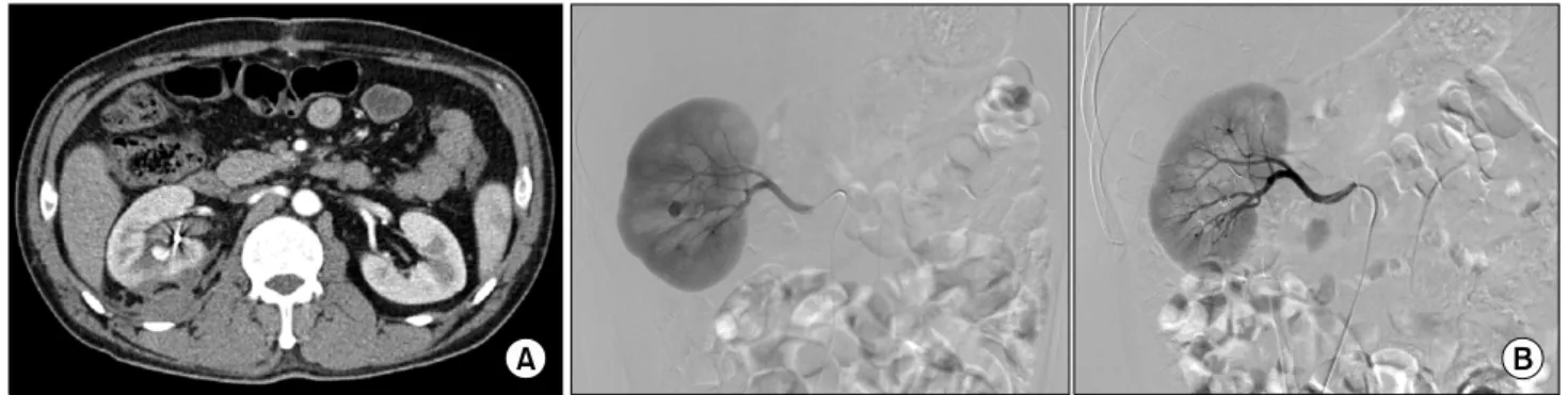
Fig. 2. (A) The embolization coil is visible around the right pelvis and a 9 mm sized pseudoaneurysm is observed. (B) The pseudoaneurysm is visible in the interlobar artery branch of the mid-artery, and embolization was performed after superselection of vessels. Devascularization of the pseudoaneurysm was observed.
Fig. 1. (A) Computed tomography with contrast in the arterial phase shows bright enhancement in the central portion of the right kidney, suspicious of a pseudoaneurysm. (B) Angiography shows a 2.5x2.0 cm sized pseudoaneurysm (black arrow) in the posterior branch of the lower pole of renal artery and early visualization of the renal veins (white arrow) during the arterial phase of the study due to arteriovenous fistula. Angiography after superselective coil embolization of the pseudoaneurysm and arteriovenous fistula.
with AVF on the right lower pole of kidney (Fig. 1A). Right renal arteriography was performed by the Seldinger technique after puncturing the right pulmonary artery. As a result, we discovered AVF with pseudoaneurysm in the posterior branch of the lower pole of the renal artery. Superselective embo- lization with a coil (TornadoⓇ Embolization Microcoils) was performed to treat the renal AVF with pseudoaneurysm through percutaneous renal angiography (Fig. 1B). Neither AVF nor pseudoaneurysm was observed after the procedure. No anti- coagulant was used before or after angiography. The patient was discharged without any complications following the embo- lization. The patient was then readmitted to the emergency room 3 weeks after the embolization for sudden gross hema- turia. Abdominal CT showed no abnormality on the embolized vessel but a newly discovered pseudoaneurysm, sized 9 mm, in the interlobar artery branch of the mid-artery (Fig. 2A).
Superselective embolization with a coil was performed by percutaneous renal angiography on the vessel with the new pseudoaneurysm (Fig. 2B). The patient showed no signs of complications during 6 months of follow-up after the second embolization.
DISCUSSION
Delayed hemorrhage is one of the most serious complications of renal trauma. It is common with laceration in the renal cortex, especially when conservatively managing a renal stab wound.2 The possibility of delayed hemorrhage occurring after renal-penetrating trauma or stab wound is reported to be 18%
to 23%.2,5 Most of the delayed hemorrhage is thought to be caused by renal AVF.9
Direct injury on the artery due to a stab wound causes
818 Korean Journal of Urology vol. 50, 816-818, August 2009
pooling of blood in the tunica adventitia, renal parenchyme, and Gerota's fascia and the development of a hematoma. When the bleeding stops, absorption of the hematoma and necrotic tissue creates a space between the tunica intima and adventitia, which forms a pseudoaneurysm. The pseudoaneurysm increases in size and may rupture into the pelvocaliceal system or perirenal space.
Most patients present with symptoms such as hematuria, flank pain, abdominal bruit on auscultation, and hypertension.
The possibility of developing a pseudoaneurysm must be considered, especially when hematuria persists in patients with penetrating renal trauma or a renal stab wound.10
Angiography is the most definitive diagnostic tool for pseu- doaneurysm and is now highly recommended due to the con- venience of treating with embolization during the procedure.6 Complications following embolization include renal abscess, postembolization syndrome, impaired renal function, pulmonary embolism caused by migration of coils, allergic reaction, and hematoma on the arterial puncture site.7,8
The present case is very rare; there are no known reports of a newly discovered pseudoaneurysm following the emboli- zation of renal AVF with pseudoaneurysm.
Vascular complications such as AVF and pseudoaneurysm should always be considered in renal trauma, especially in renal stab wound patients, and require angiography when associated symptoms are present. Complications may cause life-threate- ning conditions compared to renal injury due to blunt trauma.
Therefore, continuous follow-up after the embolization is ne- cessary to minimize the occurrence of complications. In parti- cular, delayed bleeding or hematuria following the embolization require close follow-up for possible pseudoaneurysm, which
may rarely occur, as in this case.
REFERENCES
1. Santucci RA, Wessells H, Bartsch G, Descotes J, Heyns CF, McAninch JW, et al. Evaluation and management of renal injuries: consensus statement of the renal trauma subcom- mittee. BJU Int 2004;93:937-54
2. Brandes SB, McAninch JW. Complications of renal trauma.
In: Taneja SS, Smith RB, Ehrlich RM, editors. Complications of urology. 3rd ed. Philadelphia: Saunders; 2001;205-25 3. Bernath AS, Schutte H, Fernandez RR, Addonizio JC. Stab
wounds of the kidney: conservative management in flank penetration. J Urol 1983;129:468-70
4. Kansas BT, Eddy MJ, Mydlo JH, Uzzo RG. Incidence and management of penetrating renal trauma in patients with multi- organ injury: extended experience at an inner city trauma center. J Urol 2004;172:1355-60
5. Armenakas NA, Duckett CP, McAninch JW. Indications for nonoperative management of renal stab wounds. J Urol 1999;
161:768-71
6. Benson DA, Stockinger ZT, McSwain NE Jr. Embolization of an acute renal arteriovenous fistula following a stab wound:
case report and review of the literature. Am Surg 2005;71:62-5 7. Kim HS, Ryu SB, Min BK. A case of control of renal hemor- rhage by selective renal arterial embolization. Korean J Urol 1988;29:324-8
8. Hwang HH, Cheon SH, Moon KH, Lee SK, Choo HS, Hwang JC, et al. Renal ruptures with active bleeding treated with emergency selective renal arterial embolizaton. Korean J Urol 2008;49:177-81
9. Heyns CF, Van Vollenhoven P. Selective surgical management of renal stab wounds. Br J Urol 1992;69:351-7
10. Al-Qudah HS, Santucci RA. Complications of renal trauma.
Urol Clin North Am 2006;33:41-53