Revisiting Rupture of Benign Thyroid Nodules after Radiofrequency Ablation: Various Types and Imaging Features
Sae Rom Chung1, Jung Hwan Baek1, Jin Yong Sung2, Ji Hwa Ryu3, So Lyung Jung4
1Department of Radiology and the Research Institute of Radiology, Asan Medical Center, University of Ulsan College of Medicine; 2Department of Radiology and Thyroid Center, Daerim St. Mary’s Hospital, Seoul; 3Department of Radiology, Inje University Haeundae Paik Hospital, Inje University College of Medicine, Busan; 4Department of Radiology, Seoul St. Mary’s Hospital, College of Medicine, The Catholic University of Korea, Seoul, Korea
Background: To evaluate the imaging features, clinical manifestations, and prognosis of patients with thyroid nodule rupture after radiofrequency ablation (RFA).
Methods: The records of 12 patients who experienced thyroid nodule rupture after RFA at four Korean thyroid centers between March 2010 and July 2017 were retrospectively reviewed. Clinical data evaluated included baseline patient characteristics, treatment methods, initial presenting symptoms, imaging features, treatment, and prognosis.
Results: The most common symptoms of post-RFA nodule rupture were sudden neck bulging and pain. Based on imaging features, the localization of nodule rupture was classified into three types: anterior, posterolateral, and medial types. The anterior type is the most often, followed by posterolateral and medial type. Eight patients recovered completely after conservative treatment. Four patients who did not improve with conservative management required invasive procedures, including incision and drainage or aspiration.
Conclusion: Thyroid nodule rupture after RFA can be classified into three types based on its localization: anterior, posterolateral, and medial types. Because majority of thyroid nodule ruptures after RFA can be managed conservatively, familiarity with these im- aging features is essential in avoiding unnecessary imaging workup or invasive procedures.
Keywords: Thyroid nodule; Ultrasonography; Radiofrequency ablation; Complication; Safety
INTRODUCTION
Radiofrequency ablation (RFA) is a minimally invasive tech- nique that has been used to treat various tumors and has been shown effective in patients with benign thyroid nodules [1-4]
and recurrent thyroid cancers [5-7]. Although RFA is relatively
safe for treating thyroid nodules, as shown in several systematic reviews and meta-analyses [8-10], it is technically challenging and requires extensive experience [10,11]. Understanding all possible complications of RFA is required for proper diagnosis and treatment.
The most common major complication after RFA is voice
Received: 2 September 2019, Revised: 15 November 2019, Accepted: 29 November 2019
Corresponding author: Jung Hwan Baek
Department of Radiology and the Research Institute of Radiology, Asan Medical Center, University of Ulsan College of Medicine, 88 Olympic-ro 43-gil, Songpa- gu, Seoul 05505, Korea
Tel: +82-2-3010-4348, Fax: +82-2-476-0090, E-mail: [email protected]
Copyright © 2019 Korean Endocrine Society
This is an Open Access article distributed under the terms of the Creative Com- mons Attribution Non-Commercial License (http://creativecommons.org/
licenses/by-nc/4.0/) which permits unrestricted non-commercial use, distribu- tion, and reproduction in any medium, provided the original work is properly cited.
change [8,10], caused by injury to the recurrent laryngeal or va- gus nerve, but this complication may be reduced by the use of a trans-isthmic approach and moving shot technique during RFA [8-10]. The second most common major complication is nodule rupture after RFA [8,10]. To date, however, little is known about the imaging features, clinical course, and risk factors for this ad- verse event [8,12-14]. This study therefore evaluated the imag- ing features, clinical manifestations, and prognosis of patients with thyroid nodule rupture after RFA.
METHODS
The Institutional Review Boards of the four participating thy- roid centers approved the protocol of this retrospective study (S2017-1436-0001). All patients provided written informed consent prior to RFA.
The records of 12 patients who experienced thyroid nodule rupture after RFA at four Korean thyroid centers between March 2010 and July 2017 were retrospectively reviewed. Clinical data included baseline patient characteristics, treatment method, ini- tial presenting symptoms, imaging features, treatment, and prognosis were evaluated.
Before RFA, all nodules were found to be benign by fine nee- dle aspiration or core needle biopsy [15-17]. Ultrasound (US)- guided RFA was performed by the same radiologist who per- formed the US assessment. All ablations were performed using a radiofrequency (RF) generators (Cool-Tip RF system, Covidi- en, Boulder, CO, USA; SSP-2000, Taewoong Medical, Gimpo, Korea; and M-1004, RF Medical, Seoul, Korea) and an 18- gauge internally cooled electrode (Well-Point RF Electrodes, Taewoong Medical, Goyang, Korea; VIVA, STARmed, Goy- ang, Korea; Big-Tip, RF Medical, Seoul, Korea; Cool-tip RF system, Radionics, Valleylab, Boulder, CO, USA) with 0.7-, 1-, 1.5-, and 2-cm active tips, respectively, depending on the size of the nodule and the preference of the radiologists. Local anesthe- sia consisted of injection of 2% lidocaine into the puncture site and perithyroidal area [18]. RFA was performed using the trans- isthmic approach and moving shot technique under US guid- ance [2,4,11]. If nodules contained fluid, the fluid was aspirated prior to RFA [19,20]. Adjacent structures, including nerves and vessels, were carefully monitored during the procedure to avoid complications [21]. The electrode tip was initially positioned in the deepest and farthest area of the nodule and then moved after a transient hyperechoic zone appeared within 5 to 10 seconds of turning on the RF power [2,4,11]. If a hyperechoic microbubble did not form at the electrode tip within 5 to 10 seconds, the RF
power was increased in 10 W increments. If the patient com- plained of pain during the procedure, the RF power was reduced or turned off for several seconds. The procedure was terminated when the entire visualized area of the nodule had become a tran- sient hyperechoic zone. Repeat ablations were performed when unsatisfactory volume reductions (50%) of treated nodules after 3 months and the presence of an initially untreated portion [12].
All patients suspected of nodule rupture after RFA were exam- ined by US and/or computed tomography (CT).
RESULTS
A review of medical records identified 12 patients, three men and nine women, of mean age 41 years (range, 16 to 75), who experienced post-RFA nodule rupture. Their clinical details are summarized in Table 1. Of these 12 patients, 10 had thyroid nodules with solid and cystic portions and two had solid nodule.
The mean volume of the index nodules was 17.0 mL (range, 0.19 to 74.6). One patient required three RFA procedures, two required two procedures, and nine required one ablation. The mean ablation time and mean maximal ablation power per RFA session were 13.4 minutes (range, 4.7 to 40) and 57.5 W (range, 30 to 110), respectively.
Table 2 summarizes the clinical and imaging manifestations of these 12 patients with thyroid nodule rupture. The most com- mon symptoms of post-RFA nodule rupture during follow-up were sudden neck bulging and pain. Only one patient reported fever, cough, and neck discomfort. The mean time from RFA to nodule rupture was 54.6 days (range, 11 to 156).
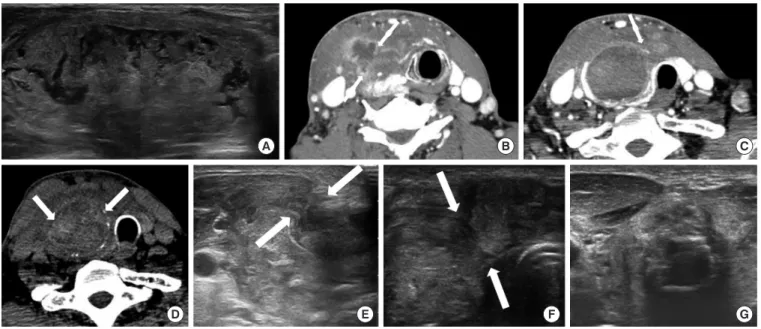
Following the development of symptoms, 10 patients were evaluated by US, one by CT, and one by US and CT. Based on their localization on imaging features, nodule rupture could be classified into three types, the most common feature being ante- rior type, observed in nine patients (75%) and characterized by extension of the nodule contents into the anterior extrathyroidal area and sometimes into the strap muscle, with discontinuity of the anterior thyroid capsule (Fig. 1). Two patients (16.7%) had the posterolateral type of nodule rupture, which was character- ized by heterogeneous fluid collection around the thyroid cap- sule (Fig. 2). One patient (8.3%) had the medial type of rupture, which was characterized on CT by soft tissue bulging at the me- dial aspect of the ablated nodule and protrusion into the lumen of the trachea (Fig. 3). This patient experienced fever, cough, and neck discomfort, unlike the other patients.
All 12 patients were initially treated conservatively, with close observation and/or oral antibiotics. Eight patients recov-
ered completely with conservative treatment after mean follow- up period of 71 days (range, 11 to 202). The mean period of use of antibiotics was 16 days (range, 7 to 34). Four patients re- quired invasive procedures, including incision and drainage or aspiration. The nature of aspirated fluid were hemorrhage in
two patients and pus in two patients. All of these four patients had the anterior type of rupture. Fig. 1 shows the US images of one patient who underwent aspiration. Sonography showed breakdown of the anterior thyroid capsule and the communica- tion between intra- and extrathyroidal lesions at the RF site. US Table 1. Baseline Characteristics of the 12 Patients with Thyroid Nodule Rupture after RFA
No. of
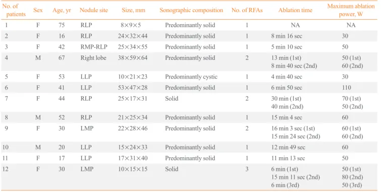
patients Sex Age, yr Nodule site Size, mm Sonographic composition No. of RFAs Ablation time Maximum ablation power, W
1 F 75 RLP 8×9×5 Predominantly solid 1 NA NA
2 F 16 RLP 24×32×44 Predominantly solid 1 8 min 16 sec 30
3 F 42 RMP-RLP 25×34×55 Predominantly solid 1 5 min 10 sec 50
4 M 67 Right lobe 38×59×64 Predominantly solid 2 13 min (1st)
8 min 40 sec (2nd) 50 (1st) 60 (2nd)
5 F 53 LLP 10×21×23 Predominantly cystic 1 4 min 40 sec 30
6 F 41 LLP 53×47×28 Predominantly solid 1 6 min 50 sec 110
7 F 44 RLP 25×17×31 Solid 2 30 min (1st)
40 min (2nd) 70 (1st)
50 (2nd)
8 M 52 RLP 21×25×34 Predominantly solid 1 15 min 4 sec 60
9 F 30 LMP 22×28×46 Predominantly solid 2 16 min 3 sec (1st)
15 min 24 sec (2nd) 60 (1st) 60 (2nd)
10 M 20 LLP 15×24×33 Predominantly solid 1 12 min 49 sec 60
11 F 17 LLP 17×31×40 Predominantly solid 1 11 min 13 sec 50
12 F 30 LMP 10×15×15 Solid 3 6 min (1st)
15 min 11 sec (2nd) 6 min (3rd)
50 (1st) 80 (2nd) 50 (3rd) RFA, radiofrequency ablation; RLP, right lower pole; NA, not applicable; RMP, right middle pole; LLP, left lower pole; LMP, left middle pole.
Table 2. Clinical and Imaging Manifestations in the 12 Patients with Thyroid Nodule Rupture after RFA Patient
no. Time to rupture
from RFA, day Symptoms Type Initial treatment Invasive procedure Outcome
1 30 NA Anterior NA - Complete recovery
2 156 Sudden neck bulging and pain Posterolateral Compression - Complete recovery
3 65 Cough, fever, neck discomfort Medial Antibiotics and observation - Complete recovery 4 80 Sudden neck bulging and pain Anterior Antibiotics and observation Aspiration Complete recovery
5 11 Sudden neck bulging and pain Anterior Observation - Complete recovery
6 48 Sudden neck bulging and pain Posterolateral Observation - Complete recovery
7 30 Sudden neck bulging and pain Anterior Antibiotics and observation Aspiration No FU
8 40 Sudden neck bulging and pain Anterior Antibiotics and observation - Complete recovery 9 57 Sudden neck bulging and pain Anterior Antibiotics and observation I&D Complete recovery 10 21 Sudden neck bulging and pain Anterior Antibiotics and observation I&D Complete recovery 11 20 Sudden neck bulging and pain Anterior Antibiotics and observation - Complete recovery
12 48 Sudden neck bulging and pain Anterior Observation - Complete recovery
RFA, radiofrequency ablation; NA, not applicable; FU, follow-up; I&D, incision and drainage.
also showed a heterogeneous mass-like lesion extending from the thyroid nodule to the superficial aspect of the strap muscle, with Doppler US showing increased vascularity. US-guided core needle biopsy showed that the mass-like lesion was an or- ganizing hematoma. Despite compression, this hematoma in- creased in size on follow-up US. US-guided aspiration resulted in the removal of dark blood material, with follow-up US show- ing that this patient recovered completely. Including this patient, all patients, except for the one lost to follow-up, experienced complete recovery.
DISCUSSION
This multicenter study, which describes 12 patients who experi- enced thyroid nodule rupture after RFA, is, to our knowledge, the first original study to classify types of post-RFA nodule rup- ture according to their localization. The anterior type is the most often, followed by posterolateral and medial type. Despite dif- ferences in their imaging features, 67% patients (eight of 12 pa- tients) with thyroid nodule ruptures were successfully managed conservatively. Knowledge about the typical clinical symptoms (sudden neck bulging and pain) and imaging features of post- Fig. 1. (A) A 67-year-old man, who presented with a bulging neck mass, had been treated with radiofrequency ablation (RFA) due to a pre- dominantly solid thyroid nodule at right thyroid gland. He had sudden bulging and pain on the right side of his neck 80 days after the RFA.
(B, C, D) Computed tomography (CT) and (E, F) ultrasound show volume expansion and discontinuity of the anterior thyroid capsule with nodule content extended to the anterior extra-thyroidal area (arrows) (anterior type). (D) Intra-nodular hyper-attenuating portions on pre- contrast CT represent intra-nodular bleeding. (G) The patient was managed with fluid aspiration and intravenous antibiotics, and the lesion gradually regressed.
Fig. 2. (A) A 24-year-old woman, who presented with a bulging neck mass, had been treated with radiofrequency ablation (RFA) due to a predominantly solid thyroid nodule at right thyroid gland. (B) One month after RFA, the nodule decreased in size. However, at 156 days af- ter RFA, the patient complained of neck pain and bulging. (C) Ultrasound shows a heterogeneous echoic lesion around the thyroid capsule (arrows) (posterolateral type). She was treated with compression and observation. (D) After a month, the lesion was completely disappeared.
A
D E F G
B C
A B C D
RFA nodule rupture can ensure the proper diagnosis of patients and reduce unnecessary treatment.
RFA is a minimally invasive technique that may serve as a safe and effective alternative to surgery in managing benign thyroid nodules that cause symptoms and cosmetic problems [1- 4,22-27]. A recent meta-analysis indicated that the mean volume change following RFA for benign thyroid nodules was 76.1%
(95% confidence interval [CI], 70.1% to 82.1%), with an abso- lute mean change in volume of 8.9 mL (95% CI, 6.6 to 11.2) [27]. RFA is also reported to improve symptom and cosmetic scores, as well as having low complication rates [1-3,22-24].
Although RFA has a lower complication rate than surgery, com- plications can still occur [8-10,13,28], with nodule rupture be- ing the second most common major complication [8,10]. Thy- roid nodule rupture after RFA is defined as the breakdown of the thyroid capsule and the communication between intra- and extrathyroidal lesions. Thyroid nodule rupture has firstly been reported after laser ablation [29]. In that study, three patients had pseudocysts with fasciitis owing to fluid leakage in the neck muscle fascia, followed by spontaneous reabsorption of these at 3 to 6 months. A subsequent report described six patients at four institutions who experienced thyroid nodule rupture after RFA
[12], and there have been occasional case reports describing the occurrence of nodule rupture [13,14].
All previous ruptures that could be evaluated using imaging modalities were anterior type [8,12]. However, although our re- view of 12 patients at four institutions found that the anterior type was the most common, two patients had the posterolateral type and one had the medial type of rupture. The anterior type typically exhibits an anterior bulging of a mass that can extend to the strap muscle layer. The posterolateral type is a hyperecho- ic lesion that surrounds the thyroid capsule rather than a bulging anterior mass. The medial type shows a discontinuity of the me- dial thyroid capsule, with the internal contents bulging toward the tracheoesophageal (TE) groove, causing a mass effect on the trachea. Because the TE groove area is a relatively limited space, unlike the anterior or posterolateral area, the medial rup- ture is the most uncommon type and its causes remain unclear.
Familiarity with these imaging characteristics is important in distinguishing among these types of nodule rupture after RFA and in avoiding additional unnecessary imaging workup or in- vasive procedures. In fact, the one patient with the posterolateral type and one with the medial type were referred to tertiary refer- ral center from local clinics because of misdiagnosis.
Fig. 3. (A) A 42-year-old female, who presented with bulging neck mass, had been treated with radiofrequency ablation (RFA) due to a pre- dominantly solid thyroid nodule at right thyroid gland. She complained of cough, neck discomfort and mild fever 65 days after RFA. (B, C) On computed tomography (CT) scan, the ablated nodule bulges to medial side and protrudes into the tracheal lumen (arrows) (medial type).
(D, E) After conservative management with antibiotics, the lesion was completely disappeared on follow-up CT and ultrasound after 2 months.
A
D E
B C
Thyroid nodule rupture after RFA is thought to be caused by delayed bleeding, as shown by the high attenuation observed on CT with volume expansion of treated nodule, as well as aspira- tion of old blood in all patients who underwent invasive proce- dures. Bleeding may result from microvascular leakage within the tumor, leading to delayed volume expansion and rupture;
tearing of the tumor wall and thyroid capsule at a weak point; or post-procedural massaging or moving of the neck. The differ- ences in imaging features observed in the three types of post- RFA nodule rupture may arise from differences in the causes of rupture. The anterior type may be caused by a more acute ex- pansion of the treated nodule with hemorrhage, whereas the posterolateral type may be caused by a more persistent oozing.
All four patients who required invasive procedures had the ante- rior type of rupture, perhaps because this type is caused by a rel- atively large volume of hemorrhage. Because hemorrhage is ba- sically aseptic and self-limiting, we recommend that these pa- tients should be managed conservatively with observation and compression, with or without oral antibiotics, rather than under- go invasive procedures [12]. Eight of the 12 patients in this study completely recovered without any invasive procedure, such as aspiration or drainage. Invasive procedures should be performed only when conservative management is unsuccessful.
In conclusion, thyroid nodule rupture after RFA can be classi- fied into three types based on its localization: anterior, postero- lateral, and medial types. Because majority of thyroid nodule ruptures after RFA can be managed conservatively, familiarity with these imaging features is essential in avoiding unnecessary imaging workup or invasive procedures.
CONFLICTS OF INTEREST
No potential conflict of interest relevant to this article was re- ported.
AUTHOR CONTRIBUTIONS
Conception or design: J.H.B. Acquisition, analysis, or interpre- tation of data: S.R.C., J.H.B., J.Y.S., J.H.R., S.L.J. Drafting the work or revising: S.R.C., J.H.B., J.Y.S., J.H.R., S.L.J. Final ap- proval of the manuscript: J.H.B.
ORCID
Sae Rom Chung https://orcid.org/0000-0003-4219-7166 Jung Hwan Baek http://orcid.org/0000-0003-0480-4754
REFERENCES
1. Baek JH, Moon WJ, Kim YS, Lee JH, Lee D. Radiofre- quency ablation for the treatment of autonomously function- ing thyroid nodules. World J Surg 2009;33:1971-7.
2. Jeong WK, Baek JH, Rhim H, Kim YS, Kwak MS, Jeong HJ, et al. Radiofrequency ablation of benign thyroid nod- ules: safety and imaging follow-up in 236 patients. Eur Ra- diol 2008;18:1244-50.
3. Kim YS, Rhim H, Tae K, Park DW, Kim ST. Radiofrequen- cy ablation of benign cold thyroid nodules: initial clinical experience. Thyroid 2006;16:361-7.
4. Baek JH, Kim YS, Lee D, Huh JY, Lee JH. Benign predom- inantly solid thyroid nodules: prospective study of efficacy of sonographically guided radiofrequency ablation versus control condition. AJR Am J Roentgenol 2010;194:1137-42.
5. Baek JH, Kim YS, Sung JY, Choi H, Lee JH. Locoregional control of metastatic well-differentiated thyroid cancer by ultrasound-guided radiofrequency ablation. AJR Am J Roentgenol 2011;197:W331-6.
6. Guenette JP, Monchik JM, Dupuy DE. Image-guided abla- tion of postsurgical locoregional recurrence of biopsy-prov- en well-differentiated thyroid carcinoma. J Vasc Interv Ra- diol 2013;24:672-9.
7. Kim JH, Yoo WS, Park YJ, Park DJ, Yun TJ, Choi SH, et al.
Efficacy and safety of radiofrequency ablation for treatment of locally recurrent thyroid cancers smaller than 2 cm. Radi- ology 2015;276:909-18.
8. Baek JH, Lee JH, Sung JY, Bae JI, Kim KT, Sim J, et al.
Complications encountered in the treatment of benign thy- roid nodules with US-guided radiofrequency ablation: a multicenter study. Radiology 2012;262:335-42.
9. Wang JF, Wu T, Hu KP, Xu W, Zheng BW, Tong G, et al.
Complications following radiofrequency ablation of benign thyroid nodules: a systematic review. Chin Med J (Engl) 2017;
130:1361-70.
10. Chung SR, Suh CH, Baek JH, Park HS, Choi YJ, Lee JH.
Safety of radiofrequency ablation of benign thyroid nodules and recurrent thyroid cancers: a systematic review and meta- analysis. Int J Hyperthermia 2017;33:920-30.
11. Kim JH, Baek JH, Lim HK, Ahn HS, Baek SM, Choi YJ, et al. 2017 thyroid radiofrequency ablation guideline: Korean Society of Thyroid Radiology. Korean J Radiol 2018;19:632- 55.
12. Shin JH, Jung SL, Baek JH, Kim JH. Rupture of benign thy- roid tumors after radio-frequency ablation. AJNR Am J
Neuroradiol 2011;32:2165-9.
13. Che Y, Jin S, Shi C, Wang L, Zhang X, Li Y, et al. Treatment of benign thyroid nodules: comparison of surgery with radio- frequency ablation. AJNR Am J Neuroradiol 2015;36:1321- 5.
14. Valcavi R, Tsamatropoulos P. Health-related quality of life after percutaneous radiofrequency ablation of cold, solid, benign thyroid nodules: a 2-year follow-up study in 40 pa- tients. Endocr Pract 2015;21:887-96.
15. Na DG, Baek JH, Jung SL, Kim JH, Sung JY, Kim KS, et al.
Core needle biopsy of the thyroid: 2016 consensus state- ment and recommendations from Korean Society of Thyroid Radiology. Korean J Radiol 2017;18:217-37.
16. Shin JH, Baek JH, Chung J, Ha EJ, Kim JH, Lee YH, et al.
Ultrasonography diagnosis and imaging-based management of thyroid nodules: revised Korean Society of Thyroid Radi- ology consensus statement and recommendations. Korean J Radiol 2016;17:370-95.
17. Baek JH. Current status of core needle biopsy of the thyroid.
Ultrasonography 2017;36:83-5.
18. Park HS, Baek JH, Park AW, Chung SR, Choi YJ, Lee JH.
Thyroid radiofrequency ablation: updates on innovative de- vices and techniques. Korean J Radiol 2017;18:615-23.
19. Park HS, Baek JH, Choi YJ, Lee JH. Innovative techniques for image-guided ablation of benign thyroid nodules: com- bined ethanol and radiofrequency ablation. Korean J Radiol 2017;18:461-9.
20. Sim JS, Baek JH, Lee J, Cho W, Jung SI. Radiofrequency ablation of benign thyroid nodules: depicting early sign of regrowth by calculating vital volume. Int J Hyperthermia 2017;33:905-10.
21. Ha EJ, Baek JH, Lee JH. Ultrasonography-based thyroidal and perithyroidal anatomy and its clinical significance. Ko- rean J Radiol 2015;16:749-66.
22. Deandrea M, Limone P, Basso E, Mormile A, Ragazzoni F, Gamarra E, et al. US-guided percutaneous radiofrequency thermal ablation for the treatment of solid benign hyper- functioning or compressive thyroid nodules. Ultrasound Med Biol 2008;34:784-91.
23. Lee JH, Kim YS, Lee D, Choi H, Yoo H, Baek JH. Radio- frequency ablation (RFA) of benign thyroid nodules in pa- tients with incompletely resolved clinical problems after ethanol ablation (EA). World J Surg 2010;34:1488-93.
24. Spiezia S, Garberoglio R, Milone F, Ramundo V, Caiazzo C, Assanti AP, et al. Thyroid nodules and related symptoms are stably controlled two years after radiofrequency thermal ab- lation. Thyroid 2009;19:219-25.
25. Hong MJ, Baek JH, Choi YJ, Lee JH, Lim HK, Shong YK, et al. Radiofrequency ablation is a thyroid function-preserv- ing treatment for patients with bilateral benign thyroid nod- ules. J Vasc Interv Radiol 2015;26:55-61.
26. Lim HK, Lee JH, Ha EJ, Sung JY, Kim JK, Baek JH. Radio- frequency ablation of benign non-functioning thyroid nod- ules: 4-year follow-up results for 111 patients. Eur Radiol 2013;23:1044-9.
27. Ha EJ, Baek JH, Kim KW, Pyo J, Lee JH, Baek SH, et al.
Comparative efficacy of radiofrequency and laser ablation for the treatment of benign thyroid nodules: systematic re- view including traditional pooling and Bayesian network meta-analysis. J Clin Endocrinol Metab 2015;100:1903-11.
28. Bernardi S, Dobrinja C, Fabris B, Bazzocchi G, Sabato N, Ulcigrai V, et al. Radiofrequency ablation compared to sur- gery for the treatment of benign thyroid nodules. Int J Endo- crinol 2014;2014:934595.
29. Valcavi R, Riganti F, Bertani A, Formisano D, Pacella CM.
Percutaneous laser ablation of cold benign thyroid nodules: a 3-year follow-up study in 122 patients. Thyroid 2010;20:
1253-61.