Effects of Deltoid Inhibition Taping on the Surface Electromyographic Activity of Shoulder Girdle Muscles During Upper Limb Elevation in Healthy Shoulders
Suhn-yeop Kim, Ph.D., P.T.
Duck-won Oh, Ph.D., P.T.
Taek-yean Kim, Ph.D., P.T.
Soo-jin Nam
Dept. of Physical Therapy, College of Health and Sports Science, Daejeon University Hwan-suk Yoo, M.Sc., P.T.
Dept. of Occupational Therapy, College of Health and Welfare, Woosong University
Abstract
1)This study aimed to examine whether McConnell taping for deltoid inhibition affects the Electromyographic (EMG) activity of shoulder girdle muscles during arm elevation. Ten young healthy men were randomly assigned to an experimental and control groups of five men each. For the ex- perimental group, we performed taping for deltoid inhibition on the skin over anterior and posterior del- toids with non-elastic specific tape, and sham-taping with non-elastic under-tape for the control group.
Surface EMG measurements were performed three times (before, during and after the tapings) at upper and lower trapezius, mid-deltoid, and serratus anterior muscles while elevating dominant arm with loading and unloading conditions. In deltoid inhibition taping group, there were significant differences in EMG ac- tivity of mid-deltoid (p<.05) and serratus anterior (p<.05) muscles during arm elevation with loading.
During arm elevation without loading, the EMG activity was significantly decreased for MD in the McConnell taping group (p<.05). The findings indicate that deltoid inhibition taping can modify the acti- vation patterns in shoulder girdle muscles as well as in deltoid muscle. in clinical setting, it may be ef- fectively used for the management of patients with shoulder dysfunction.
Key Words: Deltoid; Electromyography; McConnell taping; Shoulder.
Introduction
The restoration of movement at the scap- ulothoracic, glenohumeral, and acromioclavicular joints using taping represents a useful adjunct in pa- tient-specific integrated treatment (Yasojima et al, 2008). By promoting an earlier progression of re- habilitation, the clinical effects of taping the shoulder girdle can be significant and immediate (Yasojima et al, 2008). The physical advantages resulting from taping, including improved shoulder stability, postural correction, decreased pain, and increased function, have recently been reported to be caused by proprio- ceptive and neurophysiologic effects (Karlsson and
Andreasson, 1992) as well as mechanical effects (Thatcher and Davies, 2006).
Many studies have provided electromyographic (EMG) evidence in the application of taping (Gilleard et al, 1998; McConnell, 1986; Tobin and Robinson, 2000). In the case of neurogenic pain caused by in- flammation, the loading response to neural tissue can also be diminished by taping. This may include the neurophysiologic and mechanical benefits of taping (McConnell, 2004). The inflammation of soft tissue interferes with a variety of movements in daily ac- tivity, and it leads to subjective discomfort, in- appropriate muscle activity, and loss of dynamic control while performing functional movements.
Therefore, decreasing inflamed soft tissue loading should be considered one of the major factors that influence the functional recovery of the affected tis- sues (Grelsamer and McConnell, 1998).
Of all the joints in the human body, the shoulder has the greatest mobility, and the normal coordinated strategy of the shoulder muscles is essential to suc- cessfully perform various arm movements (Yasojima et al, 2008). During normal arm elevation, a subtle balance exists between the forces exerted by the deltoid and rotator cuff muscles. Higher loads caused by dominant activation of the deltoid may induce unwanted humeral head superior translation (Clisby et al, 2008; Sahrmann, 2002). Without proper regu- lation, this can result in a positive feedback loop, where damage leads to increased translation, which leads to further damage.
The lack of balance between the deltoid and rota- tor cuff muscles may negatively influence the gleno- humeral joint arthrokinematics of abduction, which is believed to predispose people to shoulder impinge- ment syndrome (Neumann, 2002; Reddy et al, 2000).
Changes in the activation patterns of the shoulder girdle muscles can produce a variety of clinical con- ditions ranging from minor glenohumeral instability to total shoulder dislocation. Shoulder instability can be caused by excessive glenohumeral mobility as well as poor scapular motion at the scapulothoracic joint. The uncoordinated action of scapulothoracic muscles, and hence the lack of scapular stability, has also been recognized as a cause of secondary sub- acromial impingement syndrome (Kibler, 1998).
The effectiveness of various taping techniques has been extensively studied in the knee (Bennellet al, 2006; Gilleard et al, 1998; Kowall et al, 1996) and the ankle (Alt et al, 1999; Karlsson and Andreasson, 1992; McCaw and Cerullo, 1999; Wilkerson, 1991).
Previous studies on shoulder girdle taping have mainly focused on the scapular muscles and their possible involvement in shoulder dysfunction (Ackermann et al, 2002; Cools et al, 2002; Selkowitz et al, 2007). However, to the best of the author’s
knowledge, there has been no research investigating the taping-induced changes in the activation patterns of shoulder girdle muscles in relation to the coordi- nated actions of glenohumeral and scapulothoracic muscles that are required to maintain optimal shoulder function during arm elevation. Therefore, the purpose of this study was to determine whether the McConnell taping for detoid inhibition influences the EMG activity of shoulder girdle muscles during upper limb elevation.
Methods Subjects
Ten young male subjects with ages ranging from 19 to 23 years (mean, 21.1 years), who had asympto- matic shoulders, volunteered to participate in the study. The subjects were randomly assigned to two group: 5 subjects each in the McConnell-taping group (age: 21.2±1.2 years, height: 175.8±5.2 ㎝, and weight:
71.7±8.1 ㎏) and the Sham-taping group (age:
21.0±1.4 years, height: 175.5±6.0 ㎝, and weight:
71.7±8.3 ㎏). All of the subjects were right-hand dominant. None of the subjects had a history of or- thopedic or neurological problems in the upper limb and trunk in the previous 3 months, and all were university students who did not perform resistance training for the upper limbs at the time of data collection. All subjects gave their informed consent, having previously been informed as to both the ob- jectives and the experimental procedures of the study.
Procedures
Subjects were measured as they performed arm elevationin the scapular plane (plane deviated ap- proximately 35 anterior to the frontal plane) with thumb up and elbow fully extended. Measurements of arm elevation (full arm elevation = 2 seconds) were taken under loading and unloading conditions.
The weight of the dumbbell used for loading was varied according to the subject’s body weight: sub-
Figure 1. The McConnell taping for deltoid inhibition.
Figure 2. Electrode placements.
jects with body weight below 60 ㎏, between 70 and 80 ㎏, and above 80 ㎏ used dumbbells of 2 ㎏, 2.5
㎏, and 3 ㎏, respectively. In order to avoid muscle fatigue, all subjects rested for 1minute between each trial. For the experimental group, we performed tap- ing for deltoid inhibition using non-elastic rigid tape1), and sham-taping with non-allergenic elastic adhesive under-tape2) for the control group. Surface EMG measurements were performed three times (before, during, and after the tapings) at the upper trapezius (UT), lower trapezius (LT), mid-deltoid (MD), and serratus anterior (SA) muscles of the dominant upper limb.
Deltoid Inhibition Taping2)3)
The deltoid inhibition taping was performed as recommended by Morrissey (2000), with the aim of elevating the shoulder girdle (Figure 1). All tapings for subjects were carried out by a physical therapist who had attended a special international course for the application of McConnell tape. With 30 abduction of the shoulder in the scapular plane, the application of the tape was performed with subjects sitting in a chair fitted with arm and back rests.
First, with elastic under-tape, an anchor strip was applied at the deltoid tuberosity level, encircling two-thirds of the arm’s circumference, and then an elevatory strip was applied from the anterior and pos- terior deltoid to the upper tip of the shoulder. Second, the taping for deltoid inhibition was performed over anchor tape with non-elastic rigid tape. Some folds were made by slightly pulling the anchor strips.
Surface Electromyographic Recording and Data Processing4)
The EMG data were collected simultaneously from the upper trapezius (UT), lower trapezius (LT), mid-deltoid (MD), and serratus anterior (SA) mus- cles of dominant upper limb using a four-channel portable surface EMG QEMG-43). The disposable Red Dot (3M) electrode which is used to collect EMG signal is Ag/AgCl surface electrode discs covered with conducting gel and adhesive area.
The electrodes were placed on UT, LT, MD and SA at standardized sites (Cram et al, 1998) (Figure 2). Electrodes for the UT were placed parallel to the muscle fibers of the UT, along the ridge of the shoulder, slightly lateral to and one half the distance
1) Endura-Sports tape, OPTP, Minneapolis, U.S.A.
2) Endura-Fix tape, OPTP, Minneapolis, U.S.A.
between the C7 and the acromion. The LT electrode was placed obliquely upward and laterally along a line between the intersection of the scapular spine with the vertebral border of the scapula and the T7 spinous process. The MD electrode was placed on the lateral aspect of the upper arm and approx- imately 3 ㎝ below the acromion. The SA electrode was placed midway between the lateral, inferior bor- der of the scapula and the insertion of the muscle on the anterolateral side of the thorax. A ground elec- trode was placed over the mid-portion of the lumbo- sacral junction.
Prior to electrode placement, the skin was shaved and swabbed with alcohol to reduce skin impedance (typically 10). For each muscle, 2 electrodes were placed at a distance of approximately 3 ㎝ in the di- rection of the muscle fibers. To eliminate interfer- ence, 2-poles electrode shield cables were used.
Surface EMG signals, digitalized in the EMG system, were converted with Telescan 2.89 software (LAXTHA Inc., Daejeon, Korea) on a personal computer. The sampling rate was 1024 ㎐. The EMG signal was amplified with an overall gain of 1000 and digitized using Telescan 2.89 software. Bandpass (20~450 ㎐) and notch filters (60 ㎐) were used.
The raw data were processed into the root-mean-square (RMS), and EMG signals recorded in each muscle were normalized using the RMS of 5-second maximal voluntary isometric contraction (MVIC), which was calculated for each muscle at the manual muscle testing positions (Kendall et al, 2005).
The collecting EMG signals during arm elevation were expressed as a percentage of the calculated RMS of the MVIC (%MVIC).
Statistical Analysis
The data were expressed as mean ± standard deviation. For each measurement, a two-way (group × taping condition) analysis of variance (ANOVA) was used to compare differences in the EMG data between the two groups. The alpha level for statistical sig- nificance was set as .05. When significant differences
were found, post hoc t-tests with Scheffe correction were used for multiple pairwise comparisons.
Results
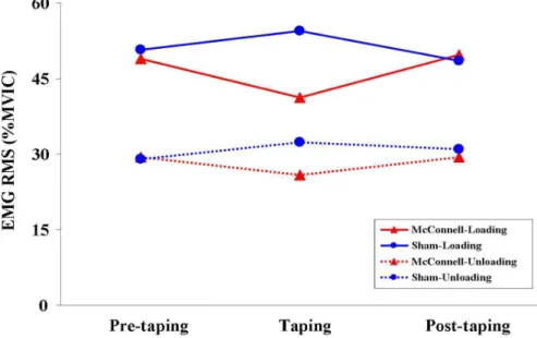
EMG RMS data for UT, LT, MD and SA muscles during arm elevation with and without loading are presented in Table 1, Table 2 and Figure 3. During arm elevation with loading, the EMG RMS values were significantly different for MD in McConnell-taping (F1,4=11.41, p<.05) and Sham-tap- ing groups (F1,4=21.48, p<.05), and the pairwise com- parisons with Scheffe correction revealed a sig- nificant difference between taping and post-taping in both groups (p<.05). Further, the EMG RMS of SA was significantly different in the McConnell-taping group (F1,4=11.24, p<.05), and the pairwise compar- isons revealed a significant difference between pre-taping and taping in both groups (p<.05). During arm elevation without loading, the EMG RMS was significantly different for MD in the McConnell-tap- ing group (F1,4=12.44, p<.05), and significant differ- ences between pre-taping and taping, and between taping and post-taping were found in the pairwise comparisons (p<.05).
Discussion
For a long time, taping has been extensively chosen by physical therapists to accomplish a variety of therapeutic purposes. Taping is a technique that aims to provide protection and support to a joint whilst continuing to permit functional movements re- quired for daily activities (Cools et al, 2002).
However, the support function of taping is lost with- in a relatively short time after application (Alt et al, 1999). There are numerous clinical assumptions on why taping may be effective in decreasing the sub- jective complaints of pain and increasing function.
Most clinicians agree that the effect of taping can
Taping Methods Pre-taping Taping Post-taping F p Mid-deltoid
(MD)
McConnell 48.98±7.30a 41.30±2.92§ 49.65±4.96 11.41 .04
Sham 50.73±12.51 54.57±11.35§ 48.51±10.68 21.48 .02
Upper trapezius (UT)
McConnell 61.15±8.22 128.75±115.38 65.54±11.02 .82 .52
Sham 61.52±13.38 52.79±18.68 57.43±25.31 1.08 .44
Lower trapezius (LT)
McConnell 52.44±34.07 42.04±21.97 45.40±27.57 1.50 .35
Sham 42.63±10.51 46.51±15.19 48.02±15.20 2.30 .25
Serratus anterior (SA)
McConnell 65.28±12.72 81.43±39.28* 72.51±16.36 11.24 .04
Sham 45.86±18.00 41.63±15.72 43.85±17.67 6.11 .09
aMean±SD.
*p<.05 in Pre-taping vs. Taping.
§p<.05 in Taping vs. Post-taping.
Table 1. Changes in the EMG RMS (%MVIC) of UT, LT, MD and SA measured at the pre-taping, taping, and post-taping stages while elevating the arm with loading
Taping Methods Pre-taping Taping Post-taping F p
Mid-deltoid (MD)
McConnell 29.37±2.05 25.82±1.49*,§ 29.43±2.38 12.44 .04
Sham 29.04±10.29 32.30±7.47 30.89±8.11 .71 .56
Upper trapezius (UT)
McConnell 34.24±5.74 49.70±26.03 38.76±7.93 .97 .46
Sham 33.24±10.67 33.50±12.07 33.47±16.25 .01 .99
Lower trapezius (LT)
McConnell 32.31±24.15 23.57±10.74 26.88±15.46 .66 .58
Sham 25.71±10.66 26.40±15.27 28.80±13.65 .66 .58
Serratus anterior (SA)
McConnell 35.22±8.70 43.90±20.19 39.84±11.45 2.63 .22
Sham 27.40±15.38 25.48±13.87 25.56±12.49 1.22 .41
aMean±SD.
*p<.05 in Pre-taping vs. Taping.
§p<.05 in Taping vs. Post-taping.
Table 2. Changes in the EMG RMS (%MVIC) of UT, LT, MD and SA measured at the pre-taping, taping, and post-taping stages while elevating the arm without loading
only partially be explained by increased mechanical stability (Alt et al, 1999; Gilleard et al, 1998; McCaw and Cerullo, 1999). Recently, clinicians’ main concern with regard to taping has focused on optimizing functional capacity by inhibiting the excessive action of synergists and antagonists, as well as facilitating less activated muscles. This is done to enhance pro- prioceptive function, to improve joint alignment, and to reduce physical loading to irritable neural tissues (Host, 1995). According to the recommendations of previous researches, this study was designed to
activation pattern of shoulder girdle muscles, based on mechanical aspects.
Taping has been applied in a variety of methods, depending on the therapeutic aims, though it is espe- cially used to control muscle action. In general, the longitudinal application of taping facilitates the mus- cle tone while the crossed application inhibits muscle tone (Morrisey, 2000). However, this opinion has been challenged by recent studies (Alexander et al, 2003; Alexander et al, 2008). The McConnell taping for deltoid inhibition used in this study can be un-
Figure 3. Changes in the EMG RMS of the mid-deltoid muscle measured at the pre-taping, taping, and post-taping stages while elevating the arm with and without loading.
ical perspective. Our assumption is that passively el- evating the shoulder girdle through taping may cause partial shortening of the deltoid muscle, which may consequently alter the contraction pattern of shoulder girdle muscles when daily activities are performed.
During normal arm elevation, unbalanced activation of the deltoid can be related to an insufficiency in the activity of the rotator cuff muscles, which leads to clinical symptoms of the shoulder (Clisby et al, 2008; Sharmann, 2002). Essentially, it should be con- sidered in the treatment of shoulder impingement syndrome. Ginn (1997) has reported the clinical ben- efits of physical therapy techniques for shoulder dys- function, which are focused on improving joint sta- bility, optimal inter-joint coordination, and muscle function. In our study, McConnell taping for deltoid inhibition was significantly related to the decrease of the EMG activity in the deltoid muscles. This find- ing suggests that the deltoid inhibition taping used in our study has clinical adaptability for patients with impingement syndrome caused by excessive deltoid activation.
Although the influence of taping on neuromuscular function is often suggested, the underlying mecha-
nism has yet to be elucidated (Morrisey, 2000). As shown in this study, EMG changes associated with taping have been reported in a variety of studies (Cowan et al, 2002; Gilleard et al, 1998). This study was developed on the assumption that taping may have some benefits in modifying the mechanical as- pects of muscle action. The EMG changes shown in this study may also be regarded as the effects in- duced from other factors, such as cutaneous sensory cues from traction on the skin, the pressure of the tape, and additional proprioceptive input to the cen- tral nervous system (Cools et al, 2002).
In a clinical setting, the lack of knowledge about scapular function makes it difficult to accurately identify the causes of shoulder dysfunction, and as a result, scapular movement has often been ignored in the treatment process (Kibler, 1998). The purpose of taping the shoulder girdle is to normalize the scap- ulohumeral rhythm by influencing on the muscle ac- tivity of the shoulder girdle and correcting an abnor- mal scapular position (Host, 1995). Selkowitz et al (2007) and Sparkes et al (2007) have reported that scapular taping altered the activities of the shoulder girdle muscles in patients with shoulder impingement
syndrome. We found that McConnell taping for del- toid inhibition decreased EMG activities of the deltoid and serratus anterior muscles during arm elevation with loading. The serratus anterior muscle is the most effective upward rotator of the scapula during arm elevation due to its larger moment arm for this action (Cools et al, 2002; Neumann, 2002). As seen in this study, the beneficial effects on the serratus anterior muscle caused by the McConnell taping for deltoid inhibition may be considered important in normalizing the scapulohumeral rhythm for patients with shoulder dysfunction. This finding is in agree- ment with previous study that investigated the effect of taping in patients with shoulder impingement syn- drome (Host, 1995).
There are many researches showing that scapular taping altered EMG activation patterns of the upper and lower trapezius in terms of scapulohumeral rhythm. Although Cools et al (2002) have reported that scapular taping did not produce EMG changes in the shoulder girdle muscles, most researchers agree that scapular taping can alter the activation patterns of the upper and lower trapezius (Lin et al, 2005;
Selkowitz et al, 2007; Sparkes et al, 2007). However, the taping applied in this study did not change the EMG patterns of the upper and lower trapezius.
Consequently, we think that such findings may be directly associated with the location of the taping.
There are limitations to this study and areas that can be further improved. As only a small sample size was recruited, and all subjects were young asymptomatic men, our findings may not be general- izable to patients with shoulder dysfunction. When measuring the effect of the taping, we included mid-deltoid and shoulder girdle muscles (the upper and lower trapezius and serratus anterior). To prop- erly predict glenohumeral joint arthrokinematics al- tered by the deltoid inhibition taping, it may be more important to identify the relationship between supra- spinatus and deltoid muscles. By the surface EMG method, the EMG activity of the supraspinatus can- not be accurately measured because this muscle lies
below the upper trapezius. Therefore, the EMG ac- tivity of the supraspinatus muscle should ideally be measured by needle EMG, whereby errors in meas- urement can be eliminated. However, despite the high reliability of needle EMG for the measurement of EMG activity, this technique has some limitations in terms of its clinical use because it is an invasive procedure, and soft tissues may get damaged be- cause of needle insertion. For this reason, many re- searchers have employed the surface EMG method to measure muscle activities, and it is a safe and con- venient method. Accordingly, the EMG measurement of the supraspinatus was excluded in our study.
Further studies on the effects of the McConnell tap- ing for deltoid inhibition will be continuously under- taken in the future.
Conclusion
The management of complex musculoskeletal dys- function and pathology at the shoulder girdle requires a multifactorial approach based on careful assessment (Morrisey, 2000). Our study demonstrated that the McConnell taping for deltoid inhibition may positively alter the activation patterns of shoulder girdle muscles. Recently, therapeutic strategies for shoulder dysfunction have been oriented toward improving mobility, reducing pain, and improving strength in combination with dynamic stability retraining. The McConnell taping for deltoid inhibition is a useful adjunct to these processes and has the specific ad- vantage of lasting well beyond the patient-therapist contact in addition to being cost-effective.
References
Ackermann B, Adams R, Marshall E. The effect of scapula taping on electromyographic activity and musical performance in professional violinists.
Aust J Physiother. 2002;48(3):197-203.
Alexander CM, McMullan M, Harrison PJ. What is the effect of taping along or across a muscle on motoneurone excitability? A study using triceps surae. Man Ther. 2008;13(1):57-62.
Alexander CM, Stynes S, Thomas A, et al. Does tape facilitate or inhibit the lower fibres of tra- pezius? Man Ther. 2003;8(1):37-41.
Alt W, Lohrer H, Gollhofer A Functional properties of adhesive ankle taping: Neuromuscular and mechanical effects before and after exercise.
Foot Ankle Int. 1999;20(4):238-245.
Bennell K, Duncan M, Cowan S. Effect of patellar taping on vasti onset timing, knee kinematics, and kinetics in asymptomatic individuals with a delayed onset of vastus medialis oblique. J Orthop Res. 2006;24(9):1854-1860.
Clisby EF, Bitter NL, Sandow MJ, et al. Relative contributions of the infraspinatus and deltoid during external rotation in patients with symp- tomatic subacromial impingement. J Shoulder Elbow Surg. 2008;17(1 suppl):87S-92S.
Cools AM, Witvrouw EE, Danneels LA, et al. Does taping influence electromyographic muscle activ- ity in the scapular rotators in healthy shoulders?
Man Ther. 2002;7(3):154-162.
Cowan SM, Bennell KL, Hodges PW. Therapeutic patellar taping changes the timing of vasti mus- cle activation in people with patellofemoral pain syndrome. Clin J Sport Med. 2002;12(6):339-347.
Cram JR, Kasman GS, Holtz J. Introduction to Surface Electromyography. Gaithersburg, Maryland, Aspen Publishers, 1998:273-291.
Gilleard W, McConnell J, Parsons D. The effect of patellar taping on the onset of vastus medialis obliquus and vastus lateralis muscle activity in persons with patellofemoral pain. Phys Ther.
1998;78(1):25-32.
Ginn KA, Herbert RD, Khouw W, et al. A random- ized, controlled clinical trial of a treatment for shoulder pain. Phys Ther. 1997;77(8):802-809.
Grelsamer RP, McConnell J. The patella: A team approach. Taxas, Pro-ED Inc., 1998.
Host HH. Scapular taping in the treatment of ante- rior shoulder impingement. Phys Ther.
1995;75(9):803-812.
Karlsson J, Andreasson GO. The effect of external ankle support in chronic lateral ankle joint instability. An electromyographic study. Am J Sports Med. 1992;20(3):257-261.
Kendall FP, McCreary EK, Provance PG, et al. Muscles:
Testing and function with posture and pain.
Baltimore, Lippincott Williams & Wilkins, 2005.
Kibler WB. The role of the scapula in athletic shoulder function. Am J Sports Med.
1998;26(2):325-337.
Kowall MG, Kolk G, Nuber GW, et al. Patellar tap- ing in the treatment of patellofemoral pain. A prospective randomized study. Am J Sports Med. 1996;24(1):61-66.
Lin JJ, Wu YT, Wang SF, et al. Trapezius muscle im- balance in individuals suffering from frozen should- er syndrome. Clin Rheumatol. 2005;24(6):569-575.
McCaw ST, Cerullo JF. Prophylactic ankle stabilizers affect ankle joint kinematics during drop landings. Med Sci Sports Exerc.
1999;31(5):702-707.
McConnell J. Taping for pain relief. In: Macdonald R, ed. Taping Techniques. Principles and practice. 2nd ed. London, Butterworth Heinemann, 2004:15-21.
McConnell J. The management of chondromalacia patellae: A long term solution. Aust J Physiother. 1986;32(4):215-223.
Morrissey D. Proprioceptive shoulder taping. Journal of Bodywork and Movement Therapy.
2000;4(3):189-194.
Neumann DA. Shoulder complex. In: Neumann DA, ed. Kinesiology of the Musculoskeletal System:
Foundations for physical rehabilitation. St. Louis, Mosby, 2002:91-132.
Reddy AS, Mohr KJ, Pink MM, et al.
Electromyographic analysis of the deltoid and rotator cuff muscles in persons with subacromial impingement. J Shoulder Elbow Surg.
2000;9(6):519-523.
This article was received September 17, 2008, and was accepted October 30, 2008.
Sahrmann SA. Diagnosis and Treatment of Movement Impairment Syndromes. St. Louis, Mosby, 2002:193-245.
Selkowitz DM, Chaney C, Stuckey SJ, et al. The ef- fects of scapular taping on the surface electro- myographic signal amplitude of shoulder girdle muscles during upper extremity elevation in in- dividuals with suspected shoulder impingement syndrome. J Orthop Sports Phys Ther.
2007;37(11):694-702.
Sparkes V, Smith M, Busse M. Scapular taping in the therapeutic management of subacromial im- pingement symptoms - Exploration of a clinical theory. Physiother Res Int. 2007;12(4):203-204 Thatcher AE, Davies GJ. Use of taping and external
devices in shoulder rehabilitation. In: Ellenbecker TS, ed. Shoulder Rehabilitation: Non-operative treatment. Thieme New York, 2006:125-130.
Tobin S, Robinson G. The effect of McConnell's vastus lateralis inhibition taping technique on vastus lateralis and vastus medialis obliquus activity. Physiotherapy. 2000;86(4):173-183.
Wilkerson GB. Comparative biomechanical effects of the standard method of ankle taping and a tap- ing method designed to enhance subtalar stability. Am J Sports Med. 1991;19(6):588-595.
Yasojima T, Kizuka T, Noguchi H, et al. Differences in EMG activity in scapular plane abduction un- der variable arm positions and loading conditions.
Med Sci Sports Exerc. 2008;40(4):716-721.