Vol. 2, No. 1, pp 9~14, 2008
Corresponding author:SukJung Kang, MD
Department of Orthopaedic Surgery, Busan Paik Hospital, Inje University,
#633-165, Gaegeum-dong, Busanjin-gu, Busan, 614-735, Korea
Tel: +82-51-890-6129, Fax: +82-51-892-6619, E-mail: [email protected]
*This work was supported by Inje University Grant of 2007 years.
The Clinical and Radiological Availability of Percutaneous Balloon Kyphoplasty as a Treatment for Osteoporotic Burst Fractures
Ki Chan An, SukJung Kang, Jang Suk Choi, and Jin Hyuk Seo
Department of Orthopedic Surgery, Busan Paik Hospital, College of Medicine, Inje University, Busan, Korea
S
Sttuuddyy DDeessiiggnn:: We retrospectively assessed the results of percutaneous balloon kyphoplasty (KP) by clinical and radiological methods.
P
Puurrppoossee:: To evaluate the outcome of KP as a treatment for osteoporotic burst fractures.
O
Ovveerrvviieeww ooff LLiitteerraattuurree:: Many surgeons are concerned about the possibility of neurological complications after percutaneous kyphoplasty for osteoporotic burst fractures, secondary to intra-canal cement leakage.
M
Meetthhooddss:: We performed KP as a treatment for osteoporotic burst fractures. We studied 12 patients/13 vertebrae. The two control groups consisted of patients who only underwent conservative treatment and those who underwent posterior instru- mentation and fusion. We measured each preoperative/postoperative vertebral kyphotic deformity angle (KDA) using simple lateral spine images and checked for leakage of cement, as well. The preoperative/postoperative visual analog scale (VAS) scores for back pain, degree of daily activity, and postoperative complications were evaluated.
R
Reessuullttss:: The mean improvement in KDA after KP was 9.7±2.2�. The mean preoperative and postoperative VAS scores for back pain were 8.3±0.4 and 3.1±0.17, respectively. Regarding the control group, the mean postoperative VAS score for the conservative group and the posterior surgery group decreased by 4.5±0.17 and 3.2±0.19, respectively. There was no statis- tically significant difference between the KP and posterior surgery groups (p=0.125). However, there was a statistically sig- nificant difference between the KP and conservative treatment groups (p=0.012).
C
Coonncclluussiioonn:: KP is safe and useful for treating osteoporotic burst fractures.
K
Keeyywwoorrddss:: Thoracolumbar spine, Osteoporotic burst fracture, Percutaneous balloon kyphoplasty (KP), Cement leakage
Introduction
Vertebroplasty and kyphoplasty (KP) are very effective treatments for osteoporotic fractures involving the anterior column of the spine. Many surgeon are concerned about the possibility of neurological complications secondary to intra- canal cement leakage, and they hesitate to perform percuta- neous vertebroplasties and KPs for osteoporotic burst frac- tures. Generally, in the case of burst fractures, conservative treatment and posterior instrumentation with bone grafting are popular treatment options. Unlike other methods, how- ever, there is no evidence to suggest that conservative treat-
ment improves pain and ambulation. We examined the clin- ical and radiological outcomes in patients undergoing per- cutaneous KP as a treatment for osteoporotic burst fractures refractory to 4 weeks of conservative treatment.
Materials and Methods
Between January 2004 and April 2006, we performed percutaneous balloon KP as a treatment for osteoporotic burst fractures in 13 vertebrae (12 senile patients, all women; age range 66~84 years; mean age, 78 years). Mini- mal follow-up duration was over 1 year. Magnetic reso-
nance imaging (MRI) and computed tomography (CT) scans were obtained to evaluate for the presence of acute vertebral fractures in each patient’s spine. They were also used to assess the relative degree of continuity of the poste- rior vertebral wall and the degree of canal encroachment, and to eliminate any other source of pain. All patients had a stable burst fracture pattern. We excluded patients with neu- rological symptoms and those with posterior column inva- sion.
These osteoporotic burst fractures had relatively acute findings on MRI or CT images. Patients complained of ini- tially experiencing difficulty in daily activities and walking and having severe tenderness around the vertebral fracture site. The patients were very old and had severe osteoporosis (under -4.6 BMD T score), along with other medical comor- bidities. They were treated with bed rest, analgesics, and braces over the course of 4 weeks. However, they still com- plained of back pain after conservative treatment. We assumed that the incidence of cement leakage would be greater before the 4-week mark, and the optimal time to correct kyphotic deformity was considered to be 4 weeks after burst fracture.
Our control group consists of 33 cases of conservative treatment and 13 cases of posterior instrumentation and bone fusion (conservative treatment group: age range 64~88 years; mean age, 80 years; and under -4.7 BMD T score;
posterior surgery group: age range 60~81 years; mean age, 74 years; and under -4.2 BMD T score). These patients were also followed for over 1 year. There were no statistical differences between the study group and the control group with respect to age and BMD (p=0.153). We retrospectively compared the percutaneous balloon KP group with the con- trol group. We performed 5 thoracic and 8 lumbar proce- dures (Table 1). Five patients had diabetes, and three
patients had cardiovascular disease. Moreover, one patient had chronic obstructive pulmonary disease. Both diabetes and cardiovascular disease were observed in three patients.
Preoperative BMD revealed the presence of osteoporosis in 12 patients (T score range, -6.2~-4.6; mean T score -4.9) (Table 2).
After skin infiltration with local anesthesia, a 1 cm para- median incision was made. On anteroposterior fluoroscopy, an 11-gauge bone biopsy needle was centered over the pedi- cle at the 10 o’clock (left pedicle) and 2 o’clock (right pedi- cle) positions. The needle was medialized through the cylin- der of the pedicle to reach the middle of the vertebral body.
A guide pin was passed through the biopsy needle into the vertebral body. The needle was then removed, and the access portal was expanded with larger cannulae. Through this cannula, an inflatable balloon was advanced. Once both balloons had been inserted, they were sequentially inflated until the fracture was realigned or maximal inflation pres- sure (220 psi) had been reached. The balloons were then removed, and the bone was stabilized with PMMA.
We radiologically assessed the kyphotic deformity angle (KDA), which was defined as the measured angle between the upper end plate and the lower end plate of the fractured vertebra. We also looked for clefts within the fractured ver- tebra and searched for cement leakage into the posterior spinal canal in the pre-operative and post-operative planes on anteroposterior and lateral radiographs. We assessed clinical symptoms by asking patients to quantify their degree of pain on the visual analog scale (VAS) before KP, on the third day after KP, 6 months after KP, and 1 year after KP. Mobility was evaluated using Chen and Lee’s semiquantitative scale: 0, walking without assistance; 1, walking with assistance; 2, wheelchair-bound; 3, activity restricted to sitting in bed; 4, activity restricted to laying flat
Table 1. Location of osteoporotic burst fracture
T12 15
L1 16
L2 12
Total 13
Table 2. Bone mineral density (T score)
-4.5 ~ -4.9 16
-5.0 ~ -5.4 14
-5.5 ~ -5.9 11
-6.0 ~ -6.5 11
Total 12
Table 3. Radiological results of kyphoplasty
Pre Op Post Op p-value
Kyphotic angle 15.9±2.4 degree 6.2±1.6 degree 0.016
Wilcoxon Signed Ranks Test, p< 0.05
in bed1. Statistical analysis was performed using the Wilcoxon signed ranks test.
Results
The overall KP results were auspicious. After completion of the procedure, the KDA improved significantly. Before balloon kyphoplasty, the mean KDA was 15.9±2.4�for stable burst fractures. After the procedure, the mean KDA was 6.2±1.6�, showing an improvement of 9.7±2.2�
(Table 3). At final follow-up, the KDA was 5.9±1.4�. The mean KDAs in the conservative treatment group at initial presentation and at final follow-up were 5.2±1.4�and 14.8
±2.1�, respectively. There was a statistically significant difference between the KP and conservative treatment groups with regard to improvement in KDA (p=0.016). The mean KDA of the open posterior surgery group before and
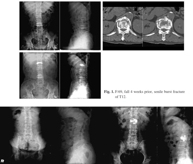
after the procedure was 19.1±2.4�and 9.1±1.8�, respec- tively; the final follow-up KDA was 8.9±1.7�. Therefore, there was no statistically significant difference between the KP and open posterior surgery groups with regard to the improvement in KDA (p=0.081). No patients had signifi- cant complications related to cement leakage into the poste- rior spinal canal (Fig. 1). One patient had cement leakage into the intervertebral disc secondary to an upper end-plate fracture, but the patient did not have neurologic symptoms (Fig. 2).
The reductions in pain from baseline to the third day and to 3 months post-operative were statistically significant (VAS scores: pre-operative, 8.3±0.4; 3 days post-opera- tive, 3.9±0.2; 3 months post-operative, 3.2±0.17; 1 year post-operative, 3.1±0.17). The average VAS score decreased by 5.2±0.3. Every patient was capable of walk- ing autonomously one day after KP. After conservative treatment, the VAS score decreased by 3.4±0.2 (VAS
Fig. 1. F/69, fall 4 weeks prior, senile burst fracture of T12.
Fig. 2. F/63, fall 10 weeks prior, senile burst fracture of L1.
score: pre-operative, 7.9±0.4; after 3 months, 4.8±0.18;
after 1 year, 4.5±0.17). Hence, there was a statistically sig- nificant difference between the KP and conservative treat- ment groups with regard to improvement in the VAS score (p=0.012). After posterior instrumentation and fusion, the VAS score decreased by 5.3±0.3 (VAS score: pre-opera- tive, 8.5±0.5; 3 days post-operative, 3.5±0.21; 3 months post-operative, 3.4±0.2; 1 year post-operative, 3.2±0.19).
Hence, there was no statistically significant difference between the KP and open posterior surgery groups with regard to improvement in VAS score (p=0.125).
The initial Chen and Lee semi-quantitative scale was grade 3 or 4 in all patients. In the KP group, 12 patients (100%) improved to grade 0 at final follow-up. In the con- servative treatment group, 12 patients (36.5%) improved to grade 0, 18 patients (54.5%) improved to grade 1, and 3 patients (9%) improved to grade 2 at final follow-up. In the posterior instrumentation and fusion group, 13 patients (100%) improved to grade 0 at final follow-up.
The average volume of bone cement injected was 5 cc.
No patient complained of symptoms suggesting pulmonary embolism. Postoperative CT scans never revealed cement leakage.
Discussion
The two most common treatment methods for osteoporot- ic burst fractures are conservative treatment and posterior instrumentation and fusion. Conservative treatment can be performed in senile patients with medical comorbidities;
however, conservative treatment may lead to cardiovascular complications, as well as decreased correction rate for kyphotic deformities. Posterior instrumentation and fusion provides solid stability; however, posterior instrumentation and fusion is not a viable surgical option for patients who lack sufficient bone mass from osteoporotic burst fractures.
Age and comorbid medical conditions may further con- tribute to the impracticality of posterior instrumentation and fusion as a treatment for osteoporotic burst fractures.
KP, as well as vertebroplasty, offers significant pain relief and fracture stabilization. The application of percutaneous vertebroplasty as a minimally invasive procedure is popular and pervasive. Nevertheless, the danger of cement leakage into the posterior spinal canal has precluded percutaneous vertebroplasty as a treatment for burst fractures. In 2004, however, Chen et al1, reported a clinically satisfactory result
by performing percutaneous vertebroplasty in six stable burst fractures without osteoporosis. Although cement leak- age into the adjacent intervertebral disc space occurred in four instances (66.7%), it did not cause the neurological symptoms associated with cement leakage into the posterior spinal canal. Despite its evident benefits with regard to pain relief and vertebral stabilization, vertebroplasty is not as effective in correcting kyphotic deformities. Balloon KP was developed in order to compensate for vertebroplasty’s disadvantages in correcting kyphotic deformities. Balloon KP minimizes the possibility of cement leakage. Hence, we performed balloon KP for the treatment of osteoporotic burst fractures.
Balloon KP was developed by applying Dr. Mark Rei- ley’s balloon tamp in percutaneous vertebroplasty2,3. The balloon tamp was developed by Reiley in 1997, with the goal of restoring vertebral body height and reducing kypho- sis. KP involves the inflation of a balloon tamp before injection of bone cement in the compressed vertebral body, in an effort to reduce kyphotic deformity. M. K. Shindle et al4, reported that balloon KP enhances the height reduction
>4.5-fold over the positioning maneuver alone and accounts for over 80% of the ultimate reduction. Because KP enables the injection of bone cement at low pressure, the procedure minimizes the possibility of cement leakage5,6,7.
Reportedly, the complication rate following balloon KP is similar to that seen in vertebroplasty. Complications deriv- ing from vertebroplasty as a treatment for osteoporotic burst fracture occur in less than 1% of patients when the proce- dure is performed by an experienced surgeon. The compli- cation rate stemming from vertebral pathologic compression fractures is 5~10%. Although complications occur slightly more frequently in KP than in vertebroplasty, we believe that such results derive from KP’s relative dearth of recog- nition. However, according to Orlando, the complication rate with respect to symptomatic cement extravasation may be lower in KP than in vertebroplasty2. We observed only one instance of cement leakage in this study. We attribute our success to 4 weeks of conservative treatment prior to the procedure and our decision to perform KP in stable burst fractures. Paul et al8, reported the following distribu- tion of cement extravasation in vertebroplasty: epidural, 32%; paraspinal, 32.5%; intradiscal, 30.5%; pulmonary, 1.7%; and foraminal, 3.3%. He further reported the follow- ing distribution of cement extravasation in KP: epidural, 11%; paraspinal, 48%; intradiscal, 38%; pulmonary, 1.5%;
and foraminal, 1.5%. In most cases (vertebroplasty, 96%;
KP, 89%), symptoms do not accompany cement extravasa- tion. Intradiscal cement leakage, which occurs relatively frequently, may cause mechanical overload to the disc and/or to the adjacent vertebrae. Lin et al9, reported that 58% of vertebral bodies adjacent to a disc with cement leak- age fractured during the follow-up period, compared with 12% of vertebral bodies adjacent to a disc without cement leakage after vertebroplasty. We only observed complica- tions accompanying cement leakage in a single case.
Frail older patients have poor tolerance for the bed rest required in non-operative management and the physiologic stress engendered by an open stabilization procedure10. Indi- cations for open surgery are rare and include major neuro- logic injury and mechanical instability. Anterior decom- pression is an effective method; however, the morbidity rate and the risk of spinal deterioration in instrumentation and fusion are very high in senile patients. Posterior decompres- sion and stabilization are perhaps better tolerated by older patients. The pedicles remain stronger than the vertebral bodies, but implant loosening remains a problem10.
We did not observe any major complications when we performed KP as a treatment for stable burst fractures. The clinical and radiological results of KP were superior to those of conservative treatment. Lyritis et al11showed that pain decreased by 33% at day 14 in vertebral compression fracture patients treated with the conservative method, and Gennari et al12, noted that pain decreased by 40% at day 30.
However, S. L. Silverman13noted the long-term sequelae associated with vertebral compression fractures: chronic back pain, muscle fatigue, reduced exercise tolerance, early satiety, insomnia, fibromyalgia, and low self-esteem. There- fore, vertebral fractures must be treated aggressively. The results of conservative treatment seen in this study were inferior to those seen in previous studies. However, the pre- vious studies addressed compression fractures, while we assessed burst fractures. We attained very satisfactory results with KP, in comparison to those attained with poste- rior instrumentation and fusion. The limitation of our study lies in the insufficient number of cases and short follow-up duration. Future studies are recommended.
Conclusion
Osteoporotic burst fractures occur most frequently at the thoracolumbar junction. Percutaneous vertebroplasty and balloon KP are minimally invasive treatments for osteo-
porotic burst fractures. However, the danger of cement leak- age into the posterior spinal canal has precluded some sur- geons from performing these two procedures.
Nevertheless, balloon KP is somewhat beneficial for strengthening the spinal wall. Because KP enables the injec- tion of bone cement at low pressure, the procedure further diminishes the possibility of cement leakage5,6,7.
Balloon KP is useful in correcting deformities, alleviating pain, and ameliorating immobility, and it is a practical treat- ment for osteoporotic burst fractures.
REFERENCES
01. Chen JF, Lee ST: Percutaneous vertebroplasty for treat- ment of thoracolumbar spine bursting fracture. Surg Neu- rol, 2004; 62: 494-500.
02. Orlando Ortiz, Gregg H Zoarski, Michael Beckerman:
Kyphoplasty. Techniques in Vascular and Interventional Radiology, Vol 5, No 4 (December), 2002; 239-249.
03. Lieberman IH, Dudeney S, Reinhardt MK, et al: Initial outcome and efficacy of “kyphoplasty” in the treatment of painful osteoporotic vertebral compression fractures. Spine 2001; 26: 1631-1638.
04. MK Shindle, MJ Gardner, J Koob, et al: Vertebral height restoration in osteoporotic compression fractures: kypho- plasty balloon tamp is superior to postural correction alone.
Osteoporos Int 2006; 17: 1815-1819.
05. Garfin SR, Yuan HA, Reiley MA: New technologies in spine; kyphoplasty and vertebroplasty for the treatment of painful osteoporotic compression fracture. Spine 2001; 26:
1511-1515.
06. Hardouin P, Fayada P, Leclet H, Chopin D: Kyphoplas- ty. Joint Bone Spine 2002; 69(3): 256-261.
07. Ledlie JT, Renfro M: Balloon kyphoplasy: one-year out- comes in vertebral body height restoration, chronic pain, and activity levels. Journal of Neurosurgery 2003; 98: 36- 42.
08. Paul A Hulme, Jorg Krebs, Stephen J Ferguson, Ulrich Berlemann: Vertebroplasty and Kyphoplasty: A Systemat- ic Review of 69 Clinical Studies. Spine 2006; 31(17):
1987-1993.
09. Lin EP, Ekholm S, Hiwatashi A, et al: Vertebroplasty:
Cement leakage into the disc increase the risk of new frac- ture of adjacent vertebral body. Am J Neuroradiology 2004; 25: 175-180.
10. Eeric T, Alan H, Alexander R, Vaccaro: Percultaneous
vertebral augmentation. The Spine Journal 2004; 4: 218- 229.
11. Lyritis GP, Magiasis B, Eliopoulos A, Tsekoura M, Ioakimidis D: Analgesic effect of salmon calcitonin in cases of osteoporotic vertebral fractures. Int Osteoporosis 1990; 1392-1395.
12. Gennari C, Agnusdei D, Camporeale A: Use of calci- tonin in the treatment of bone pain associated with osteo- porosis. Calcif Tissue Int 1991; 49 (suppl 2): S9-S13.
13. SL Silverman: The clinical consequences of vertebral compression fracture. Bone 1992; 13: S27-S31.