초음파를 이용한 미추 경막외 차단술의 결과 비교 - 추간판 탈출증과 척추관 협착증 -
인하대학교 의과대학 정형외과학교실
김영태∙조규정∙안치훈
서 론
요추 경막외 차단술은 추간판 탈출증 및 척추관 협착증에 의한 요통과 하지 방사통을 치료하기 위해 널리 사용 된다.1-3)경막 외 주사의 방법으로는 경신 경공 경막외 주사(transforaminal), 미추(caudal)
Comparison of the Results of Ultrasound-guided Caudal Epidural Block - Herniated Intervertebral Disc vs Spinal Stenosis -
Young-Tae Kim, M.D., Kyu-Jung Cho, M.D., Chi-Hoon Ahn, M.D.
Department of Orthopedic Surgery, College of Medicine, Inha University, Incheon, Korea
Purpose: Ultrasound-guided epidural caudal block for low back pain and radiating pain is often performed in the treat- ment of outpatients. However, this procedure has a failure rate of up to 25% even when it performed by an experienced physician. The authors investigate the effectiveness of Ultrasound-guided epidural caudal block in patients related to disc herniation or spinal stenosis.
Materials and Methods: Ultrasound-guided caudal epidural block was performed in 55 outpatients with LBP and radiat- ing pain. Patient was placed in the prone position and sonographic image of sacral hiatus was obtained using linear probe. A 22-gauge needle was advanced into the sacrococcygeal membrane under ultrasound guidance and then med- ication was injected into the caudal epidural space. There were 31 cases of disc herniation, and 24 cases of spinal steno- sis. Patients were evaluated by Visual Analog Scale (VAS) pain score at pre-treatment, post-treatment, 2 weeks and 4 weeks by telephone interviews.
Results: 53 of the 55 cases (96.4%) of needle insertion into the sacral canal under ultrasound guidance were successful.
Gender was not significantly different between disc herniation group and spinal stenosis group. But there was a signifi- cant age difference between disc herniation group (42.3±10.8), and spinal stenosis group (62.8±15.1) [p<0.001]. The VAS score at pre-treatment, post-treatment, 2 weeks, 4 weeks in disc group were 6.84, 3.1, 1.8 & 1.77. The VAS score at pre-treatment, post-treatment, 2 weeks, 4 weeks in spinal stenosis group were 6.88, 3.58, 4.33 & 4.88. The VAS score in both groups was significantly improved after the procedure (p<0.001). Over time, the two groups were statistically sig- nificant differences in VAS score after adjusting for age (p<0.001).
Conclusion: Ultrasound-guided caudal epidural block seems to provide a high success rate and a significantly better response in disc group than spinal stenosis group
Key Words: Ultrasound, Caudal epidural block, Disc herniation, Spinal stenosis
통신저자: 조 규 정
인천광역시 중구 신흥동 인항로 27 인하대학교 의과대학 정형외과학교실 Tel: 032-890-3043, Fax: 032-890-3047 E-mail: [email protected]
대한정형외과초음파학회지: 제 7 권 제 2 호 2014
경막외 주사, 추궁판 사이(interlaminar) 경막외 주 사가 있다. 특히, 미추 경막외 주사의 경우 해부학적 구조물이 쉽게 촉지되어 외래 환자의 치료에 자주 사용되고 있지만, 임상 경험이 풍부한 의사에 의해 시술되더라도 방사선 투시기를 이용하지 않는 경우 20~38%의 부정확한 needle의 위치가 보고되고 있다.4,5) 이에 Chen 등은 초음파 유도하의 미추 경 막외 주사를 시행하는 경우, 정확한 needle의 위치 를100% 확보할 수 있다고 주장하였다.6)
요추 경막외 차단술의 효과에 대해서는 아직까지 논란이 있다. 일부의 저자들은 요통 및 방사통의 치 료에 요추부 경막외 차단술이 증등도 이상의 단기 효과가 있음을 보고하였고,7-10) 다른 저자들은 위약 (placebo) 주사와 비교할 때 효과의 차이가 크지 않음을 보고하였다.11-13)
요통과 방사통은 다양한 원인에 의해 발생할 수 있지만, 젊은 연령에서는 추간판 탈출증이 흔하며, 연령이 증가함에 따라 척추관 협착증이 흔한 것으로 알려져 있다.14) 두 질환에 대한 경막외 차단술의 결 과는 주사 약제와 접근법에 따라 논문마다 조금씩 다르며, 초음파를 이용한 시술에 대한 결과 보고는
거의 없는 실정이다.15,16) 최근, 척추 통증 분야에서 도 다양한 초음파 유도하 중재술이 시도되고 있는 점을 고려하여 저자들은 초음파 유도하 미추 경막외 차단술의 유효성을 추간판 탈출증 환자군과 척추관 협착증 환자군으로 나누어 결과를 비교하였다.
대상 및 방법 1. 연구 대상
2013년 10월부터 2014년 1월까지 요통과 하지 방사통을 호소하는 환자 중 2주 이상의 약물치료 및 물리치료에도 증상의 호전이 없고, 내원 당시 통증 의 강도가 시각통증척도(VAS) 6 이상인 환자와 요 추부 병변이 한 분절인 경우를 대상으로 하였고. 2 개월 이전에 경막외 차단술을 받은 경우, 이전에 요 추부 수술을 한 경우, 두 분절 이상의 요추부 병변이 있는 경우, 기왕력으로 당뇨를 가지고 있는 경우는 제외하였다.
진단은 환자의 병력, 이학적 검사, 자기공명영상 등 을 통해 시행하였으며, 추간판 탈출증 환자군이 32명
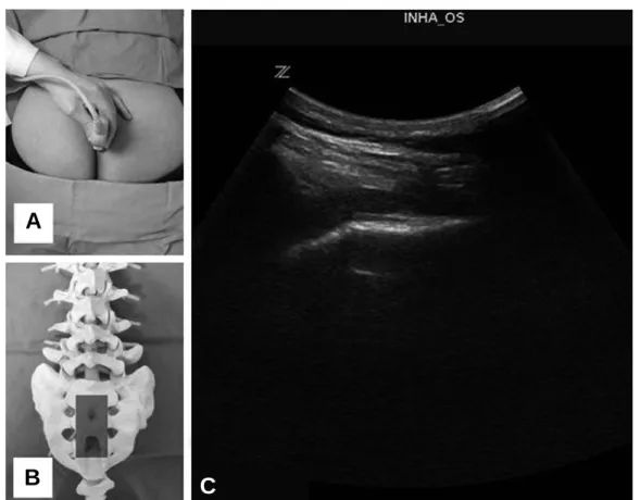
Fig. 1. The transverse view of sacral hiatus (A) photo, (B) skeleton, (C) ultrasound.
A
B C
이었으며, 척추관 협착증 환자군이 24명이었다.
자기공명영상 소견상 추간판 탈출증 환자군 32명 중, protrusion은 21명, extrusion은 9명, seques- tration은 2명이었으며, 척추관 협착증 환자군 24명 중 central stenosis는 8명, lateral recess steno- sis는 11명, foraminal stenosis는 5명이었다.
2. 시술 방법
모든 시술은 동일한 한 명의 술자에 의해 진행되 었으며, 환자를 복와위 자세로 눕힌 후 천추 열공 부 위를 방사형 탐지자(round probe)를 이용하여 가 로축상 영상을 얻었다(Fig. 1).
이 후 탐지자를 90도 회전시켜 천골의 극돌기와 일직선 상에 위치시켜 천추 열공의 세로축상 영상을 확인하고 바늘을 삽입할 위치와 각도를 정하였다.
그 후 천추 열공 주위를 무균 소독하고 소독포로 덮 은 뒤 초음파 유도하 미추 경막외 차단술을 진행하 였다(Fig. 2).
초음파로 천추 열공과 천-미추막의 세로축상 영 상을 얻은 후 22 G 또는 24 G spinal needle을 천
추관의 바닥면과 평행하게 밀어넣어 바늘이 천-미 추막을 통과하는 것을 초음파 영상으로 관찰하면서 천추관 내로 삽입하였다(Fig. 3).
이 후 실린지로 음압 검사와 공기저항 검사를 한 후, 0.2% Ropivacaine 10 ml과 Dexamethason 1 Fig. 2. The longitudinal view of sacral hiatus (A) photo, (B) skeleton, (C) ultrasound.
A
B C
Fig. 3. The needle insertion into caudal epidural space.
대한정형외과초음파학회지: 제 7 권 제 2 호 2014
ml를 이 후 경막외 공간으로 주입하였다.
3. 결과 판정
임상적 결과의 평가는 시술 당일 및 시술 후 1주 일 후는 전화 인터뷰를 이용하였으며, 시술 2주일 후 및 시술 1달 후는 외래 추시를 통해 통증이 호전 되는 정도를 시각통증척도로 평가하였다. 시술 2주 일 후에 시행한 외래 추시에서 시각통증척도가 최소 2이상 호전이 없고, 시각통증척도가 5이상인 경우 에는 시술을 재시행하였다.
4. 통계 방법
연구에 수집된 자료는 SPSS 12.0 프로그램을 이 용 하 여 분 석 하 였 다 . 인 구 학 적 인 비 교 는 Chi- square test 및 independent t-test를 이용하였 고, 시술 전과 시술 후 통증 정도의 비교는 paired t-test를 이용하였다. 추시 기간에 따른 통증 정도 의 변화는 mixed model을 이용하여 분석하였으며, p<0.05인 경우 통계적으로 유의하다고 판정하였다.
결 과 1. Baseline data
추간판 탈출증 환자 32명 중 1명이 미추 경막외
차단술 시행 후 증상의 호전이 없고, 통증이 악화되 어 시술 후 2일에 추궁판 및 디스크 절제술을 시행 하여 추간판 탈출증 환자 31명, 척추관 협착증 환자 24명에 대해 분석하였다.
두 군간에 성별(sex), 키(weight), 몸무게(height) 는 차이가 없었으나, 나이(age)는 추간판 탈출증군 (42.3±10.8세)이 척추관 협착증군(62.8±15.1 세)과 비교할 때 유의한 차이가 있었다(p<0.001).
통증의 양상 및 연관된 spine pathology level은 두 군간에 차이가 없었다(Table 1).
2. 임상적 통증 평가
초음파 유도하 미추 경막외 차단술은 55명 중 53 명에서 바늘이 성공적으로 삽입되어 96.4%의 성공 률을 보였다. 추간판 탈출증 환자군에서 1명, 척추 관 협착증 환자군에서 1명의 경우, 초음파 유도하 바늘 삽입이 되지 않아서 방사선 투시 상 바늘을 삽 입한 후 미추 경막외 차단술을 시행하였다.
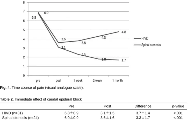
허리 통증 및 방사통의 변화는 추간판 탈출증군에 서 시행 전 시각통증척도가 6.8에서 차단술 시행 후 3.1으로 호전되었으며, 척추관 협착증 군에서 시행 전 시각통증척도가 6.9에서 시행 후 3.6으로 통계 적으로 유의하게 호전되었다(Table 2).
추시 상 변화는 추간판 탈출증 군에서 시행 1주 후 시각통증척도가 2.3, 시행 2주 후 1.8, 시행 4주 후 1.7로 나타났으며, 척추관 협착증 군에서는 시행
Table 1. Baseline demographic characteristics
HIVD (n=31) Spinal stenosis (n=24) p-value
Sex <0.460
Men 12 (38.7%) 07 (29.2%)
Women 19 (61.3%) 17 (70.8%)
Weight (kg) 66.1±7.5 64.5±4.3 <0.487
Height (cm) 162.6±25.5 159.3±32.4 <0.574
Age (year) 042.3±10.9 062.8±15.2 <0.001
Pain distribution
Both 07 06 <0.245
Right 14 11
Left 10 07
Involved segment
L34 03 02 <0.451
L45 17 12
L5S1 11 10
VAS 06.8±0.9 06.9±0.9 <0.257
1주 후 시각통증척도가 3.8, 시행 2주 후 4.3, 시행 4주 후 4.8으로 나타났다. 이러한 변화의 양상을 baseline data에서 두 군간의 나이가 다르다는 것 을 고려하여 나이를 보정 후 통계 분석을 하였으며, 두 군 간의 시각통증척도 차이의 변화는 통계적으로 유의하였다(Fig. 4).
3. 합병증
55명의 환자 중 미추 경막외 차단술 시행 후 경막 외 혈종 및 감염과 같은 합병증은 관찰되지 않았다.
추간판 탈출증 군에서 2명과 척추관 협착증 군에서 1명의 경우 미추 경막외 차단술 시행 후 2주 외래 추시에서 시각통증척도가 최소 2 이상 호전이 없고, 시각통증척도가 5 이상으로 확인되어 시술을 재시 행하였다.
재시술 후 추간판 탈출증 환자 1명은 증상이 호전 되었으며, 미추 경막외 차단술 재시술로도 증상의 호전이 없었던 추간판 탈출증 환자 1명과 척추관 협 착증 환자 1명에 대해서는 방사선 투시하 선택적 신
경 차단술을 시행하였고 증상의 호전을 확인하였다.
고 찰
본 연구 결과 초음파를 이용한 미추 경막외 차단 술은 55명의 환자 중 53명에서 천골관 내로 성공적 으로 바늘이 위치하여 96.4%의 높은 성공률을 보 였으며, 추간판 탈출증 및 척추관 협착증에서 발생 하는 급성기의 요통과 방사통에 효과적인 것으로 확 인되었다. 하지만, 외래 추시 상 통증 변화 양상은 추간판 탈출증 환자군과 척추관 협착증의 환자군에 서 유의한 차이가 관찰되었다.
추간판 탈출증 환자군의 경우, 급성기 요통과 방 사통이 미추 경막외 차단술로 호전된 후 유지 되거 나 더욱 호전되는 양상으로 나타났으나, 척추관 협 착증 환자군의 경우, 요통과 방사통이 미추 경막외 차단술로 호전된 이후, 점차 다시 악화되는 양상으 로 확인되었다.
저자들은 이러한 양상의 변화를 추간판 탈출증과 척추관 협착증에서 발생하는 통증의 특성이 다르기
Table 2. Immediate effect of caudal epidural block
Pre Post Difference p-value
HIVD (n=31) 6.8±0.9 3.1±1.5 3.7±1.4 <.001
Spinal stenosis (n=24) 6.9±0.9 3.6±1.6 3.3±1.7 <.001
Fig. 4. Time course of pain (visual analogue scale).
대한정형외과초음파학회지: 제 7 권 제 2 호 2014
때문으로 생각한다. 추간판 탈출증에 의한 요통과 방사통은 디스크의 수핵(nucleus pulposus) 에서 분비된 IL-6, IL-8, PGE2 등과 같은 pro- inflammatory cytokine이 주위 신경 말단(nerve ending)을 자극하여 발생하는 침해성 통증(noci- ceptive pain) 양상이며,17-19)척추관 협착증에 의한 통증은 chronic mechanical compression 및 venous congestion과 TNF-α와 같은 cytokine에 의해 주위 신경의 dysfunction에 의해 발생하는 신 경병증 통증(neuropathic pain)의 양상으로 알려 져 있다.20-22) 따라서 일반적으로 경막외 차단술에 사용되는 국소 마취제와 steroid가 prostaglandin 의 합 성 을 억 제 시 키 며 , 염 증 반 응 을 억 제 하 고 nociceptive C fiber를 blocking하는 효과가 있다 는 것을 고려할 때 통증 발생 기전이 상이한 추간판 탈출증 환자군과 협착증 환자군에서 다른 반응을 보 이는 것으로 판단된다.23-25)
Ohtori 등은 80명의 척추관 협착증 환자를 대상 으로 한 최근 연구에서 국소 마취제와 TNF-a inhibitor를 경신경공(transforaminal) 경막외 투 여해서 국소 마취제와 steroid를 투여한 군에서 보 다 좋은 결과를 보고하여 신경병증 통증(neuro- pathic pain)에 생물학 제제의 사용 효과의 가능성 을 보여 주었다.26)
본 연구와 최근의 연구 결과를 고려할 때, 추간판 탈출증과 척추관 협착증에서 발생하는 통증의 양상 이 상이함과 질환의 자연 경과를 숙지하여 좀 더 효 과적인 중재적 시술에 대한 고민이 필요할 것으로 판단된다.
초음파는 방사선 투시를 하지 않고 실시간으로 바 늘의 움직임과 목표 구조물에 대해 시각화할 수 있 어 정확도를 올릴 수 있다는 장점이 있다. 최근 이러 한 유용성이 확대되면서 신경 차단술 시 방사선 투 시 검사를 대신할 수 있는 방법으로 고려되고 있으 며, 초음파 영상을 통해 미추 경막외 차단술을 시행 한 성공적인 연구들이 보고되고 있다.6,27) 본 연구에 서도 96.4%의 정확한 바늘의 위치를 확인하였으 며, 이는 blind technique을 이용한 미추 경막외 차 단술의 실패율이 38%까지 보고되는 것을 고려할 때 의미 있는 결과라고 사료된다.4,5) 초음파는 시술 자간 결과의 변화가 크고, 골성 구조에 대해서는 관 찰되는 부분의 한계가 있어 초음파 유도하 미추 경 막외 차단술의 성공률을 높이기 위해서는 시술 전
천추 열공에 대한 충분한 해부학적 지식이 필요할 것으로 판단된다.
본 연구에서 시술 후 4주로 추시 기간이 짧았다는 것과 미추 경막외 차단술을 시행 후 NSAIDs 투여 와 물리 치료가 시행되어 독립적인 미추 경막외 차 단술의 효과 평가가 이루어지지 못했다는 것, 그리 고 시술의 평가 항목이 환자 주관적이었다는 것이 본 연구의 제한점으로 사료된다.
결 론
초음파 유도하 미추 경막외 차단술은 높은 성공률 을 보이며 척추관 협착증 환자군에 비해 추간판 탈 출증 환자군에서 더 효과적인 것으로 사료된다.
참고문헌
01. Schaufele MK, Hatch L, Jones W.
Interlaminar versus transforaminal epidural injections for the treatment of symptomatic lum- bar intervertebral disc herniations. Pain Physician. 2006;9:361-6.
02. Young IA, Hyman GS, Packia-Raj LN, et al.
The use of lumbar epidural/transforaminal steroids for managing spinal disease. J Am Acad Orthop Surg. 2007;15:228-38.
03. Cooper G, Lutz GE, Boachie-Adjei O, et al.
Effectiveness of transforaminal epidural steroid injections in patients with degenerative lumbar scoliotic stenosis and radiculopathy. Pain Physician. 2004;7:311-7.
04. Manchikanti L, Cash KA, Pampati V, et al.
Evaluation of fluoroscopically guided caudal epidural injections. Pain Physician. 2004;7:81- 92.
05. Dashfield A, Taylor M, Cleaver J, et al.
Comparison of caudal steroid epidural with tar- geted steroid placement during spinal endoscopy for chronic sciatica: a prospective, randomized, double-blind trial. Br J Anaesth. 2005;94:514-9.
06. Chen CP, Tang SF, Hsu TC, et al. Ultrasound guidance in caudal epidural needle placement.
Anesthesiology. 2004;101:181-4.
07. Koes BW, Scholten RJ, Mens JM, Bouter LM. Efficacy of epidural steroid injections for
low-back pain and sciatica: a systematic review of randomized clinical trials. Pain. 1995;63:279- 88.
08. Watts RW, Silagy CA. A meta-analysis on the efficacy of epidural corticosteroids in the treat- ment of sciatica. Anaesth Intensive Care.
1995;23:564-9.
09. Luijsterburg PA, Verhagen AP, Ostelo RW, van Os TA, Peul WC, Koes BW. Effectiveness of conservative treatments for the lumbosacral radicular syndrome: a systematic review. Eur Spine J. 2007;16:881-99.
10. Conn A, Buenaventura RM, Datta S, Abdi S, Diwan S. Systematic review of caudal epidural injections in the management of chronic low back pain. Pain Physician. 2009;12:109-35.
11. Carette S, Leclaire R, Marcoux S, et al.
Epidural corticosteroid injections for sciatica due to herniated nucleus pulposus. N Engl J Med.
1997;336:1634-40.
12. Ng L, Chaudhary N, Sell P. The efficacy of cor- ticosteroids in periradicular infiltration for chronic radicular pain: a randomized, double- blind, controlled trial. Spine. 2005;30:857-62.
13. Karppinen J, Malmivaara A, Kurunlahti M, et al. Periradicular infiltration for sciatica: a randomized controlled trial. Spine. 2001;26:
1059-7.
14. Rivest C, Katz JN, Ferrante FM, Jamison RN. Effects of epidural steroid injection on pain due to lumbar spinal stenosis or herniated disks:
a prospective study. Arthritis Care Res. 1998;
11:291-7.
15. Ng LC, Sell P. Outcomes of a prospective cohort study on peri-radicular infiltration for radicular pain in patients with lumbar disc herniation and spinal stenosis. Eur Spine J. 2004;13:325-9.
16. Lee JH, Moon J, Lee SH. Comparison of effec- tiveness according to different approaches of epidural steroid injection in lumbosacral herniated disk and spinal stenosis. J Back Musculoskeletal Rehabil. 2009;22:83-9.
17. Kang JD, Georgescu HI, McIntyre-Larkin L, Stefanovic-Racic M, Evans CH. Herniated cervical intervertebral discs spontaneously pro- duce matrix metalloproteinases, nitric oxide,
interleukin-6, and prostaglandin E2. Spine.
1995;20:2373-8.
18. Kang JD, Georgescu HI, McIntyre-Larkin L, et al. Herniated lumbar intervertebral discs spontaneously produce matrix metalloproteinas- es, nitric oxide, interleukin-6, and prostaglandin E2. Spine. 1996;21:271-7.
19. Takahashi H, Suguro T, Okazime Y, et al.
Inflammatory cytokines in the herniated disc of the lumbar spine. Spine. 1996;21:218-24.
20. Myers RR, Wagner R, Sorkin LS. Hyperalgesic actions of cytokines on peripheral nerves. In:
Watkins LR, Maier SF, eds. Cytokines and Pain.
Basel, Switzerland: Birkhauser. 1999:133-57.
21. Olmarker K, Nutu M, St∅∅rkson R. Changes in spontaneous behavior in rats exposed to experi- mental disc herniation are blocked by selective TNF-alpha inhibition. Spine. 2003;28:1635-4219.
22. Sekiguchi M, Kikuchi S, Myers RR.
Experimental spinal stenosis: relationship between degree of cauda equina compression, neuropathology, and pain. Spine. 2004;29:1105- 11.
23. Saal JA, Saal JS, Herzog RJ. The natural his- tory of lumbar intervertebral disc extrusions treated nonoperatively. Spine. 1990;15:683-6.
24. Vad VB, Bhat AL, Lutz GE, et al.
Transforaminal epidural steroid injections in lumbosacral radiculopathy: a prospective ran- domized study. Spine. 2002;27:11-6.
25. Onda A, Yabuki S, Kikuchi S, et al. Effects of lidocaine on blood flow and endoneurial fluid pressure in a rat model of herniated nucleus pul- posus. Spine. 2001;26:2186-91.
26. Ohtori S, Miyagi M, Eguchi Y, et al. Epidural administration of spinal nerves with the tumor necrosis factor-alpha inhibitor, etanercept, com- pared with dexamethasone for treatment of sciat- ica in patients with lumbar spinal stenosis: a prospective randomized study. Spine. 2012;37:
439-44.
27. Roh JH, Kim WO, Yoon KB, Yoon DM. The success rate of caudal block under ultrasound guidance and the direction of the needle in the sacral canal. Korean J Pain. 2007;20:40-5.
대한정형외과초음파학회지: 제 7 권 제 2 호 2014
목적: 미추 경막외 차단술은 요통과 하지 방사통에 효과적으로 외래환자의 치료에 자주 사용되고 있으나 임상 경험이 풍 부한 의사에 의해 시술되어도 25%의 실패율이 보고되고 있다. 저자들은 초음파 유도하 미추 경막외 차단술의 유효성을 추간판 탈출증 환자군과 척추관 협착증 환자군으로 나누어 결과를 비교하였다.
대상 및 방법: 요통과 하지 방사통을 호소하여 본원 외래에 내원한 55명의 환자를 대상으로 초음파 유도하 미추 경막외 차단술을 시행하였다. 환자를 복와위 자세로 눕힌 후 방사형 탐지자(round probe)를 이용하여 22게이지 바늘이 천-미 추막을 통과한 것을 확인 후 약물을 투여하였다. 추간판 탈출증 환자군은 31명이었으며, 척추관 협착증 환자군은 24명이 었다. 임상적 평가는 시각통증척도(VAS)를 이용하여 통증의 변화를 평가하였으며, 시행 전, 시행 후, 시행 2주 후, 시행 4주 후 전화 인터뷰와 외래 방문 시 조사하였다.
결과: 초음파 유도하 미추 경막외 차단술은 55예 중 53예에서 바늘이 성공적으로 삽입되어 96.4%의 성공률을 보였다.
추간판 탈출증 환자군과 척추관 협착증 환자군에서 성별의 차이는 없었으나 나이는 추간판 탈출증군에서 42.3±10.8세 로 척추관 협착증군의 62.8±15.1세와 비교할 때 유의한 차이가 있었다(p<0.001).
시각통증척도는 추간판 탈출증 군에서 시행 전 6.8, 시행 후 3.1, 시행 2주 후 1.8, 시행 4주 후 1.77로 나타났으며, 척 추관 협착증 군에서 시행 전6.9, 시행 후 3.6, 시행 2주 후 4.3, 시행 4주 후 4.9로 나타났다. 시행 전에 비해 시행 후 시각통증척도는 양 군에서 유의하게 호전되었으며(p<0.001), 나이를 보정한 후의 결과, 시간에 따라 양 군간 시각통증척 도의 차이는 통계적으로 유의하였다(p<0.001).
결론: 초음파 유도하 미추 경막외 차단술은 높은 성공률을 보이며 척추관 협착증 환자군에 비해 추간판 탈출증 환자군에 서 더 효과적인 것으로 사료된다.
색인단어: 초음파, 미추 경막외 차단술, 추간판 탈출증, 척추관 협착증.
국문초록