Cyclophosphamide 연관 중증 심부전을 동반한 Cardiomyopathy 환자에서 치료 후 회복된 증례
박정민ㆍ한승민ㆍ한정우ㆍ유철주
연세대학교 의과대학 소아과학교실
A Case of Successfully Treated Severe Heart Failure due to Cyclophosphamide Induced Cardiomyopathy
Jung Min Park, M.D., Seung Min Hahn, M.D., Ph.D., Jung Woo Han, M.D., Ph.D. and Chuhl Joo Lyu, M.D., Ph.D.
Division of Pediatric Hematology and Oncology, Yonsei University College of Medicine, Seoul, Korea
Cyclophosphamide-induced cardiotoxicity is an uncommon complication especially in patients who have never undergone mediastinal irradiation or cardiotoxic chemotherapy and do not have underlying cardiac diseases. Here, we describe the case of a 19-year-old female with chronic myeloid leukemia. She was previously treated with oral tyrosine kinase inhibitors and developed cardiomyopathy after receiving infusion of 60 mg/kg intravenous cyclophosphamide for two days with a conditioning regimen for allogenic hematopoietic stem cell transplantation. Severe thickening of the left ventricle and re- duced ejection fraction without triggering agents were characteristic for cyclo- phosphamide-induced cardiomyopathy. Her NT-pro BNP and troponin T concentrations surged to >70,000 pg/mL (0=130 pg/mL) and 2,031 pg/mL (0-14 pg/mL), respectively, during the course of the therapy and multiple organ failure seemed imminent evidenced by unresponsive decline in blood pressure. However, with close monitoring and persis- tent conservative management which consisted of intravenous hydration, continuous he- modialysis, and mechanical ventilation, her condition recovered.
pISSN 2233-5250 / eISSN 2233-4580 https://doi.org/10.15264/cpho.2018.25.1.71 Clin Pediatr Hematol Oncol 2018;25:71∼75
Received on March 28, 2018 Revised on April 5, 2018 Accepted on April 11, 2018
Corresponding Author: Chuhl Joo Lyu Division of Pediatric Hematology and Oncology, Yonsei University College of Medicine, 50-1 Yonsei- ro, Seodaemun-gu, Seoul, 03722, Korea
Tel: +82-2-2228-2060 Fax: +82-2-393-9118 E-mail: [email protected]
ORCID ID: orcid.org/0000-0001-7124-7818
Key Words: Cyclophosphamide, Cardiomyopathy, Chemotherapy
Introduction
Cardiac dysfunction related to chemotherapy has become an important cause of morbidity and mortality in patients [1]. Common cardiovascular complications include left ven- tricular dysfunction, myocardial ischemia, hypertension, thromboembolism, QT prolongation, and bradycardia [2].
Since many of these adverse effects significantly impact pa-
tient’s outcome, they have been an enormous concern for both cardiologists and oncologists.
Cyclophosphamide is a nitrogen mustard alkylating agent
with potent antineoplastic, immunosuppressive, and im-
munomodulatory properties. Despite its use in cancer ther-
apy and pretransplant stem cell conditioning regimens, the
toxicity profile using distinctive dosing regimens has not
been clarified [3-5]. Herein, we report the case of cyclo-
phosphamide-induced cardiotoxicity occurring without an
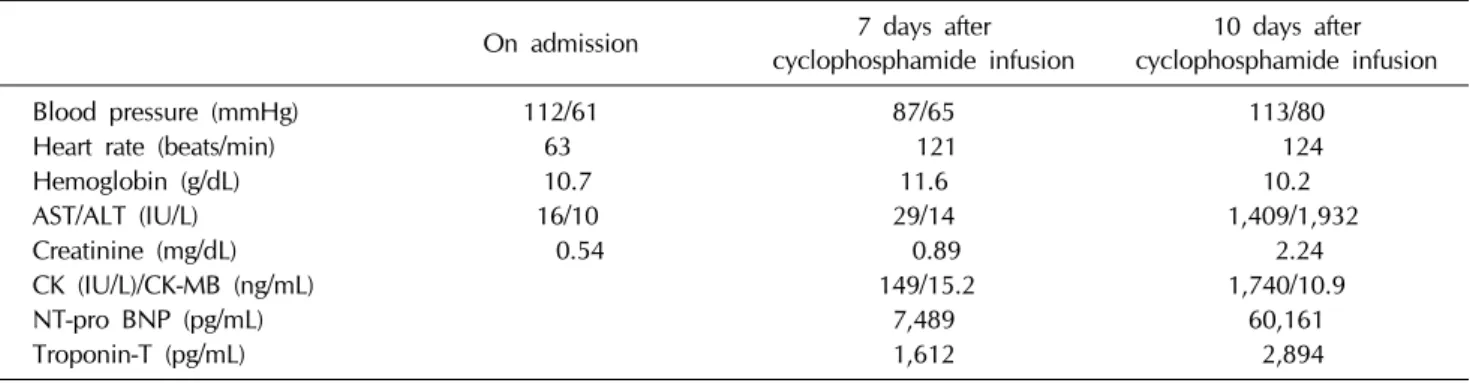
Table 1. Vitalsigns and laboratory characteristics of the day of admission, day 7 and day 10 after cyclophosphamide infusion
On admission 7 days after
cyclophosphamide infusion
10 days after cyclophosphamide infusion
Blood pressure (mmHg) 112/61 87/65 113/80
Heart rate (beats/min) 63 121 124
Hemoglobin (g/dL) 10.7 11.6 10.2
AST/ALT (IU/L) 16/10 29/14 1,409/1,932
Creatinine (mg/dL) 0.54 0.89 2.24
CK (IU/L)/CK-MB (ng/mL) 149/15.2 1,740/10.9
NT-pro BNP (pg/mL) 7,489 60,161
Troponin-T (pg/mL) 1,612 2,894
underlying heart disease or a history of previous radiation or cardiotoxin use. In addition, we introduce the manifes- tations, laboratory findings, and echocardiography of the patient and suggest the screening markers for the early de- tection and treatment approach for heart failure due to cyclophosphamide.
Case Report
A 19-year-old woman with chronic myelocytic leukemia was admitted to our hospital for sibling hematopoietic stem-cell transplantation on January 8
th, 2018. In February 2016, she was diagnosed as having chronic myelocytic leuke- mia revealing a BCR/ABL1 gene rearrangement (b2a2 type) with a triple translocation, 46,XX,t(3;9;22)(p21;q34;q11.2), on bone marrow. Results were negative for the JAK2 V617F mutation. The patient started taking imatinib mesylate (Gleevec) but was switched to dasatinib (Sprycel) after a year because of the non-reactive BCR/ABL1 quantitation in whole blood. However, Spyrcel caused her severe skin problems, such as multiple erythematous papules on the forehead, and she underwent a dermatologic consult. Due to an unsatisfactory treatment response and adverse effects of tyrosine kinase inhibitors, the medical faculty searched for a suitable hematopoietic stem cell transplantation donor and found her brother suitable despite one locus mismatch of DRB1.
At the time of admission, the patient’s general condition was fine, and her vital signs were normal. Her laboratory findings and chest X-ray did not show any abnormality. She was 162.6 cm in height and 59 kg in weight. Physical mea-
surements and laboratory values are summarized in Table 1. Basal electrocardiography showed normal sinus rhythm with a heart rate of 84 beats per minute. The baseline two-dimensional echocardiography indicated normal-sized cardiac chambers with normal global left ventricular systolic function with a left ventricular (LV) ejection fraction of 62%.
Apart from cardiologic evaluations, other pre-trans- plantation baseline studies demonstrated no abnormalities, including pulmonary function test (FEV
197%, FVC 98%), X-ray of paranasal sinuses, and dental evaluations.
The regimen of treosulfan/cyclophosphamide/etoposide
was selected as conditioning chemotherapy. The patient
was intravenously started on 12 mg/m
2treosulfan for 3
days, 15 mg/kg etoposide for 2 days, and 60 mg/kg cyclo-
phosphamide for 2 days with Uromitexan (Mesna) pro-
tection for hemorrhagic cystitis. Intrathecal methotrexate
(15 mg) injection was administered. Sibling bone marrow
transplantation was performed (CD34 2.3×10
6/kg) on
January 17th with no complications. On the third day after
transplantation (day 3), she had a syncope after standing
up in a bathroom, and her vital signs were blood pressure
96/70 mmHg, heart rate 89/min, and percutaneous oxygen
saturation 99%. Then, the arterial blood gas analysis, chest
X-ray, and EKG revealed no difference, so we considered
it to be a vasovagal syncope or orthostatic hypotension. On
day 4 in the morning (7 days after cyclophosphamide in-
fusion), her blood pressure suddenly dropped to 87/65
mmHg and was not responsive to normal saline (20 mg/kg)
loading. Chest X-ray showed mild cardiomegaly with CT ra-
tio of 0.60 compared to the previous X-ray with a ratio of
0.43 (Fig. 1). Electrocardiography showed sinus rhythm
Fig. 1. Comparison of chest
radiography between the day of admission (A) and day 7 after cyclophosphamide infusion (B).The CT ratios were 0.43 (A) and 0.60 (B), respectively.
Fig. 2. Comparison of echocardiography at the baseline and at day 7 after cyclophosphamide infusion. (A) Baseline echocardiography
shows normal left ventricular septum and chamber size. (B) Echocardiography on day 7 shows severe thickness of left ventricular septum and increased pericardial effusion.with a short PR interval and rightward axis deviation with a heart rate of 73 beats per minute. Urgent 2D-echocardiog- raphy found severe thickening of the myocardium with an LV mass of 154 g/m
2and a 10-12 mm-sized pericardial effu- sion without the evidence of tamponade physiology. The LV ejection fraction was 50.0% (Fig. 2). The level of tropo- nin T was 134 pg/mL (0-14 pg/mL) and that of NT-pro BNP was 8,134 pg/mL (0-130 pg/mL); these levels drastically ele- vated within two days to 2,031 pg/mL and >70,000 pg/mL, respectively. The patient did not complain of upper respira- tory or gastrointestinal symptoms, viral rash, or other sys- temic symptoms indicative of an infection. Drug-induced cardiomyopathy was suspected. Intravenous furosemide was started to reduce preload, and inotropic agents (milrinone, dopamine, and epinephrine) were administered
to raise blood pressure. Blood pressure was maintained at systolic BP of over 90 mmHg; however, tachycardia with a heart rate of 160 beats per minute persisted, and the pa- tient complained of severe chest discomfort and nausea.
Meanwhile, oliguria due to low cardiac output developed
and continuous renal replacement therapy (CRRT) was
administered. Shortly after the initiation of CRRT, the pa-
tient became unconscious and developed supraventricular
tachycardia which was resolved by performing synchron-
ized direct-current (DC) cardioversion (Fig. 3). We in-
tubated her and she underwent intensive care including
mechanical ventilator, CRRT, arterial blood pressure mon-
itoring, and daily echocardiography with proper intra-
venous fluid replacement for the following days. On day
7, we extubated her, and despite the remaining pericardial
Fig. 3. Electrocardiography showing supraventricular tachycardia terminated by synchronized DC cardioversion.
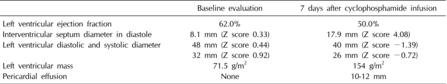
Table 2. Comparison of the echocardiography indices at the baseline and day 7 after cyclophosphamide infusion
Baseline evaluation 7 days after cyclophosphamide infusion
Left ventricular ejection fraction 62.0% 50.0%
Interventricular septum diameter in diastole 8.1 mm (Z score 0.33) 17.9 mm (Z score 4.08) Left ventricular diastolic and systolic diameter 48 mm (Z score 0.44)
32 mm (Z score 0.92)
40 mm (Z score −1.39) 26 mm (Z score −0.72)
Left ventricular mass 71.5 g/m2 154 g/m2
Pericardial effusion None 10-12 mm