156 https://e-jcvi.org
A 63-year-old woman presented with exertional dyspnea. She had received 6 cycles of adriamycin- based chemotherapy 5 years prior and an additional 6 cycles of the same regimen due to cancer metastasis. Treatments were completed 3 weeks before onset of dyspnea. The cumulative dose of adriamycin was 563 mg/m
2. Her B-type natriuretic peptide level was 4,116 pg/mL (reference:
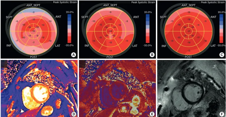
< 100 pg/mL), and transthoracic echocardiography revealed severe dysfunction [left ventricular ejection fraction (LVEF) 33%, global longitudinal strain (GLS) −8.3%]. We started heart failure medications, and her symptoms improved. Follow-up echocardiography showed gradual improvement: LVEF changed from 33% to 52% to 61% (Movie 1-3) and GLS from −8.3% to
−13.0% to −17.1% (Figures 1A-C). Subsequently, beta-blocker dose was halved due to low blood pressure and no further complaints of dyspnea. One month later, however, she presented at an emergency department with resting dyspnea with decreased LVEF of 33%. After the stabilization period, we performed echocardiography; surprisingly, the LVEF again recovered to 58.6% and GLS to −17.5%. Cardiac magnetic resonance imaging (3T, Verio, Siemens, Enlangen, Germany) showed elevated native T1 value (1,394–1,411 ms, reference: 1,278 ± 30 ms), T2 value (55–59 ms, reference: 40.5 ± 2.5 ms), and extracellular volume fraction (ECV, 32.5-33.3%, reference:
27.4 ± 2.4%), although we did not detect late gadolinium enhancement (Figures 1D-F). These findings suggest diffuse interstitial fibrosis combined with edema and inflammation, which might explain the myocardial vulnerability. Consistent with recent studies,
1-4)our case shows the usefulness of multi-modality imaging for evaluation of myocardial vulnerability.
SUPPLEMENTARY MATERIALS
Movie 1
Initial echocardiography.
Click here to view
Movie 2
Follow-up echocardiography performed after 3 months of heart failure management.
Click here to view J Cardiovasc Imaging. 2019 Apr;27(2):156-157
https://doi.org/10.4250/jcvi.2019.27.e15 pISSN 2586-7210·eISSN 2586-7296
Images in
Cardiovascular Disease
Received: Dec 13, 2018 Revised: Jan 6, 2019 Accepted: Jan 14, 2019 Address for Correspondence:
Mi-Hyang Jung, MD, PhD
Division of Cardiology, Department of Internal Medicine, Chuncheon Sacred Heart Hospital, Hallym University, 77 Sakju-ro,
Chuncheon 24253, Korea.
E-mail: [email protected] Copyright © 2019 Korean Society of Echocardiography
This is an Open Access article distributed under the terms of the Creative Commons Attribution Non-Commercial License (https://
creativecommons.org/licenses/by-nc/4.0/) which permits unrestricted non-commercial use, distribution, and reproduction in any medium, provided the original work is properly cited.
ORCID iDs Mi-Hyang Jung
https://orcid.org/0000-0003-0224-5178 Jung Im Jung
https://orcid.org/0000-0001-8264-9388 Sang Min Park
https://orcid.org/0000-0001-6521-303X Ho-Joong Youn
https://orcid.org/0000-0002-0435-3570 Kyung-Soon Hong
https://orcid.org/0000-0002-9118-8912 Conflict of Interest
The authors have no financial conflicts of interest.
Mi-Hyang Jung
1, Jung Im Jung , MD, PhD
2, Sang Min Park , MD, PhD
1, Ho-Joong Youn , MD, PhD
3, and Kyung-Soon Hong , MD
11 Division of Cardiology, Department of Internal Medicine, Chuncheon Sacred Heart Hospital, Hallym University, Chuncheon, Korea
2Department of Radiology, Seoul St. Mary's Hospital, The Catholic University of Korea, Seoul, Korea
3Cardiovascular Center, Seoul St. Mary's Hospital, The Catholic University of Korea, Seoul, Korea
A Case of Reversible but Highly
Vulnerable Adriamycin-induced
Cardiomyopathy: A Multi-modality
Imaging Approach
Movie 3
Follow-up echocardiography performed after 6 months of heart failure management.
Click here to view
REFERENCES
1. Kim H, Chung WB, Cho KI, et al. Diagnosis, treatment, and prevention of cardiovascular toxicity related to anti-cancer treatment in clinical practice: an opinion paper from the working group on Cardio- Oncology of the Korean Society of Echocardiography. J Cardiovasc Ultrasound 2018;26:1-25.
PUBMED | CROSSREF
2. Schelbert EB, Messroghli DR. State of the art: clinical applications of cardiac T1 mapping. Radiology 2016;278:658-76.
PUBMED | CROSSREF
3. Hong YJ, Park HS, Park JK, et al. Early detection and serial monitoring of anthracycline-induced cardiotoxicity using T1-mapping cardiac magnetic resonance imaging: an animal study. Sci Rep 2017;7:2663.
PUBMED | CROSSREF
4. Jordan JH, Vasu S, Morgan TM, et al. Anthracycline-associated T1 mapping characteristics are elevated independent of the presence of cardiovascular comorbidities in cancer survivors. Circ Cardiovasc Imaging 2016;9:e004325.
PUBMED | CROSSREF
157 https://e-jcvi.org https://doi.org/10.4250/jcvi.2019.27.e15
Reversible Adriamycin-induced Cardiac Toxicity
A B C
D E F
ANT_SEPT
−2
−6
−10
−7
−10
−12
−12
−10
−12 −10 −13
−9 −8
−10
−10
−7
−2
−9
−10
−15
−19
−21
−11
−11
−8
−15 −14 −10
−15 −12
−14
−17
−12
−14
−12
−11
−16
−21
−23
−16
−17
−11
−23 −18 −11
−21 −21
−21
−18
−16
−15 Peak Systolic Strain
ANT
20.0%
−20.0%
LAT POST
INF SEPT
ANT_SEPT Peak Systolic Strain ANT
20.0%
−20.0%
LAT POST
INF SEPT
ANT_SEPT Peak Systolic Strain ANT
20.0%
−20.0%
LAT POST
INF SEPT
Figure 1. (A) Global longitudinal strain (GLS) at initial presentation (-8.3%). (B) GLS after 3 months of heart failure management (-13.0%). (C) GLS after 6 months of heart failure management (-17.1%). (D) Native T1 mapping of left ventricle (LV) (1,394 msec). (E) Post-contrast T1 mapping of LV (453 msec); extracellular volume was then calculated (32.5%). (F) Late gadolinium enhancement imaging.