大韓獸醫學會誌 (2015) 第 55 卷 第 3 號 Korean J Vet Res(2015) 55(3) : 209~211 http://dx.doi.org/10.14405/kjvr.2015.55.3.209
209
<Case Report>
Hypertrophic cardiomyopathy secondary to severe right and left ventricular outflow tract obstruction in a Maltese dog
Won-Kyoung Yoon
1, Sang-Il Suh
2, Yeon-Su Oh
3, Changbaig Hyun
2,*
1
Guardian Angel Animal Hospital, Anyang 431-081, Korea
2
Section of Small Animal Internal Medicine, and
3Department of Veterinary Pathology, College of Veterinary Medicine, Kangwon National University, Chuncheon 201-100, Korea
(Received: June 1, 2015; Revised: August 5, 2015; Accepted: August 12, 2015)
Abstract : An 8-year-old spayed female Maltese (2.5 kg of body weight) presented with the primary complaint of loud heart murmur and exercise intolerance. Diagnostic imaging revealed severe pulmonic stenosis (peak velocity 5.2 m/
s) with right ventricular hypertrophy. The dog revisited after 2 years, at which time, diagnostic imaging revealed severe biventricular hypertrophy, dynamic left ventricular outflow tract obstruction, left atrial dilation and pulmonary hypertension with worsened pre-existing pulmonic stenosis. Postmortem investigation revealed hypertrophic cardiomyopathy and regional myocardial infarction. The case was diagnosed as hypertrophic cardiomyopathy secondary to severe right and left ventricular outflow tract obstruction.
Keywords : dog, dynamic obstruction, hypertrophic cardiomyopathy, pulmonic stenosis, ventricular outflow tract obstruction
Hypertrophic cardiomyopathy (HCM) is a primary myo- cardial disease causing diastolic dysfunction from abnor- mally thickened interventricular septum and ventricular free wall [3, 4]. HCM is most common heart disease in cats where has rarely been reported in dogs [4]. Primary HCM is mostly occurred by genetic defects in proteins consisting of cardiac sarcomere in cats and humans [1, 5]. Secondary HCM can be occurred by abnormally thickened intramural coro- nary arteries, subendocardial ischemia, and other cardiac structural abnormalities including stenosis in ventricular out- flow tract [2, 5-7]. This case report described a rare case of HCM secondary to severe right and left ventricular outflow tract obstruction in a dog.
An 8-year-old spayed female Maltese (2.5 kg of body weight) was presented with the primary complaint of loud heart murmur and exercise intolerance. Physical examina- tion found grade V/VI left basal systolic ejection quality murmur and pink/moist mucosa in lips. Systolic blood pres- sure measured by Model 811-B Doppler Ultrasonic Flow Detector (Parks Medical Electronics, USA) was 130 mmHg.
Electrocardiogram (ECG) studies revealed normal sinus rhythm with right ventricular enlargement (presence S wave in lead I, II, III and V4-V6; ≥ 0.6 mV of S wave in V3).
Complete blood count (CBC) and serum chemistry profiles have no significant abnormalities. Thoracic radiography revealed mild cardiomegaly (vertebral heart scale [VHS] 11),
bulging on main pulmonary artery, and increased cardiac sternal contact, suggesting right sided cardiac enlargement.
The lung fields were under-circulated. Echocardiography found a mosaic pattern systolic jet turbulent flow (5.27 m/s, pressure gradient 111.1 mmHg) at pulmonic valve, suggest- ing severe pulmonic stenosis (PS). Further echocardiography also revealed narrowing of right ventricular outflow tract (RVOT; aortic to pulmonary ratio 1.3), although the pulmonic valve itself was intact. There was fibrotic ring around pulmo- nary infundibulum, suggesting infundibular type PS. Based on diagnostic imaging studies, the case was diagnosed as severe PS. Although the balloon valvuloplasty was indicated for treatment, the owner refused to proceed. The dog was released with prescription of enalapril (0.5 mg/kg, PO, q12h;
Dongkwang Pharm, Korea) and diltiazem (1 mg/kg, PO, q12h;
CJ HealthCare, Korea). According to referring veterinarian, the clinical condition was better after therapy.
The dog revisited 2 years after the first presentation of acute respiratory distress and coughing. Physical examina- tion found grade V/VI left basal systolic ejection and right apical decrescendo quality murmurs. Respiration rate was rapid and shallow (80 breaths per min). The dog was depressed and panting. Systolic blood pressure measured by Model 811-B Doppler Ultrasonic Flow Detector (Parks Medical Electronics) was 100 mmHg. ECG studies revealed rapid normal sinus rhythm (~170 beats per min) with right ventric-
*Corresponding author
Tel: +82-33-250-8681, Fax: +82-33-244-2367 E-mail: [email protected]
210
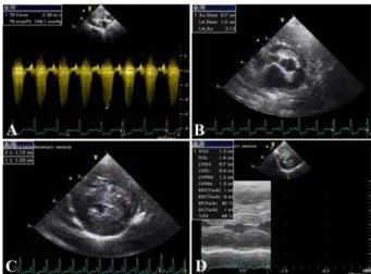
Won-Kyoung Yoon, Sang-Il Suh, Yeon-Su Oh, Changbaig Hyunular enlargement. Laboratory tests revealed mildly increased blood urea (33 mg/dL; reference range 7 −27 mg/dL). Tho- racic radiography revealed severe generalized cardiomegaly (VHS 13) and under-circulated lung fields. There were also increased cardiac sternal contact, marked dorsal displace- ment of trachea and marked distention of caudal vena cava suggesting right sided cardiac enlargement. Echocardiography found worsened systolic jet (5.76 m/s, pressure gradient 132.7 mmHg; Fig. 1A) concomitant with diastolic regurgitant jet (2.47 m/s, pressure gradient 24.4 mmHg, 200 ms pressure half time), suggesting severe pulmonic hypertension. Inter- estingly, further echocardiography revealed thickening of anterior cusps of mitral valve by degenerative change and narrowing of left ventricular outflow tract (LVOT; Fig. 1B).
Color Doppler echocardiogram taken at left apical long axis of five chamber view revealed bidirectional systolic jets into LVOT and mitral annulus due to systolic anterior motion (SAM) of mitral valve by thickening and narrowing of LV chamber (Fig. 1C). Continuous wave Doppler study at the same plane found 3.66 m/s (pressure gradient 53.6 mmHg) systolic jet at aorta, suggesting LVOT obstruction (Fig. 1D).
Further continuous wave Doppler study taken at right paraster- nal long axis of four chamber view found 5.20 m/s (pressure gradient 108.1 mmHg) systolic regurgitant jet flowing back
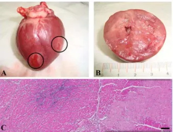
to right atrium at tricuspid valve, suggesting severe tricuspid regurgitation (Fig. 2A). The 2D and M-mode echocardio- graphy also found left atrial dilation (left atrial to aorta ratio 2.12 : 1; Fig. 2B) and severe thickening of interventricular septum and left ventricular free wall suggesting HCM by long-lasting pressure overload at both ventricular chambers (Fig. 2C and D). Based on diagnostic imaging studies, the dog was then diagnosed as HCM complicated with severe congenital PS and acquired left ventricular outflow tract obstruction. Emergency intensive care with oxygen, pimoben- dan (0.2 mg, q4h, IV, Vetmedin; Boehringer Ingelheim, Ger- many), furosemide (4 mg/kg, PRN, IV, Lasix; Handok, Korea) and diltiazem (1 mg/kg, PO, q12h; CJ HealthCare, Korea) was immediately proceeded. After stabilizing clinical condi- tion, the dog was then released with prescription of enalapril (0.5 mg/kg, PO, q12h), pimobendan (0.4 mg, q12h, PO), furosemide (1 mg/kg, q12h, PO) and diltiazem (1 mg/kg, PO, q12h). The clinical condition was manageable for 2 months after this medication. However, the dog died suddenly by cardiac arrest. Because the owner accepted the request of postmortem examination, the necropsy has done by the stan- dard method. Grossly, heart was enlarged. There were two infarcted regions in left and right ventricular area (Fig. 3A).
On the cross section of heart, there were marked thickening
Fig. 1. Echocardiography of this case at the first presentation.(A) Continuous wave Doppler study taken at right parasternal short axis of pulmonary artery. There was 5.76 m/s (pressure gradient 132.7 mmHg) systolic jet at pulmonary artery, suggest- ing that the stenosis was deteriorated with time. (B) The 2D- echocardiogram taken at left apical long axis of five chamber view. There was thickening of anterior cusp (arrow) of mitral valve by degenerative change (Inset: Gross finding on anterior mitral valvular cusp). In addition, there was narrowing of left ventricular outflow tract (LVOT). (C) Color Doppler echocar- diogram taken at left apical long axis of five chamber view.
There were bidirectional systolic jets into LVOT and mitral annulus due to systolic anterior motion (SAM) of mitral valve by thickening and narrowing of LV chamber. (D) Continuous wave Doppler study taken at left apical long axis of five cham- ber view found 3.66 m/s (pressure gradient 53.6 mmHg) sys- tolic jet at aorta, suggesting dynamic LVOT obstruction.
Fig. 2. Echocardiography of this case after 2 years of the first presentation. (A) Continuous wave Doppler study taken at right parasternal long axis of four chamber view found 5.20 m/s (pressure gradient 108.1 mmHg) systolic regurgitant jet flowing back to right atrium at tricuspid valve, suggesting severe tri- cuspid regurgitation. (B) The 2D-echocardiogram taken at right parasternal short axis of aortic valve found left atrial dilation (left atrial to aorta ratio 2.12 : 1; enlarged rectangle). (C) The 2D-echocardiogram taken at right parasternal short axis of pap- illary muscle found severe thickening of interventricular septum (11.5 mm at diastole) and left ventricular free wall (12.2 mm at diastole) suggesting hypertrophic cardiomyopathy by long last- ing pressure overload at both ventricular chambers. (D) The M mode-echocardiogram taken at right parasternal short axis of papillary muscle level revealed severe thickening of interven- tricular septum (13.0 mm at distole) and left ventricular free wall (12 mm at distole).
Hypertrophic cardiomyopathy secondary to severe right and left ventricular outflow tract obstruction in a Maltese dog
211
of left and right ventricular free wall and interventricuar sep- tum (Fig. 3B). Myocardial infarction was diffusely dispersed through myocardium (Fig. 3B). On histological section of the heart, the myofibers appeared to be hypertrophic, with focal areas of hypereosinophilia and acute coagulative necrosis (Fig. 3C). In these areas fibers were somewhat fragmented and accompanied by a minimal cellular infiltrate which appears to be neutrophilic. The section was characterized by regionally extensive infarction of the muscle, accompanied by early neutrophilic cellular inflammation and mild calcifi- cation (Fig. 3C). Interfiber hemorrhage is also noted focally.
There were degenerative changes in mitral valvular cusps, probably by aging process. There were evidence of infarction and early abscessation possibly from chronic HCM, or embo- lus formation resulting in blood vessel occlusion and down- stream ischemic degeneration and necrosis of the affected cardiac tissue as similarly noticed in other case [7]. However, there was no evidence for infection or neoplasia.
The dog presented here initially had severe PS which was characterized by right ventricular (RV) hypertrophy and right atrial dilation. Marked RV hypertrophy usually resulted in interventricular septal flattening and narrowing of LV cham- ber [1]. These changes often leaded to misdiagnosis of LV hypertrophy. However, in this case of dog, surprisingly the interventricular septal flattening was not severe, considering
the severity of RV pressure overload by long-lasting and severe PS. The dynamic pressure gradient across the LV out- flow tract from HCM in this dog might be the cause of aor- tic stenosis (dynamic LVOT obstruction). The SAM of the mitral valve from the narrowing of LVOT by thickening (hypertrophy) of interventricular septum have attributed to this dynamic obstruction of LVOT in cats with HCM [2].
The initiation of hypertrophic changes in both ventricles might be from the severe RV pressure overload of RVOT obstruction (PS). Subsequently, chronic RV pressure over- load was resulted in hypertrophy in interventricular septum, which caused to dynamic obstruction of LVOT. The LVOT obstruction induced marked pressure overload in the LV chamber and pressure gradient between LV and LVOT caus- ing systolic jet in this case. The degenerative change in ante- rior cusp of mitral valve might be contributed to severe SAM at the LVOT. The pressure overload from LVOT obstruction in the LV in this dog might be the cause of marked hypertro- phy in LV free wall. Those changes impaired ventricular fill- ing and increased filling pressure in both chambers causing tricuspid regurgitation and pulmonic hypertension in the right cardiac chambers and LA dilation in the left chamber in this dog. Regional myocardial infarction in this dog reflected severe chronic pressure overload in both ventricles.
The case described here had diagnostic features of HCM including thickening of LV free wall and interventricular sep- tum, SAM, dynamic LVOT obstruction and diastolic dys- function. However, the HCM in this case might not be primary HCM which has been widely reported in cats [1]. The HCM was secondary to the dynamic obstruction of LVOT occurred by degenerative valvular change of anterior cusp of mitral valve and interventricular hypertrophy from pre-existing PS.
To our best knowledge, this is the first case of HCM second- ary to other cardiac defects in dogs.
References
1. Abbott JA. Feline hypertrophic cardiomyopathy: an update.
Vet Clin North Am Small Anim Pract 2010, 40, 685-700.
2. De Majo M, Britti D, Masucci M, Niutta PP, Pantano V. Hypertrophic obstructive cardiomyopathy associated to mitral valve dysplasia in the Dalmatian dog: two cases. Vet Res Commun 2003, 27 (Suppl 1), 391-393.
3. Lee SG, Moon HS, Hyun C. Pulmonic stenosis corrected by balloon valvuloplasty in a Maltese dog. Korean J Vet Res 2007, 47, 333-336.
4. Liu SK, Maron BJ, Tilley LP. Canine hypertrophic cardiomyopathy. J Am Vet Med Assoc 1979, 174, 708-713.
5. Maron BJ. Hypertrophic cardiomyopathy: a systematic review. JAMA 2002, 287, 1308-1320.
6. Pang D, Rondenay Y, Hélie P, Cuvelliez SG, Troncy E.
Sudden cardiac death associated with occult hypertrophic cardiomyopathy in a dog under anesthesia. Can Vet J 2005, 46, 1122-1125.
7. Washizu M, Takemura N, Machida N, Nawa H, Yamamoto T, Mitake H, Washizu T. Hypertrophic cardiomyopathy in an aged dog. J Vet Med Sci 2003, 65, 753-756.
Fig. 3. Gross and histopathological findings of this case. (A) Grossly, heart was enlarged. There were two infarcted regions in left and right ventricular area (circles). (B) The cross section of heart. There were marked thickening of left and right ven- tricular free wall and interventricuar septum. Myocardial inf- arction was diffusely dispersed through myocardium. (C) Histological section of the heart (H&E stain, ×400). The myo- fibers appeared to be hypertrophic, with focal areas of hyper- eosinophilia and acute coagulative necrosis. In these areas fibers were somewhat fragmented and accompanied by a minimal cel- lular infiltrate which appears to be neutrophilic. The section was characterized by regionally extensive infarction of the muscle, accompanied by early neutrophilic cellular inflammation and mild calcification. Interfiber hemorrhage is also noted focally.
Scale bar = 100µm.