J Korean Soc Radiol 2016;74(5):291-298 http://dx.doi.org/10.3348/jksr.2016.74.5.291
INTRODUCTION
A sizable segment of the population will undergo coronary artery stent implantation in their lifetime. They require frequent follow-up for stent patency; hence, noninvasive, low radiation dose imaging technique to rule out in-stent re-stenosis is desir- able (1). Coronary CT angiography (CCTA) has been used for this purpose, and the evaluation of larger (≥ 3 mm) coronary ar- tery stents is considered appropriate according to current guide- lines (1-7). Two recent meta-analyses demonstrate the high di- agnostic value of CCTA with sensitivities and specificities > 90%
when non-evaluable stents are excluded (1, 8, 9). However, these studies also confirm CCTA’s long recognized limitations of bl- ooming artifact, which can cause a decrease in diagnostic im-
age quality or even render studies non-diagnostic (1, 4). There- fore, it is important to reduce both noise and blooming artifact for a better quality CCTA image, in the evaluation of coronary artery stents.
Improved computing power has led to the introduction of it- erative reconstruction into the clinical arena, and its beneficial effects throughout the body are reported (1). Improved image quality, reduction in image noise, and the potential for radiation dose savings are demonstrated, as compared with traditional filtered back projection (FBP) (1, 10-12). Several studies showed that iterative reconstruction techniques could reduce image noise and blooming artifacts when heavy calcifications or stent are present in coronary arteries (1, 13). Adaptive statistical iterative re- construction (ASIR) techniques are reported to significantly im-
Coronary Stent on Coronary CT Angiography: Assessment with Model-Based Iterative Reconstruction Technique
모델기반 반복재구성법을 이용한 관상동맥 스텐트의 컴퓨터단층촬영 평가
Eunchae Lee, MD, Yeo Koon Kim, MD, Eun Ju Chun, MD, Sang Il Choi, MD*
Department of Radiology, Seoul National University Bundang Hospital, Seongnam, Korea
Purpose: To assess the performance of model-based iterative reconstruction (MBIR) technique for evaluation of coronary artery stents on coronary CT angiography (CCTA).
Materials and Methods: Twenty-two patients with coronary stent implantation who underwent CCTA were retrospectively enrolled for comparison of image quality between filtered back projection (FBP), adaptive statistical iterative reconstruction (ASIR) and MBIR. In each data set, image noise was measured as the standard devi- ation of the measured attenuation units within circular regions of interest in the ascending aorta (AA) and left main coronary artery (LM). To objectively assess the noise and blooming artifacts in coronary stent, we additionally measured the stan- dard deviation of the measured attenuation and intra-luminal stent diameters of total 35 stents with dedicated software.
Results: All image noise measured in the AA (all p < 0.001), LM (p < 0.001, p = 0.001) and coronary stent (all p < 0.001) were significantly lower with MBIR in comparison to those with FBP or ASIR. Intraluminal stent diameter was significantly higher with MBIR, as compared with ASIR or FBP (p < 0.001, p = 0.001).
Conclusion: MBIR can reduce image noise and blooming artifact from the stent, leading to better in-stent assessment in patients with coronary artery stent.
Index terms
Image Reconstruction Noise
Stent
Received July 27, 2015 Revised November 11, 2015 Accepted December 7, 2015
*Corresponding author: Sang Il Choi, MD Department of Radiology, Seoul National University Bundang Hospital, 82 Gumi-ro 173beon-gil, Bundang-gu, Seongnam 13620, Korea.
Tel. 82-31-787-7609 Fax. 82-31-787-4011 E-mail: drsic@hanmail.net
This is an Open Access article distributed under the terms of the Creative Commons Attribution Non-Commercial License (http://creativecommons.org/licenses/by-nc/3.0) which permits unrestricted non-commercial use, distri- bution, and reproduction in any medium, provided the original work is properly cited.
prove the imaging evaluation of coronary artery stents and have the potential to substantially reduce the radiation dose (1, 14).
Recently developed model-based iterative reconstruction (MBIR) technique is a raw data based 3-dimensional image re- construction method (15). It does not involve blending with FBP images, and is mathematically more complex and accurate than ASIR techniques (16). Several studies showed that MBIR imag- es, as compared with ASIR and FBP images, have equivalent or better image quality with a substantial decrease in image noise and potential to reduce radiation dose on CT images of cardiac, liver, lung and cervicothoracic region (15-18).
However, to the best of our knowledge, there is no data regard- ing the potential usefulness of MBIR for evaluation of coronary artery stent. Therefore, the purpose of this study was to assess the performance of MBIR technique for evaluation of coronary ar- tery stent using CCTA in comparison to ASIR and the traditional FBP algorithm.
MATERIALS AND METHODS
Patients
The study was approved by our Institutional Review Board that waived the need for individual informed patient consent.
The study population consisted of consecutive 33 patients of post-percutaneous coronary intervention status with stent im- plantation who underwent CCTA within 12 months for risk as- sessment due to prior positive ischemic equivalents (1-3). Ex- clusion criteria were images with poor quality due to motion artifact, small caliber of vessel diameter less than 2.5 mm, in-stent
restenosis on previous follow-up conventional angiography, and insufficient medical records. After exclusion of 11 patients, CT im- ages of 22 patients (13 men, 9 women; mean age 66.27 ± 10.16 years, 35 coronary stents) were analyzed (Tables 1, 2).
Image Acquisition
CCTA were performed using a 256-slice MDCT scanner (Brilliance iCT; Philips Medical Systems, Best, the Netherlands).
The parameters were detector configurations: 256 overlapping slices of 0.625-mm thickness and a dynamic z-focal spot; gantry rotation time, 0.27 seconds; tube voltage, 120 kVp; and 800–1050 mAs (depending on patient habitus). Prior to CCTA, all patients with a baseline heart rate over 70 beats per minute (bpm) re- ceived intravenous esmolol (10–30 mg; Jeil Pharm, Seoul, Ko- Table 1. Patient Demographics and CCTA Scan Parameters
Parameter Data
Age (yr) 66.27 ± 10.16
Male vs. female 13 vs. 9
Height (cm) 162.75 ± 9.59
Weight (kg) 67.38 ± 11.01
Body mass index (kg/m2) 25.39 ± 3.04
Heart rate 69.05 ± 14.55
(49–111)
Tube potential (kVp) 120
Tube current-time product (mAs) 750.16 ± 131.84
CT dose index volume (mGy) 44.30 ± 13.49
Dose-length product (mGy*cm) 801.94 ± 298.06 Prospective triggered vs. retrospective ECG gated 19 vs. 3 Data are means ± standard deviations, the range are in parentheses.
CCTA = coronary computed tomography angiography, ECG = electrocardi- ography
Table 2. Types of Stents in the Study Population
Stent Name Manufacturer Material Eluting Drug Stent Diameter (mm)
2.5 2.75 3.0 3.5 4.0 Total No.
Xience Abbott CoCr Everolimus 0 1 1 1 0 3
Endeavor Medtronic CoCr Phosphorylcholine +
ABT578
0 0 2 1 1 4
Resolute Medtronic Cobalt-based metal Zotarolimus 0 0 0 0 1 1
Promus-element Boston Scientific CoCr Everolimus 1 0 1 0 0 2
Cypher Cordis Stainless steel Serolimus 0 0 3 2 0 5
Vision Abbott CoCr No 0 0 0 2 0 2
Novori Terumo Stainless steel Biolimus A9 1 0 1 0 0 2
Taxus Boston Scientific Stainless steel Paclitaxel 2 2 1 2 1 8
Unknown 0 0 5 3 0 8
Total No. 4 3 14 11 3 35
CoCr = cobalt chromium, No. = number
rea). Nitroglycerine (0.6 mg) was immediately administered sublingually to all patients before contrast injection, with the ex- ception of patients with a contraindication to nitroglycerin (19, 20). A bolus of 70 mL of iomeprol (Iomeron 400; Bracco, Milan, Italy) was injected intravenously (4 mL/s), followed by a 50-mL saline chaser. A region of interest (ROI) was placed in the de- scending thoracic aorta, and image acquisition was automatical- ly initiated 7 seconds after a selected threshold [150 Hounsfield units (HU)] had been reached with bolus tracking. Details of data acquisition are described previously (19). Patients with heart rates < 70 bpm underwent CCTA using a prospectively- triggered axial technique, and patients with heart rates > 70 bpm underwent a retrospectively-gated helical technique. Details of MBIR reconstruction are described previously (17). After a pilot study with 15 cases, soft-tissue kernel was selected as the opti- mal type to reconstruct the CT images.
Measurement of Image Noise and Stent Lumen Diameter
All images were reviewed and interpreted on dedicated work- station by two radiologists. In each data set, one observer mea- sured image noise, which was defined as the standard deviation (SD) of the measured attenuation (in HU) within circular ROIs in the ascending aorta and left main coronary artery (LM). The size of the ROIs was adapted to account for anatomic differences of our patients, representing an area that was not too small as to be affected by pixel variability and not so large as to approach the edges of the vessel. However, between the different recon- struction approaches, the ROI size was kept constant within each patient. We compared these parameters between the three reconstruction settings.
We also measured the stent lumen diameter in portions free of stenosis on curved multi-planar images acquired with the FBP, ASIR, and MBIR algorithm, in order to objectively assess the reduction of blooming artifact in coronary stent by each re- construction method. Using dedicated software, in-stent lumen maximal diameter was calculated automatically. The window width and level was set at 1500 and 300 HU, respectively.
Statistical Analysis
Statistical analysis was performed using dedicated statistical software (SPSS 12.0, SPSS Inc., Chicago, IL, USA). Noise and
in-stent diameters were expressed as mean ± SD and compared using paired t-test and repeated measure ANOVA. A p-value of <
0.05 was considered statistically significant. Interobserver agree- ment was determined by intra-class correlation coefficient.
RESULTS
Patient demographics and scan parameters on CCTA were provided in Table 1. Image noise with MBIR in the ascending aorta was significantly lower, as compared to those with FBP and
50.00
40.00
30.00
20.00
10.00
0.00
Noise_AA (HU)
FBP
56 47 66
ASIR MBIR
Reconstruction_method
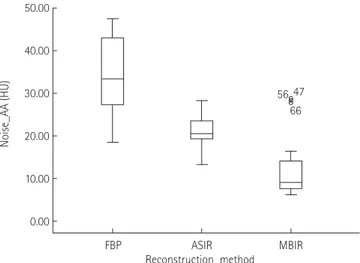
Fig. 1. Image noise measured in the ascending aorta is significantly lower by MBIR, as compared to ASIR and FBP (all p < 0.001).
AA = ascending aorta, ASIR = adaptive statistical iterative reconstruc- tion, FBP = filtered back projection, HU = Hounsfield units, MBIR = mod- el-based iterative reconstruction
200.00
150.00
100.00
50.00
0.00
Noise_LM (HU)
FBP
44 22
66
ASIR MBIR
Reconstruction_method
Fig. 2. Image noise measured in the left main coronary artery is sig- nificantly lower by MBIR, as compared to ASIR and FBP (p < 0.001, p = 0.001).
ASIR = adaptive statistical iterative reconstruction, FBP = filtered back projection, HU = Hounsfield units, LM = left main coronary artery, MBIR = model-based iterative reconstruction
ASIR (all p < 0.001; 34.07 ± 8.67 HU with FBP, 21.15 ± 4.16 HU with ASIR, and 10.59 ± 10.59 HU with MBIR) (Fig. 1). Interob- server agreements were between 0.616 and 0.896.
Image noise with MBIR in the LM was significantly lower, as compared to those with FBP and ASIR (p < 0.001, p = 0.001).
The mean values of image noise in each data set were 55.32 ±
30.60 HU with FBP, 41.71 ± 24.04 HU with ASIR, and 34.80 ± 23.93 HU with MBIR (Fig. 2). Interobserver agreements were between 0.958 and 0.977.
Image noise with MBIR within coronary stent was signifi- cantly lower, as compared to those with FBP and ASIR (all p <
0.001) (Fig. 3). The mean values of image noise were 39.39 ±
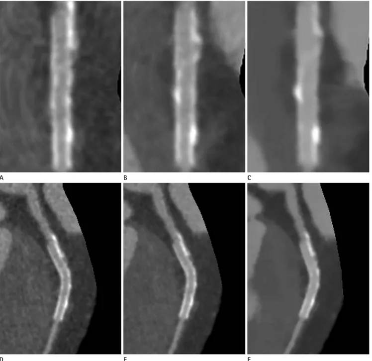
Fig. 3. Straight view of curved MPR images with FBP (A), ASIR (B), and MBIR (C) technique. Curved MPR images with FBP (D), ASIR (E), and MBIR (F) technique. Note the reduction of noise and blooming artifact in coronary stent is better on images with MBIR (C, F) than those with ASIR (B, E) and FBP (A, D).
ASIR = adaptive statistical iterative reconstruction, FBP = filtered back projection, MBIR = model-based iterative reconstruction, MPR = multi- planar reconstruction
A
D
B
E
C
F
17.46 HU with FBP, 29.53 ± 13.82 HU with ASIR, and 19.96 ± 12.19 HU with MBIR (Fig. 4). Interobserver agreements were between 0.720 and 0.907.
In-stent diameters with MBIR showed significantly higher value, as compared to those with ASIR and FBP (p < 0.001, p = 0.001), which indicated that the reduction of blooming artifact was best on images with MBIR than those with ASIR and FBP (Fig. 3). The mean values of intraluminal stent diameter were 1.67 ± 0.45 with FBP, 1.75 ± 0.43 with ASIR, and 1.80 ± 0.43 with MBIR
(Fig. 5). Interobserver agreements were between 0.990 and 0.994.
DISCUSSION
The main findings of the present study were as follows: 1) All image noise was significantly lower with MBIR in comparison to those with FBP or ASIR. 2) Intraluminal stent diameter was significantly higher with MBIR, as compared with FBP or ASIR.
Our study showed that the application of MBIR in patients with coronary stent could improve the image quality, as compared to FBP or ASIR, in terms of reduced noise and blooming artifact.
Results reported from previous studies likewise indicated that MBIR could provide improved image quality in cardiovas- cular imaging. Scheffel et al. (15) reported that MBIR can pro- vide reduced image noise and increased contrast-to-noise ratio compared to those using ASIR and FBP in CCTA of ex vivo hu- man heart (15). Oda et al. (21) also reported improved image quality in low-dose cardiac CT with MBIR (20% of the standard tube current) than those using full dose or low dose FBP. Be- sides, other studies showed similar or improved image quality and equivalent lesion detection in abdominal (17, 22) and chest (18) CT using MBIR, as compared with FBP or ASIR.
Interestingly, although Scheffel et al. (15) were unable to show the effective suppression of blooming artifact in the eval- uation of calcified plaque with MBIR than other reconstruction methods, our study showed significant reduction of blooming artifact in the evaluation of coronary stent. This discrepancy is possibly due to different methods of analysis; while Scheffel et al. (15) only used qualitative method with four-point scale to qualify blooming artifact, we adopted quantitative method by measuring the intraluminal stent diameters using dedicated soft- ware. Moreover, other studies reported that ASIR technique pro- vides improved in-stent lumen visualization by reducing noise or blooming artifact in the evaluation of coronary stent using CCTA at a lower radiation dose. However, they are either studies with phantom only (14) or those that do not include MBIR (1, 14).
This study is meaningful, since it is the first to evaluate in-stent image quality in CCTA with MBIR.
From a clinical viewpoint, the expected increased accuracy and reliability in diagnosing in-stent stenosis could be extended to clinical applicability in follow-up-patients with implanted Fig. 4. Image noise measured in the stent is significantly lower by
MBIR, as compared to ASIR and FBP (all p < 0.001).
ASIR = adaptive statistical iterative reconstruction, FBP = filtered back projection, HU = Hounsfield units, MBIR = model-based iterative re- construction
100.00
80.00
60.00
40.00
20.00
0.00
Noise_stent (HU)
FBP 9
57
ASIR MBIR
Reconstruction_method
Fig. 5. In-stent diameters are significantly higher by MBIR, as com- pared to ASIR and FBP, which means that the reduction of blooming artifact is better by MBIR (p < 0.001, p = 0.001) than ASIR and FBP.
ASIR = adaptive statistical iterative reconstruction, FBP = filtered back projection, HU = Hounsfield units, MBIR = model-based iterative re- construction
3.00
2.50
2.00
1.50
1.00
Diameter_stent (HU)
FBP
3 44 79
9
ASIR MBIR
Reconstruction_method
coronary stent. Also, improvement of image quality with MBIR suggests the potential to reduce radiation dose while preserving similar image quality level, as compared to those of FBP or oth- er iterative reconstructions.
There are several limitations to be considered in our study.
First, we did not provide the diagnostic accuracy of MBIR in the evaluation of in-stent restenosis using CCTA; hence, further study will be needed. Second, due to the limited size of the study population, we did not conduct subgroup analysis according to the diameter of coronary stent, which could possibly have bi- ased the results. Third, despite improved image quality and in- stent lumen visualization, we did not assess the diagnostic accu- racy of disease such as in-stent restenosis in coronary stent on CCTA using MBIR. Fourth, potential radiation dose reduction using MBIR can only be assumed and is without definite evi- dence, because the image quality of MBIR was compared with other reconstruction methods at the same radiation dose set- tings. Further studies are needed to compare the image quality of MBIR in reduced radiation dose setting, with those of other re- construction techniques in conventional radiation dose settings.
Fifth, our study did not provide intraobserver agreement. We considered that calculation of intraobserver agreement would be unhelpful, because the observers measured noise and diam- eter using dedicated software. Sixth, we did not include signal to noise ratio and contrast to noise ratio in this study.
In summary, the results of our study suggested that MBIR could provide significant improvement of image quality, as com- pared to FPB or ASIR by reducing noise and blooming artifact in the evaluation of coronary stent on CCTA. MBIR has the potential to enhance diagnostic accuracy of in-stent stenosis or reduce the radiation dose in the evaluation of coronary stent.
Acknowledgments
This work was supported by the National Research Founda- tion of Korea (NRF) grant funded by the Korea government (MEST) (No. 2011-0023624).
REFERENCES
1. Ebersberger U, Tricarico F, Schoepf UJ, Blanke P, Spears JR, Rowe GW, et al. CT evaluation of coronary artery stents with iterative image reconstruction: improvements in im-
age quality and potential for radiation dose reduction. Eur Radiol 2013;23:125-132
2. Kim YJ, Yong HS, Kim SM, Kim JA, Yang DH, Hong YJ.
Guideline for Appropriate Use of Cardiac CT in Heart Dis- ease. J Korean Soc Radiol 2014;70:93-109
3. Taylor AJ, Cerqueira M, Hodgson JM, Mark D, Min J, O’Gara P, et al. ACCF/SCCT/ACR/AHA/ASE/ASNC/NASCI/SCAI/SCMR 2010 appropriate use criteria for cardiac computed to- mography. A report of the American College of Cardiology Foundation Appropriate Use Criteria Task Force, the Soci- ety of Cardiovascular Computed Tomography, the Ameri- can College of Radiology, the American Heart Association, the American Society of Echocardiography, the American Society of Nuclear Cardiology, the North American Society for Cardiovascular Imaging, the Society for Cardiovascular Angiography and Interventions, and the Society for Car- diovascular Magnetic Resonance. J Am Coll Cardiol 2010;
56:1864-1894
4. Andreini D, Pontone G, Bartorelli AL, Mushtaq S, Trabatto- ni D, Bertella E, et al. High diagnostic accuracy of prospec- tive ECG-gating 64-slice computed tomography coronary angiography for the detection of in-stent restenosis: in- stent restenosis assessment by low-dose MDCT. Eur Radiol 2011;21:1430-1438
5. Kerl JM, Schoepf UJ, Vogl TJ, Ackermann H, Vogt S, Costello P, et al. In vitro evaluation of metallic coronary artery stents with 64-MDCT using an ECG-gated cardiac phantom: rela- tionship between in-stent visualization, stent type, and heart rate. AJR Am J Roentgenol 2010;194:W256-W262 6. Sheth T, Dodd JD, Hoffmann U, Abbara S, Finn A, Gold HK,
et al. Coronary stent assessability by 64 slice multi-detector computed tomography. Catheter Cardiovasc Interv 2007;
69:933-938
7. Zhang X, Yang L, Ju H, Zhang F, Wu J, He B, et al. Prevalence and prognosis of coronary stent gap detected by multi-de- tector CT: a follow-up study. Eur Radiol 2012;22:1896-1903 8. Sun Z, Almutairi AM. Diagnostic accuracy of 64 multislice
CT angiography in the assessment of coronary in-stent re- stenosis: a meta-analysis. Eur J Radiol 2010;73:266-273 9. Kumbhani DJ, Ingelmo CP, Schoenhagen P, Curtin RJ, Flamm
SD, Desai MY. Meta-analysis of diagnostic efficacy of 64-slice computed tomography in the evaluation of coro-
nary in-stent restenosis. Am J Cardiol 2009;103:1675-1681 10. Gosling O, Loader R, Venables P, Roobottom C, Rowles N,
Bellenger N, et al. A comparison of radiation doses be- tween state-of-the-art multislice CT coronary angiogra- phy with iterative reconstruction, multislice CT coronary angiography with standard filtered back-projection and invasive diagnostic coronary angiography. Heart 2010;96:
922-926
11. Prakash P, Kalra MK, Ackman JB, Digumarthy SR, Hsieh J, Do S, et al. Diffuse lung disease: CT of the chest with adap- tive statistical iterative reconstruction technique. Radiolo- gy 2010;256:261-269
12. Sagara Y, Hara AK, Pavlicek W, Silva AC, Paden RG, Wu Q.
Abdominal CT: comparison of low-dose CT with adaptive statistical iterative reconstruction and routine-dose CT with filtered back projection in 53 patients. AJR Am J Roentgenol 2010;195:713-719
13. Renker M, Nance JW Jr, Schoepf UJ, O’Brien TX, Zwerner PL, Meyer M, et al. Evaluation of heavily calcified vessels with coronary CT angiography: comparison of iterative and filtered back projection image reconstruction. Radiol- ogy 2011;260:390-399
14. Funama Y, Oda S, Utsunomiya D, Taguchi K, Shimonobo T, Yamashita Y, et al. Coronary artery stent evaluation by combining iterative reconstruction and high-resolution ker- nel at coronary CT angiography. Acad Radiol 2012;19:1324- 1331
15. Scheffel H, Stolzmann P, Schlett CL, Engel LC, Major GP, Károlyi M, et al. Coronary artery plaques: cardiac CT with model-based and adaptive-statistical iterative reconstruc- tion technique. Eur J Radiol 2012;81:e363-e369
16. Katsura M, Sato J, Akahane M, Matsuda I, Ishida M, Yasaka K, et al. Comparison of pure and hybrid iterative recon-
struction techniques with conventional filtered back pro- jection: image quality assessment in the cervicothoracic re- gion. Eur J Radiol 2013;82:356-360
17. Chang W, Lee JM, Lee K, Yoon JH, Yu MH, Han JK, et al. As- sessment of a model-based, iterative reconstruction algo- rithm (MBIR) regarding image quality and dose reduction in liver computed tomography. Invest Radiol 2013;48:598- 606
18. Kim H, Park CM, Song YS, Lee SM, Goo JM. Influence of radiation dose and iterative reconstruction algorithms for measurement accuracy and reproducibility of pulmonary nodule volumetry: a phantom study. Eur J Radiol 2014;83:
848-857
19. Lee MS, Chun EJ, Kim KJ, Kim JA, Vembar M, Choi SI. Re- producibility in the assessment of noncalcified coronary plaque with 256-slice multi-detector CT and automated plaque analysis software. Int J Cardiovasc Imaging 2010;
26(Suppl 2):237-244
20. Chun EJ, Lee W, Choi YH, Koo BK, Choi SI, Jae HJ, et al. Ef- fects of nitroglycerin on the diagnostic accuracy of elec- trocardiogram-gated coronary computed tomography an- giography. J Comput Assist Tomogr 2008;32:86-92 21. Oda S, Utsunomiya D, Funama Y, Katahira K, Honda K,
Tokuyasu S, et al. A knowledge-based iterative model re- construction algorithm: can super-low-dose cardiac CT be applicable in clinical settings? Acad Radiol 2014;21:104- 110
22. Shuman WP, Green DE, Busey JM, Kolokythas O, Mitsumori LM, Koprowicz KM, et al. Model-based iterative recon- struction versus adaptive statistical iterative reconstruc- tion and filtered back projection in liver 64-MDCT: focal lesion detection, lesion conspicuity, and image noise. AJR Am J Roentgenol 2013;200:1071-1076
모델기반 반복재구성법을 이용한 관상동맥 스텐트의 컴퓨터단층촬영 평가
이은채 · 김여군 · 전은주 · 최상일*
목적: 관상동맥 컴퓨터단층촬영술을 이용한 스텐트 평가에 있어, 기존의 조절통계 반복 재구성법 및 여과 후 역투사법과 비교하여 모델기반 반복 재구성법의 유용성을 평가하고자 하였다.
대상과 방법: 기존에 경피적 관상동맥 혈관성형술로 스텐트 시술을 시행한 환자들 중 관상동맥 컴퓨터단층촬영술을 시행 한 22명의 환자의 총 35개의 스텐트를 대상으로 하였다. 세 가지 재구성법으로 만들어진 각각의 이미지에서, 상행대동맥 과 좌측 주관상동맥에 관심영역을 위치시켜 감쇄계수의 표준편차를 측정하여 이미지 노이즈를 평가하였다. 총 35개의 스 텐트에서 노이즈와 색번짐 인공음영을 평가하기 위하여 감쇄계수의 표준편차와 스텐트의 내경을 측정하였다.
결과: 이미지의 노이즈와 스텐트 내부 노이즈는 모델기반 반복 재구성법을 이용한 이미지가 조절통계 반복 재구성법 및 여
과후 역투사법에서보다 상행대동맥과 좌측 주관상동맥 모두에서 작게 측정되었다(p < 0.001). 스텐트 내경의 평가에 있
어 모델기반 반복 재구성법을 이용한 이미지가 조절통계 반복 재구성법 및 여과 후 역투사법에서보다 유의하게 크게 측정 되었다(p = 0.001).
결론: 모델기반 반복 재구성법은 스텐트 내부의 이미지 노이즈와 색번짐 인공음영을 감소하여 주므로, 관상동맥 컴퓨터단 층촬영술을 시행한 환자에서 스텐트 내부를 평가하는 데 도움이 될 수 있다.
분당서울대학교병원 영상의학과