자동유방초음파의 임상적 의의
전북대학교 의과대학 외과학교실 유방ㆍ갑상선외과
강상율, 이승주, 윤현조, 정성후
Clinical Significance of Automated Breast Ultrasound
Sang Yull Kang, Seung Ju Lee, Hyun Jo Youn, Sung Hoo Jung
Division of BreastㆍThyroid Surgery, Department of Surgery, Chonbuk National University Medical School, Jeonju, Korea
Received April 17, 2017 Revised May 3, 2017 Accepted May 7, 2017
Breast ultrasound is a well-established diagnostic tool that is coupled with mammography for evaluation of breast abnormalities. This technique is widely available, painless, well-tol- erated and does not involve ionizing radiation. However, it has several faults. Because of its dependence on operator techniques for hand-held ultrasound (HHUS), the skill and knowl- edge of the operator influence the diagnostic accuracy, while poor standardization and re- producibility reduce the diagnostic yield. To overcome these problems, state-of-the-art equipment such as an automated breast ultrasound (ABUS) has been introduced. Automated breast ultrasound acquires a series of consecutive B-mode pictures and reconstructs three-di- mensional datasets of the entire breast volume. These data can then be sent to a separate workstation for analysis by a radiologist. Automated breast ultrasound can produce ob- jective and responsible images and is expected to minimize operator dependency. Several studies have demonstrated the feasibility of ABUS and shown equal performance when com- paring ABUS with HHUS. Moreover, some physicians suggested that it could be used not only for follow-up of benign lesions, but also for screening of breast cancer. However, ABUS also possesses certain limitations, including difficulty in clearly visualizing tissue in the axilla and behind the nipple, corrugation artifacts, and absence of flow information. Based on these limitations, other physicians have claimed that ABUS cannot currently displace HHUS and still requires further evaluation. Here, we review previous studies of ABUS and discuss its clinical significance as it relates to breast lesions.
Keywords: Automated, Hand, Breast, Ultrasonography
Correspondence to:Hyun Jo Youn
Division of BreastㆍThyroid Surgery, Department of Surgery, Chonbuk National University Medical School, Jeonju 54907, Korea
Tel: +82-63-250-2389 Fax: +82-63-271-6197 E-mail: [email protected]
서 론
유방암은 전 세계적으로 여성에서 가장 흔한 빈도로 발 생하는 암으로 치료 기술의 발전에도 불구하고 사망률은 감소하지 않고 있다.(1) 그러나 국내에서는 최근 유방암에 대한 관심 증가와 초음파를 포함한 영상 기기의 발전으로 조기에 유방암을 발견하여 치료하는 경우가 늘어나면서
생존율도 증가하는 추세이다.(2) 이와 같이 조기에 유방 암을 발견하는 것은 유방암으로 인한 사망률을 줄일 뿐만 아니라 수술 후 항암화학요법 등의 보조 요법(adjuvant therapy)을 축소할 수 있기 때문에 매우 중요하며, 따라서 유방암의 조기 발견을 위해 적절한 영상의학 검사를 시행 하는 것이 필수적이다.(3)
유방촬영술(mammography)은 오랜 동안 유방암의 기
REVIEW ARTICLE
J Surg Ultrasound 2017;4:12-17
JSU
Journal of Surgical Ultrasound초 검사로 여겨졌으며 선별 검사(screening test)로써 유 일하게 사망률을 낮출 수 있는 검사로 알려져 있다.(4) 그 러나 치밀 유방(dense breast)에서의 낮은 민감도 (sensitivity), 높은 위양성률(false positive rate)로 인한 불필요한 조직검사와 환자의 불안 유발, 방사선 노출 및 검사 시 심한 통증 등의 제한점으로 선별 검사 시행을 감소 시킬 수 있는 단점이 있다.(5,6)
이러한 유방촬영술의 단점을 보완하기 위해 유방 초음 파(ultrasound)를 추가로 시행하며, 유방촬영술과 유방 초음파를 함께 사용하는 경우 유방암을 포함한 유방 병변 (lesion)의 발견을 유의하게 증가 시킬 수 있다.(7,8) 하지 만 전통적인 수동 유방 초음파 검사(hand-held ultra- sound; HHUS)의 경우 높은 검사자(operator) 의존도, 낮 은 재현성(reproducibility), 표준화(standardization)의 부재, 긴 검사 시간 등의 단점이 존재해 새로운 초음파 양 식들의 개발이 요구된다.(4)
최근 소개되고 있는 자동유방초음파(Automated Breast Ultrasound; ABUS)는 영상의학과 전문의가 아닌 방사선 사(radiographer)가 촬영할 수 있기 때문에 상대적으로 덜 숙련된 인력으로 표준화되고 일관성 있는 영상을 얻을 수 있는 장점이 있어 HHUS의 단점을 보완할 수 있는 새로 운 기술로 각광받고 있다.(9-11)
이에 저자들은 문헌 고찰을 통해 유방암을 포함한 유방 병변의 진단에 있어 ABUS의 유용성을 알아봄으로써 실제 임상 적용에 도움이 되고자 한다.
본 론
1. ABUS의 소개
1980년 Maturo 등(12)에 의해 ABUS가 처음 소개된 이 후 초창기에는 주로 실험적인 접근 방법으로 연구가 이루 어졌다. 그 후 ABUS에 관한 많은 임상 결과가 발표되었고 그 유용성이 입증되면서 현재는 전 세계적으로 세 종류의 기기가 널리 사용되고 있다. 2008년 SonoCiné가 유방촬 영술의 보조적인 사용 적응증으로 미국 식약청(U.S.
Food and Drug Administration; FDA)의 허가를 처음 받 았고, 이후 Siemens Healthcare사의 Automated Breast Volume Scanner (ABVS)가 유방의 3차원 입체 영상을 얻 는 시스템을 구축하였다. 가장 최근인 2012년에 FDA 승 인을 받은 U-System의 somo.v는 ABVS와 마찬가지로 3
차원 영상을 제공하며 치밀 유방을 가진 무증상 여성의 유 방암 선별 검사를 위한 기기로 공식 인정을 받았다.(13) 본 연구는 이 중에서 현재까지 가장 많은 임상 연구가 발표된 ABVS를 대상으로 살펴보고자 한다.
1) ABUS의 구성
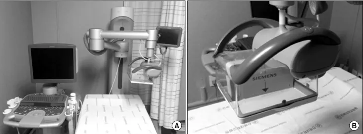
지금까지 ABUS에 관하여 발표된 많은 연구들은 Siemens Healthcare사의 ACUSON S200 (Siemens Medical Solution, Mountain View, CA)을 사용하였다.
이 시스템은 주파수 5-14 MHz, 길이 15.4 cm의 탐촉자 (probe)로 6 cm 깊이, 16.8 cm 범위 영역을 자동으로 스 캔(scan)하여 0.5 mm 두께의 고해상도 영상을 만들어낸 다(Fig. 1). 최적화된 영상을 얻기 위해 tissue harmonic imaging, advanced SieClearTM spatial compounding, dynamic tissue contrast enhancement, inferior-nipple image correction 그리고 3D image brightness au- to-correction과 같은 다양한 기술을 이용한다. 얻어진 영상들은 ABUS에서 작업단말기(workstation)로 자동으 로 전송된 후 전체 유방을 볼 수 있도록 3차원 영상으로 변 환되어 다면재구성(multiplanar reconstruction) 방식으 로 보여진다(Fig. 2).
2) ABUS의 검사 방법
환자는 편안히 누운(supine) 자세에서 양손을 머리 위 로 올린 상태로 압박판으로 유방을 압박하여 고정한 후 넓 은 탐촉자가 압박판 위를 가로지르며 자동으로 영상을 획 득한다. 일반적으로 양측 유방의 횡단면(transverse plane), 종단면(longitudinal plane), 관상면(coronal plane)의 세 영상을 기본으로 얻는다(Fig. 3). 환자의 유방 크기에 따라 검사자는 적절한 스캔 수를 결정하며 검사하는 동안 환자가 숨을 참을 필요는 없다. 실시간으로 다면재구성을 이용한 3차원적 영상 구성이 가능하며, 한쪽 유방을 검사 하는데 약 3-5분, 한 명의 환자를 검사하는데 약 10-15분 의 시간이 소요된다.
3) ABUS의 장점
ABUS는 HHUS에 비해 검사자 오류가 적은 자동화되고 표준화된 영상을 제공하며 지연 판독(delayed inter- pretation)이 가능하고 재구성된 이미지의 보존과 전달이 용이해 원격 협진(remote consultation)이 가능한 장점 이 있다.(14,15) 또한 ABUS는 재현성이 높으며 전체 유방 을 포함하는 넓은 영역의 영상을 보여주기 때문에 병변을 간과할 가능성이 적다.(16) ABUS의 관상면 영상에서는
Fig. 2. Multiplanar reconstruction of the volume data displayed on
the automated breast volume scanner.Fig. 3. Three standard images using automated breast volume
scanner. Cononal view (left), longitudinal (right, upper) and transverse views (right, lower) are synchronously visualized on the screen.Fig. 1. Installation of the ACUSON S2000 automated breast volume scanner (ABVS). (A) The overview of ABVS system. (B) The transducer that
has been designed for the ABVS.악성 병변을 감별하는데 중요한 ‘뒷당김 현상(retraction phenomenon)’을 좀 더 명확히 관찰할 수 있는 장점이 있 으며(Fig. 4), 피부부터 흉벽까지 해부학적 구조를 연속적 이고 체계적으로 볼 수 있기 때문에 유방 수술 시 효과적인 정보를 제공해 준다.(17)
4) ABUS의 단점
유두 아래와 겨드랑이 병변의 명확한 관찰이 어려우며 환자가 너무 마르거나 팔을 위로 충분히 올리지 못하는 경 우 전체 유방을 완벽하게 촬영하기가 어렵다.(14) 환자의 호흡으로 인한 ‘주름 결함(corrugation artifact)’이 발생
할 수 있으며 유관을 따라 스캔할 수 없다는 단점이 있다.
또한 혈류(blood flow) 정보를 얻을 수 없어 악성 병변을 감별하기 위해 Color Doppler flow imaging (CDFI) 또는 Pulsed Doppler Unit (PDU)와 같은 추가 영상을 필요로 한다.(18) 검사자에 의한 실시간 판독이 아닌 지연 판독을 시행하기 때문에 검사 시 문진을 통한 특정 부위의 자세한 검사를 추가로 시행할 수 없고 의심되는 병변이 검사 이후 에 발견될 경우 2차(second-look) 초음파를 요하는 빈도 가 높다.
Fig. 4. Retraction phenomenon in standard automated breast
volume scanning images. Three standard view of a right breast with an invasive ductal carcinoma lesion. Glandular tissue surrounding the lesion shows loss of its normal distribution pattern. This phenomenon (red arrow) is easier to visualize in the coronal as opposed to other planes.2. ABUS와 HHUS의 비교
앞서 기술한 바와 같이 ABUS는 여러 장점과 단점들을 가지고 있기에 지금까지 기존의 HHUS를 대체할 수 있는 새로운 기기인지에 관한 많은 논란이 있어왔다.
ABUS와 HHUS의 정확도를 비교한 초창기 예비 조사 (pilot study)의 연구 결과를 살펴보면 Wojcinski 등(19) 은 ABUS의 100% 민감도와 52.8%의 특이도, 66.0%의 정 확도로 HHUS와 동등한 유용성을 보고하였고, Kim 등(9) 은 두 검사 방법 사이에 Breast Imaging Reporting and Data System (BI-RADS) 범주(category) 2와 3에서는 66.2%, 범주 4와 5에서는 100%의 일치율을 보여 ABUS가 향후 유방 병변을 진단하는데 유용한 방법이 될 수 있을 것으로 예상하였다.
이후 ABUS의 병변 발견율과 진단 정확도가 HHUS와 유사하거나 오히려 높다는 연구 결과가 많이 보고 되었는 데 Lin 등(20)은 ABUS의 진단 정확도로 100%의 민감도 와 95.0%의 특이도를 보고하였고, 5566명의 대규모 환자 를 대상으로 한 Choi 등(21)의 보고에 따르면 유방암의 진 단 정확도(97.7% vs. 96.5%)와 특이도(97.8% vs. 96.7%) 에서 모두 ABUS가 HHUS에 비해 우월한 결과를 보였고 재검률(recall rate)도 ABUS (2.57/1,000예)가 HHUS (3.57/1,000예)에 비해 통계적으로 낮은 결과를 보였다.
유방에 형성되는 석회화를 동반하는 병변은 양성 및 악
성 모두에서 발견될 수 있는데 특히 악성 석회화의 경우 크기가 작은 미세석회화가 많아 초음파 상에서 후방음향 감소(posterior acoustic shadow)를 관찰하기 어려워 유 방촬영술에 비해 큰 제약점으로 인식되어 왔다.(22) 그러 나 최근에는 해상도가 좋은 초음파 기기의 도입과 배경 음 영에 의한 발견 의존도가 줄어들어 유방 초음파의 석회화 병변 발견 정확도가 증가하고 있는데,(23) ABUS는 HHUS에 비해 횡단면, 종단면, 관상면 뿐 만 아니라 다양 한 각도의 영상을 선택적으로 볼 수 있어서 석회와의 위치 와 수 그리고 모양을 좀 더 정확하게 알 수 있어 유방의 석 회화 병변을 진단하는데 매우 유용한 것으로 알려져 있 다.(24,25)
요약하면 ABUS는 병변의 경계를 좀 더 잘 볼 수 있는 관상면 영상을 추가로 얻을 수 있고 각각의 구획 단면 (sectional plane)을 관찰할 수 있기 때문에 HHUS에 비 해 좀 더 높은 정확도를 보일 수 있다.(16,18)
3. 판독자간의 일치율
ABUS는 검사자 의존도를 최소화 할 수 있는 장점이 있 지만 이는 병변의 위치와 크기 그리고 특징을 판독자들이 얼마나 일관성있게 판독할 수 있느냐는 또 다른 중요한 문 제점을 낳았다.
ABUS와 HHUS의 판독자간의 일치율(interobserver agreement)을 비교한 많은 연구에서 ABUS가 HHUS에 비해 병변의 모양, 경계, 에코발생(echogenicity), 후방음 향과 BI-RADS 최종 평가 등의 모든 항목에서 동등하거나 우월한 일치율을 보고하였다.(26-28) 특히, Shin 등(11) 은 ABUS 영상을 다섯 명의 판독자가 분석하였을 때 병변 의 발견율, 특징 및 병변의 위치, 크기 등을 보고하는데 있 어 판독자간의 일치율이 높다고 보고하였다. 이러한 결과 는 ABUS가 다양한 각도의 삼차원적 영상을 표준화된 다 중절편영상(multislice imaging)으로 얻을 수 있고 ‘뒷당 김 현상’을 포함한 구조적 왜곡(architectural distortion) 을 좀 더 잘 관찰할 수 있는 장점이 있기 때문이다. 그러나 이와 달리 병변의 모양과 경계 측면에서 ABUS의 판독자 간 일치율이 낮다는 보고도 있었다.(29) 최근에 판독자간 의 일치율과 관련된 체계적 고찰(systemic review)을 시 행한 Meng 등(30)에 따르면 ABUS는 양성과 악성 유방 병 변을 감별하는 민감도(92.0%)와 특이도(84.9%)는 높지 만 판독자간의 신뢰도(reliability)는 각 검사마다 매우 이
질적(heterogeneous)이라고 보고해 ABUS의 판독자간의 일치율에 관해서는 잘 고안된 좀 더 많은 전향적 연구가 필요할 것으로 생각한다.
4. 유방암 선별검사로서 ABUS의 유용성
우리나라를 포함한 여러 나라에서 유방암의 선별 검사 방법으로 40세 이상의 여성에서 1-2년 간격의 유방촬영 술을 권고하고 있다.(31) 유방촬영술은 유방암의 사망률 을 낮추는 유일한 선별 검사 방법으로 알려져 있지만 유방 암 발생위험도가 높고 젊은 여성에서 많이 관찰되는 치밀 유방에서는 정확도가 떨어지는 단점이 있다.(4,5) 이러한 제한점은 외국에 비해 젊은 여성에서의 유방암 발생률이 높은 우리나라에서 더욱 중요한 문제이다.(32)
유방 초음파는 유방 병변의 진단 정확도를 높이기 위한 유방촬영술의 보완적인 영상 검사로 여겨져 왔으며 아직 논란은 있지만 최근에는 특히 치밀 유방에서 유방암 선별 검사로서의 유용성에 관하여 많이 보고되고 있다.(7,33) ABUS가 HHUS와 비교해 동등한 진단 정확도를 보인다는 연구 결과들이 보고된 이후 양성 병변들의 추적관찰 뿐만 아니라 유방암의 선별검사로서 ABUS의 유용성을 알아보 려는 연구가 많이 이루어졌다.
Zang 등(15)은 ABUS가 HHUS에 비해 검사자 의존도 가 낮고 검사 시간이 짧아 선별 검사로서 유리하다고 하였 고, Kelly 등(34)은 특히 치밀 유방을 가진 여성에서 유방 암의 선별 검사로 유방촬영술에 ABUS를 추가하면 정확도 를 높이고 재검률을 낮출 수 있다고 보고하였다. ABUS는 특히 유방촬영술의 민감도가 떨어지는 치밀 유방 조직을 갖는 여성에서 유방암 선별 검사로서 유방촬영술과 함께 시행한다면 기존의 HHUS보다 더욱 정확한 결과를 보일 것으로 예상된다.
5. ABUS의 미래
지금까지 ABUS의 정확성 및 유용성에 관한 많은 보고 에도 불구하고 기존의 HHUS를 대체할 수 있는 방법으로 ABUS를 권고하고 있지는 않다.(14) 즉, HHUS를 대신해 ABUS를 사용함으로써 유방암의 발견율을 높이거나 불필 요한 조직검사를 줄일 수 있다는 명확한 연구 결과는 아직 보고되지 않았다. 그러나 앞으로 영상 기술이 더욱 발전하 고 명확한 진단 기준이 표준화 된다면 ABUS의 유용성은 훨씬 향상될 것으로 예상된다. 향후 대규모의 다기관, 전
향적 연구를 통한 타당성 검토가 이루어 진다면 ABUS의 적절한 임상 적응증을 확립할 수 있을 것이다.
결 론
ABUS는 일관성 있고 재현성이 우수하며 검사자 의존 도가 낮은 고화질의 3차원 영상을 제공한다. 판독자간의 높은 일치율이 입증된다면 ABUS는 유방암을 포함한 유방 병변의 정확한 관찰을 위한 기존의 유방촬영술과 HHUS 의 효과적인 보조 기기가 될 수 있다. ABUS가 기존의 HHUS를 대체할 수 있는지에 대해서는 향후 좀 더 많은 연 구가 필요할 것으로 생각한다.
REFERENCES
1. Youlden DR, Cramb SM, Dunn NA, Muller JM, Pyke CM, Baade PD. The descriptive epidemiology of female breast cancer: an international comparison of screen- ing, incidence, survival and mortality. Cancer Epidemiol 2012;36:237-48.
2. Park EH, Min SY, Kim Z, Yoon CS, Jung KW, Nam SJ, et al. Basic facts of breast cancer in korea in 2014:
the 10-year overall survival progress. J Breast Cancer 2017;20:1-11.
3. Chow LW, Yip AY, Ng EL. Prevention of oncological diseases: primary and secondary prevention. Int J Biol Markers 2012;27:e337-43.
4. Drukteinis JS, Mooney BP, Flowers CI, Gatenby RA.
Beyond mammography: new frontiers in breast cancer screening. Am J Med 2013;126:472-9.
5. Kolb TM, Lichy J, Newhouse JH. Comparison of the performance of screening mammography, physical examination, and breast US and evaluation of factors that influence them: an analysis of 27,825 patient evaluations. Radiology 2002;225:165-75.
6. Feig SA. Adverse effects of screening mammography.
Radiol Clin North Am 2004;42:807-19, v.
7. Berg WA, Blume JD, Cormack JB, Mendelson EB, Lehrer D, Böhm-Vélez M, et al. Combined screening with ultrasound and mammography vs mammography alone in women at elevated risk of breast cancer.
JAMA 2008;299:2151-63.
8. Flobbe K, Bosch AM, Kessels AG, Beets GL, Nelemans PJ, von Meyenfeldt MF, et al. The additional diag- nostic value of ultrasonography in the diagnosis of breast cancer. Arch Intern Med 2003;163:1194-9.
9. Kim YW, Kim SK, Youn HJ, Choi EJ, Jung SH. The clinical utility of automated breast volume scanner: a pilot study of 139 cases. J Breast Cancer 2013;
16:329-34.
10. Chang JM, Moon WK, Cho N, Park JS, Kim SJ.
Radiologists' performance in the detection of benign and malignant masses with 3D automated breast ul- trasound (ABUS). Eur J Radiol 2011;78:99-103.
11. Shin HJ, Kim HH, Cha JH, Park JH, Lee KE, Kim JH.
Automated ultrasound of the breast for diagnosis: in- terobserver agreement on lesion detection and characterization. AJR Am J Roentgenol 2011;197:747- 54.
12. Maturo VG, Zusmer NR, Gilson AJ, Smoak WM, Janowitz WR, Bear BE, et al. Ultrasound of the whole breast utilizing a dedicated automated breast scanner.
Radiology 1980;137:457-63.
13. Giger ML, Inciardi MF, Edwards A, Papaioannou J, Drukker K, Jiang Y, et al. Automated breast ultra- sound in breast cancer screening of women with dense breasts: reader study of mammography-negative and mammography-positive cancers. AJR Am J Roentgenol 2016;206:1341-50.
14. Tozaki M, Isobe S, Yamaguchi M, Ogawa Y, Kohara M, Joo C, et al. Optimal scanning technique to cover the whole breast using an automated breast volume scanner. Jpn J Radiol 2010;28:325-8.
15. Zhang Q, Hu B, Hu B, Li WB. Detection of breast le- sions using an automated breast volume scanner system. J Int Med Res 2012;40:300-6.
16. Kotsianos-Hermle D, Hiltawsky KM, Wirth S, Fischer T, Friese K, Reiser M. Analysis of 107 breast lesions with automated 3D ultrasound and comparison with mammography and manual ultrasound. Eur J Radiol 2009;71:109-15.
17. Watermann DO, Földi M, Hanjalic-Beck A, Hasenburg A, Lüghausen A, Prömpeler H, et al. Three-dimen- sional ultrasound for the assessment of breast lesions. Ultrasound Obstet Gynecol 2005;25:592-8.
18. Wang HY, Jiang YX, Zhu QL, Zhang J, Dai Q, Liu H, et al. Differentiation of benign and malignant breast lesions: a comparison between automatically generated breast volume scans and handheld ultrasound exa- minations. Eur J Radiol 2012;81:3190-200.
19. Wojcinski S, Farrokh A, Hille U, Wiskirchen J, Gyapong S, Soliman AA, et al. The Automated Breast Volume Scanner (ABVS): initial experiences in lesion detection compared with conventional handheld B-mode ultrasound: a pilot study of 50 cases. Int J Womens Health 2011;3:337-46.
20. Lin X, Wang J, Han F, Fu J, Li A. Analysis of eighty- one cases with breast lesions using automated breast volume scanner and comparison with handheld ultra- sound. Eur J Radiol 2012;81:873-8.
21. Choi WJ, Cha JH, Kim HH, Shin HJ, Kim H, Chae EY, et al. Comparison of automated breast volume scan- ning and hand- held ultrasound in the detection of breast cancer: an analysis of 5,566 patient evaluations.
Asian Pac J Cancer Prev 2014;15:9101-5.
22. Huang CS, Wu CY, Chu JS, Lin JH, Hsu SM, Chang
KJ. Microcalcifications of non-palpable breast lesions detected by ultrasonography: correlation with mammog- raphy and histopathology. Ultrasound Obstet Gynecol 1999;13:431-6.
23. Moon WK, Im JG, Koh YH, Noh DY, Park IA. US of mammographically detected clustered microcalcifi- cations. Radiology 2000;217:849-54.
24. Xiao Y, Zhou Q, Chen Z. Automated breast volume scanning versus conventional ultrasound in breast cancer screening. Acad Radiol 2015;22:387-99.
25. Xiao YM, Chen ZH, Zhou QC, Wang Z. The efficacy of automated breast volume scanning over conventional ultrasonography among patients with breast lesions.
Int J Gynaecol Obstet 2015;131:293-6.
26. Zhang J, Lai XJ, Zhu QL, Wang HY, Jiang YX, Liu H, et al. Interobserver agreement for sonograms of breast lesions obtained by an automated breast vol- ume scanner. Eur J Radiol 2012;81:2179-83.
27. Abdullah N, Mesurolle B, El-Khoury M, Kao E. Breast imaging reporting and data system lexicon for US:
interobserver agreement for assessment of breast masses. Radiology 2009;252:665-72.
28. Lee HJ, Kim EK, Kim MJ, Youk JH, Lee JY, Kang DR, et al. Observer variability of Breast Imaging Reporting and Data System (BI-RADS) for breast ultrasound.
Eur J Radiol 2008;65:293-8.
29. Kim SH, Kang BJ, Choi BG, Choi JJ, Lee JH, Song BJ, et al. Radiologists' performance for detecting le- sions and the interobserver variability of automated whole breast ultrasound. Korean J Radiol 2013;14:
154-63.
30. Meng Z, Chen C, Zhu Y, Zhang S, Wei C, Hu B, et al.
Diagnostic performance of the automated breast vol- ume scanner: a systematic review of inter-rater reli- ability/agreement and meta-analysis of diagnostic accuracy for differentiating benign and malignant breast lesions. Eur Radiol 2015;25:3638-47.
31. Siu AL; U.S. Preventive services task force. Screening for breast cancer: U.S. preventive services task force recommendation statement. Ann Intern Med 2016;164:
279-96.
32. Yoo KB, Kwon JA, Cho E, Kang MH, Nam JM, Choi KS, et al. Is mammography for breast cancer screen- ing cost-effective in both Western and Asian coun- tries?: results of a systematic review. Asian Pac J Cancer Prev 2013;14:4141-9.
33. Wang FL, Chen F, Yin H, Xu N, Wu XX, Ma JJ, et al.
Effects of age, breast density and volume on breast cancer diagnosis: a retrospective comparison of sen- sitivity of mammography and ultrasonography in China's rural areas. Asian Pac J Cancer Prev 2013;
14:2277-82.
34. Kelly KM, Dean J, Lee SJ, Comulada WS. Breast can- cer detection: radiologists' performance using mam- mography with and without automated whole-breast ultrasound. Eur Radiol 2010;20:2557-64.