대 한 밤사선 의 학회 지 1995 ; 33( 5) : 699- 704
두경부 종양과 경부 임파절 전이 :갈륨 스캔과 CT소견의 비교1
권삼옥 · 김종민 · 배상균2 . 김상석 · 오경승 · 조영덕
목 적 :두경부종앙과 경부 임파절 전이를 발견하는데 있어서 갈륨 단순평면촬영, 갈륨 단일광자방 출전산화단층촬영 영상과전산화단층촬영간의 상대적인 진단적 가치를 평가하고자 하였다.
대상 및 방법 : 병리조직학적으로 확진된 두경부의 펀평상피암 32예, 임파종 3여1, 미분화암 2예, 선암 1 여|를 대상으로 하였다.5mCi의 Ga -67 citrate를 이용한 갈륨 단순평면촬영과 갈륨 SPECT 영상을 얻 었고, 임파절의 장경을 30mm 와 20mm 기준으로 갈륨 단순평면촬영, 갈륨 SPECT, CT 각각의 발견률을 비교하였다.
결 과:두경부종양 38예 중 갈륨 단순평면촬영상 29예(76.3%), 갈륨 SPECT에서 37여 1(97.3%), CT 에서 32여 1(84.2%)가 발견되었고, 펀평상피암 32예중 갈륨 단순평면촬영상 25예, 갈륨 SPECT에서는 32예 모두 앙성을 보였으며, 미분화암 2여|는 갈륨 단순평면촬영과 갈륨 SPECT에서 모두 발견되었고,
악성임파종 3여|는 갈릅 단순평면촬영에서 2예, 갈륨 SPECT에서 모두 양성을 보였으며, 선암 1 여|는 갈 륨 섭취를 보이지 않았다. 갈륨 단순평면촬영상 종앙을 발견할 수 없었던 9예중 8예는 장경이 30mm 미
만의 종앙이었고 나머지 1 예가 선암이었는데 잠경이 72mm 였으며, 갈륨 SPECT에서는 선암 1예를 제 외한 모든 두경부 종앙에서 앙성을 보였다. CT에서는 구강내, 성대, 비인두에 위치한 6여|의 펀평상피암 이 인공음영으로 인해 발견되지 않았다.51 개의 경부 임파절중 4개가 CT에서 음성이었고, 장경 30mm 를 기준으로 하였을때 30mmOl 상 9개중 갈륨 단순평면촬영상 7개, 갈륨 SPECT에서는 9개 모두 앙성 이었으며, 장경 20mm 를 기준으로 하였을때는 20mmOl 상 21 개중 갈륨 단순평면촬영과 갈륨 SPECT에 서 앙성으로 나온 경우가 각각 16개였다.
결 론 :CT는갈륨스캔보다전이된 경부임파절의 위치와크기 분석을용이하게 하였으며 구강내와 성대에 위치한 원발 종앙중 인접 인공 음영에 의해 병소를 찾을 수 없었던 경우에는 갈륨 SPECT가 도 움이 되었으며, 갈륨 SPECT는 갈륨 단순평면촬영보다 해상력이 더 좋아서 30mmOl 상의 임파절을 발 견하는데유용하였다.
서 료응 ‘-
방사성 갈륨이 뼈와 연부조직종양에 섭취되는 것이 알려 진 후(1) 많은 저자들이 이 핵종에 대한 연구를 시행해 왔 으며, 갈륨은 종양 친화성을 가진 양이온으로 알려져 있고 특히 엄파종, 미분화 갑상선암, 흑색종, 폐암, 간암 등에서 유용하다고 보고되어 있다 (2). 두경부 종양의 진단 및 병 기 결정을 위한 갈륨스캔의 유용성에 대해서는 논란이 있 으나 (3-5) 이는 모두 단순평면촬영의 결과이고, 임파종, 폐 암 등의 연구에서 단층촬영술 즉 SPECT의 도업으로 평면 영상에 비해 더 나은 공간해상력으로 병변의 존재유무와
]고신대학교 의과대학 진 단방사선과학교실
2고신대학교 의과대학 핵의학교실
이 논문은 1995년 8월 1일 접수하여 1995년 10월 21 일에 채택되었음
파급정도를 더 잘 보여줄 수 있다는 것이 보고되고 있다 (6-7). 저자들은 두경부 종양의 원발병소와 경부 임파절 침 범 유무를 규명함에 있어 갈륨 스캔이 유용한지를 알아 보고, 갈륨 단순평면촬영 (planar ), 갈륨 단일광자방출 전 산화단층촬영 (SPECT) 영 상과 전산화단층촬영 (CT) 간의 상대적 언 진단적 가치를 평가하고자 하였다.
대상및방법
병리조직학적으로 확진된 두경부종양 38예를 대상으로 하였는데 편평상피암 32예, 임파종 3예, 미분화암 2예, 선 암 1예였고, 원발병소 부위는 후두 13예, 하언두(hypopha rynx) 6예, 혀 7예, 편도 5예, 비 인두 2예, 구강저 2예, 상악 동 1 예, 비강 1 예, 외이도 1 예였다.
갈륨 스캔과 CT의 시행 간격은 35예가 15일 이내에, 나
%
ι 0
대 한 방 사 선 의 학 회 지 1995: 33( 5) : 699-704
머지 3예가 2개월 이내였고, 갈륨 스캔은 5mCi 의 Ga-67 citrate를 정맥주사 후 72시간 뒤에 93, 184, 296ke V의 에 너지 창을 선택하여 갈륨 단순평면촬영과 갈륭 SPECT 영 상을 BASICAM (Simens, 독일)과 SOPHY single head rotating gamma camera(Sophamedical
,
프랑스)로 얻은 후 Hamming-Hann filter를 이용하여 여과 후 역투사법 으로 영상재구성을 하여 약 6.1mm 두께의 횡단면, 관상단 면, 시상단면상을 얻었다. 사용한 CT기기는 ShimadzuSCT-3000T 및 SCT-2000T( 일본)였고 조영제의 정맥내
투여후 횡단면 영상을 얻였으며 필요에 따라 관상면 (cor
onal) 영상을 얻였고 절편간격은 5-7mm, 절편두께는 5 mm로하였다.
갈륨 스캔상 두경부에서 갈륨이 정상적으로 섭취될 수 있는 침샘, 눈물샘, 비점막, 뼈 이외의 콧에 섭취될 때 양성 으로 하였으며, CT상 전이된 임파절의 기준 (8) 은 상부 내 경정맥 임파절과 악하 임파절인 경우 최대 직경이 1.5cm 이상인 경우로 하였고 전술한 부위 이외의 경부 임파절은 l.Ocm 이상인 경우, 그리고 중심부 저음영과 주변부 조영 증강이 있는 경우에는 크기에 관계없이 이환된 것으로 하 였으며, 측정된 임파절의 장경을 30mm와 20mm 기준으 로 갈륨 단순평 면촬영, 갈륨 SPECT, CT 각각의 발견률
a b
a b
을 비교하였다. 그리고 갈륨 스캔과 CT 소견은 독립적, 후 향적으로분석하였다.
결 과
두경부암 원발병소 38예중 갈륨 단순평면촬영상 29예
(76.3%), 갈륨 SPECT에서 37예 (97.3%) , CT 에서 32예
(84.2%) 가 발견되었고, 편평상피암 32예중 갈륨 단순평면 촬영상 25예, 갈륨 SPECT에서는 32예 모두 양성을 보였 으며 (Fig. 1), 미분화암 2예는 갈륨 단순평면촬영과 갈륨 SPECT에서 모두 발견되였고, 악성엄파종 3예는 갈륨 단 순평면촬영에서 2예, 갈륨 SPECT에서 모두 양성을 보였 으며, CT에서 발견되였던 선암 1 예는 갈륨 섭취를 보이지 않았다 (Fig.2). 갈륭 단순평면촬영상 종양을 발견할 수 없 었던 9예중 8예는 장경이 30mm미만의 종양이었고 그 중 편평상피암이 7예, 악성 임파종이 1 예였으며, 나머지 1 예 는 72mm의 선암이였고, 갈륨 SPECT에서는 72mm의 선 암 1예에서 음성을 보였다.CT에서는 6예의 편 i영상피암을 발견할 수 없었는데, 갈륨섭취를 보였던 구강설에 위치한 2예는 인접 치아보철물에 의한 인공음영에 의해 종양발 견이 불가능하였고 (Fig. 3), 2예는 성대에 위치하여 동적
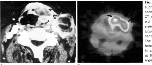
Fig. 1. Squamous cell carcinoma in supraglottic area and cervical Iymph node
CT scan of the neck at the level of hy-
。 id bone(a) reveals 30 X 40 X 60 mm sized left upper and middle internal jugular Iymph node enlargement with central low attenuation(curved arrow) The supraglottic tumor( arrow) shows heterogeneous density. Ga-67 SPECT in axial plane(b) shows strong uptake at the primary tumor( arrow) and enl- arged Iymph node(curved arrow)
Fig. 2. A 63-year-old male with、adeno
carcinoma in left tonsil
CT scan at the level of tongue base(a) shows soft tissue mass involving left tonsil( arrow), but Ga-67 SPECT scin- tigraphy in axial plane(b) reveals n 。 abnormal uptake in the area of left ton- sil(arrow)
- 700-
인공 음영 (rnotion artifact) 에 의해 종양이 발견되지 않았 으며 (Fig.4) , 각각 1 예의 구강저와 버인두종양은 인접 인 공음영 없이 발견되지 않았다.
전이된 경부 임파절은 51 개였는데 3개가 상부 내경정맥 임파절과 악하 임파절이었고 장경이 15rnrn 이하로 CT에 서 음성으로평가되였으나그중 1개가갈륨단순평면촬영 과 SPECT에서 모두 양성이었고, 1개는 갈륨 단순평면촬 영상 음성, 갈륭 SPECT에서 양성이였으며 나머지 1 개는 갈륨 SPECT에서 양성이였으나 CT 스캔에서 그 부분이 포함되지않아 임파절의 장경을 측정할 수 없었고, CT상 양성인 임파절 47개중 장경 30rnrn를 기준으로 하였을때 (Table 1) 30rnrn미만 38개중 갈륨 단순평면촬영상 6개,
갈륨 SPECT에서는 197B 가 양성이었으며, 30rnrn이상 9개 중 갈륨 단순평면촬영상 7개, 갈륨 SPECT에서는 9개 모 두 양성이었다. 장경 20rnm를 기준으로 하였을때 (Table 2) 20rnm미만 26개중 갈륨 단순평면촬영상 2개, 갈륨 SPECT에서는 11 개가 양성으로 나왔으며, 20rnrn이상 21 개중 갈륨 단순평면촬영상 양성으로 나온 경우가 167B, 갈 륨 SPECT상 양성으로 나온 경우가 167B 였다. 수술 또는
a
a
‘ . " 증gi
-븐튼튿뀔
b
b
권삼옥 외 ’ 두경부 종앙과 경부 임파절 전이
생검을 시행했던 임파절 5개에서 종양세포를 확인 할 수 있었고그중 3개가갈륭스캔과 CT 모두에서 양성이었고
(Fig. 5), 1 개가 갈륨 단순평면촬영상 음성이었고 갈륨
SPECT와 CT에서 양성이었으며, 나머지 l 개가 갈륨 단순 평면촬영과 갈륨 SPECT에서 음성, CT에서 양성이었다.
고 찰
두경부종양은 편평상피암이 가장 많고 원발종양의 위치 에 따라 임파절 전이 빈도가 달라지며 경부임파절 전이가 있는 편평상피암의 경우 원발종양의 위치에 관계없이 5년 생존율이 30%미만이고 동측 경부에 하나의 임파절 전이 가 있을때 5년 생존율은 50%이며 양측 경부에 임파절 전 이가 있을 때는 25%여서 경부엄파절 침범 유무가 편평상 피암의 예후에 가장중요한요인이 된다 (9).
신체에는 대략 8007B 의 임파절이 있고 약 3007B 가 경부 에 분포하며 4개 혹은 5개의 집단으로 나눌 수 있고 이들 은 서로 연결되어 있다 (1이 .10억개의 악성종양 세포가 l rnrn3의 멍어리를 만들기 때문에 종양이 임파절을 침범하
c
Fig. 3. A 33-year-old male with squa- mous cell carcinoma in right aspect 01 the tongue.
CT scan at the level 01 the tongue(a) shows beam hardening artilact produ- ced by dental amalgam. Ga-67 SPECT scintigraphy in axial plane(b) demon- strates an obvious increased uptake at the area 01 the tumor(arrow). * The hi- ghest degree 01 Ga-67 uptake is ma- pped as white, lollowed by red, yellow, green, and blue
Fig. 4. A 84-year-old man had a glottic tumor, biopsy proven squamous cell carcinoma
The lesion is not well delined in CT scan(a) at the level 01 the vocal cord. Ga-67 planar image in right anterior oblique view(b) and SPECT in axial plane(c) show strong uptake at the tumor(arrows)
701 -
대 한 밤사 선 의 학회 지 1995; 33(5): 699- 704
는 것은 아주 미세적인 일이어서 조직 검사 이외의 방법으 로 엄파절내의 작은 종양을 발견하는 것은 불가능하고 방 사선과의 실제적 임무는 육안적인 임파절 전이 유무를 입 증하는 것이다 (8). 그러므로 CT나 MR로 발견할 수 있을 민큼 종양이 충분히 크면 종양의 병기 결정에 대한 기준이 필요하게 된다. 임파절 크기에 대한 기준은 CT와 MRI가 동일하며, 일반적으로 알려진 정상 임파절의 장경은 상부 내경정맥 임파절과 하악 임파절인 경우 1.5cm이고 그 이 외의 경부 임파절은 1.0cm이며 이보다 큰 임파절인 경우 80%가 전이된 임파절이다. CT와 MRI는 병리학적으로 커진 임파절을 찾는데 거의 유사한 능력을 가진다고 하며,
임파절의 중심부 저음영이 가장 중요한 기준이 되고, 임파 절외로의 파급 유무 또한 편평상펴암의 예후에 중요한 요 인이 된다 (9). Yousem등 (11) 에 의하면 CT가 MRI보다 중심부 괴사와 종양의 임파절외로 파급을 발견하는데 더 우수하다고 하였다. CT상 전이된 경부 임파절에 대한 여 러 기준이 있지만 저자들은 Som(8) 에의한 CT 기준을 기 본으로 하였고, CT상 측정할 수 있는 경부 임파절을 기준 으로 갈륨 단순평면촬영과 갈륨 SPECT소견을 비교하였 다.
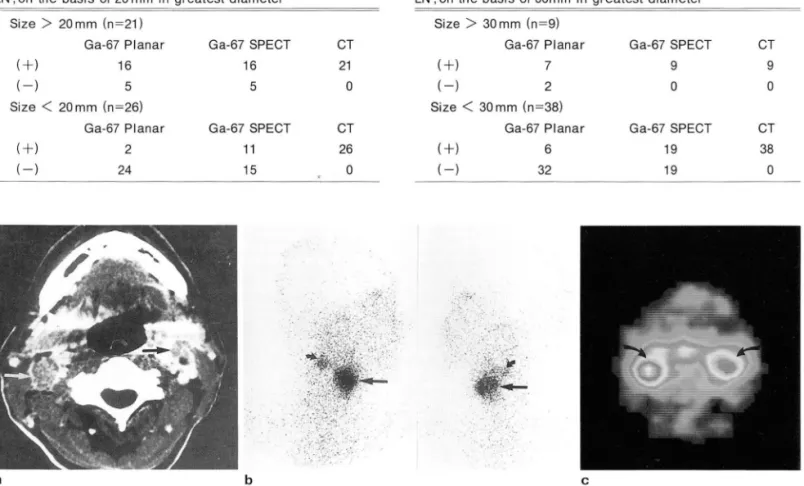
Table 1. Gallium Scan versus CT Lesion Detection of Cervical LN ; on the basis of 20 mm in greatest diameter
갈륨 스캔은 두경부종양을 발견하는데 있어 또다른 방 법 으로, 1960년대 에 Ga-67 citrate가 종양 특이 방사성 추 적자 (tracer) 로 시-용된 이후 감마 카메라의 발달과 SPE CT의 도입으로 종양발견률이 증가하여 현재에는 호지킨 병과 다른 임파종의 병기 결정과 치료 판정에 이용되고 있 고 그 이외에도 골, 연부조직 육종, 흑색종, 간암,두경부종 양에도 이용된다. Ga-67 citrate가 종양에 섭취되는 기전 은 갈륨이 제이철 이온 (ferric ion) 과 유사한 동태를 보여 서 정맥 주사하면 트란스페린( transferrin) 에 결함되고 갈 륨-트란스페련 복합체로서 혈액을 순환하며, 활동척으로 성장하는 종양 세포 표면에 있는 트란스페린 수용체에 결 합하여 세포내로 섭취가 된다(12). 혈관밖에서는 락토페 련 Oactoferrin)과 결합하고 백혈구가 만들어 내는 락토페 린과 세균이 만드는 시데로포어에 의해서 종양이외의 염 증병소에도 집적되며 눈물샘과 침샘, 골수, 비장, 그리고 유방은 정상 상태에서도 락토페련을 분비하므로 갈륨이 섭취된다 (2). 방사선 치료를 한 후 일시적 또는 영구적으 로 갈륨이 섭취가 되지 않고, 화학요법 후에도 일시적으로 갈륨섭취가감소되므로치료후 4-6주뒤에 갈륨스캔을 실시해야 한다(1 2). Teates등(13)에 의하면 두경부 종양
Table 2. Gallium Scan versus CT Lesion Detection of Cervical LN ; on the basis of 30mm in greatest diameter
Size
>
20 mm (n=21) Size>
30 mm (n=9)Ga-67 Planar Ga-67 SPECT CT Ga-67 Planar Ga-67 SPECT CT
(+) 16 16 21 (+) 7 9 9
(-) 5 5 0 (-) 2 0 0
Size
<
20mm (n=26) Size<
30 mm (n=38)Ga-67 Planar Ga-67 SPECT CT Ga-67 Planar Ga-67 SPECT CT
(+) 2 11 26 (+) 6 19 38
(- ) 24 15 0 (-) 32 19 0
‘ •
a b c
Fig. 5. A 50-year-old man had a supraglottic squamous cell carcinoma, pathologically proven cervical Iymph node metastasis a. CT scan of the neck at suprahyoid region, axial section through low floor of mouth, reveals 20
x
20 X 15mm sized right upper internal jugular Iymph node enlargement and 12x18x15mm sized left upper internal jugular Iymph node enlargement(arrows) b. Ga-67 planar images in both oblique view show strong uptake of supraglottic carcinoma(arrow) and both upper jugular Iymph nodes(curved arrows)c. SPECT in axial plane shows strong uptake at the both upper jugular Iymph nodes metastasis(curved arrows)
m
에서 병변의 발견률은 예민도가 56%에 불과하며 3cm이 상일때 70%에서 갈륨 섭취를 보였다고 한다. 실제 두경부 에서 정상적으로 갈륨이 타액션과 비점막등에 섭취되어 병변과의 감별을 어렵게 하나 이들은 2-4mCi 의 소량의 Ga-67 citrate를 사용하였고 단순평면촬영만을 얻었기때 문에 종양 발견에 있어서 어느 정도의 한계점을 가지고 있 었다. 갈륨스캔에서도 SPECT를 하면 단순평면촬영보다 더 해상력이 좋아지고, 횡단면, 관상면, 종단면으로 영상을 만들 수 있어서 해부학적 위치를 더 정확하게 알 수 있다.
저자들의 경우 두경부암 원발병소를 찾는데 있어 갈륨 SPECT가 97.3%로 84, 2%의 CT, 76.3%의 갈륨 단순평 면 촬영보다 예민했는데 그 이유는 구강내나 성대에 위치한 종양의 경우 언접 인공 음영에 의해 CT상 원발 병소를 찾 을 수 없었기 때문이며 이 경우 갈륭 SPECT가 도움이 되 였다. 갈륨 단순평면촬영상 종양을 발견할 수 없었던 9예 중 8예는 장경이 30mm이하의 종양이어서 종양의 크기와 갈륨 섭취와는 상관관계가 있었다. 그리고 종양의 종류에 따라갈륨의 섭취 유무가달라져서 비록 l 예에 불과하지만 선암은 장경이 72mm이였으나 갈륨 섭취를 볼 수 없었다.
경부임파절의 장경을 30mm 기준으로 하였을 때 갈륨 SPECT에서는 30mm이상인 9개 모두가 양성으로 나왔으 며, 20mm를 기준으로 하였을 때는 20mm이상인 21 개중 167B 가 갈륨 SPECT에서 양성으로 나와서 장경이 30mm 이상일 때 갈륨SPECT에서의 종양발견율이 좋았다.
본 연구는 원발병소에 대해서는 조직학적으로 진단되였 으나, 대부분의 경부 임파절에서는 조직학적으로 확인되 지 않아서 CT상 크기 기준으로 갈륨 스캔과의 결과를 비 교할 수 밖에 없였다. 그래서 CT상 비정상적으로 크며 갈 륨 SPECT에서는 음성인 경우 양성 증식( beni gn h yper - plasia) 혹은 갈륨 SPECT의 위 음성 인 가능성 을 생 각해 야 할 것이며, CT상 크기 기준에는 미달하지만 갈륨 SPECT 상 양성인 경우는 전이병소 혹은 염증성 명소의 가능성을 모두 염두에 두어야 할 것이다.
결론적으로 CT는 갈륨 스캔보다 전이된 경부 임파절의 위치와 크기 분석을 용이하게 하였으며, 구강내와 성대에 위치한 원발 종양중 인접한 인공 음영에 의해 병소를 찾을
권삼옥 외 . 두경부 종앙과 경부 입파절 전이
수 없었던 경우에는 갈륨 SPECT가 도움이 되어서 갈륨 SPECT는 CT와 상호 보완적 인 역할을 하였다. 한편 갈륨 SPECT는 갈륨 단순평 면촬영보다 해상력 이 더 좋아서 30 mm이상의 임파절을 발견하는데 유용하였다 .
. :;;J
C그 고 τc C그 헌
1. Edwards CL , Hayes RL. Tumor scanning with Ga-67 citrate. J Nucl Med 1969;10:103-105
2. Backerman C, Hoffer PB, Bitran JD. The role 01 gallium-67 in the clinical evaluation 01 cancer. Semin Nucl Med 1984;14 296-323
3. Kashima MK, McKusik KA, Malmud S. Gallium-67 scanning in patients with head and neck cancer. Laryngoscopy 1974; 84: 1078
4. Konrblut AD, Silberstein EB, Saenger EL. Scintiscanning with gallium citrate-67: Diagnosis 01 head and neck malignant neoplasms. Arch Otolaryngo/1974; 100: 201
5. Smith NJ, Teates CD, EI-Mahdi AM. The value 01 gallium-67 scanning in the evaluation 01 head and neck malignancy Layngoscopy 1975; 85: 778
6. Front 0, Israel 0, Epelbaum R, et al. Ga-67 SPECT belore and after treatment 01 Iymphoma. Radiology 1990; 175: 515- 519
7. 0’Donnell JK, Go RT, Cordasco EM, et al. Improved evalu- ation 01 pulmonary disease with gallium-67 emission tom-
。graphy. C/eve Clin Q 1985; 52: 525-531
8. Som PM. Detection 01 metastasis in cervical Iymph nodes: CT and MR criteria and dillerential diagnosis. AJR 1992; 158 961-969
9. Madison MT, Remley KB, Latchaw RE, Mitchell SL. Radio- logic diagnosis and staging 01 head and neck squamous cell carcinoma. Radiol Clin North Am 1994;32:163-181
10. Som PM. Lymph nodes 01 the neck. Radiology 1987; 165:
593-600
11. Yousem DM, Som PM, Hackney DB, Schwaibold F, Hendrix RA. Central nodal necrosis and extracapsular neoplastic spread in cervical Iymph nodes: MR imaging versus CT Radiology 1992; 182 : 753-759
12. Hoffer P. Galli um : Mechanism. J Nucl Med 1980; 21 : 282-285 13. Teates CD, Preston DF, Boyd CM. Gallium-67 citrate imaging
in head and neck tumors. report 01 cooperative group. J Nucl Med 1980;21 ‘ 622-627
m
대 한 밤 사 선 의 학 회 지 1995: 33(5) : 699-704
Journal of the Korean Radiological Society 1995: 33(5): 699- 704
Head and Neck Tumors and Neck Node Metastasis:
Comparison of Ga-67 Scan and CT Findings'
Sam Ok Kwon, M.D., Jong Min Kim, M .D., Sang Kyun Bae, M.D.2, Sang Suk Kim, M.D., Kyeung Seung Oh, M.D., Young Duk Joh, M.D.
1 Department of Diagnostic Radiology, Kosin Medical College, Pusan, Korea
2 Department of Nuclear Medicine, Kosin Medical College, Pusan, Korea
Purpose: To assess relative diagnostic value of Ga-67 planar, Ga-67 SPECT, and CT images for detection of head and neck tumors and cervical Iymph node metastasis.
Materials and Methods: Thirty eight patients of pathologically proven head and neck tumors including squamous cell carcinomas(n=32), malignant lymphomas(n=3), undifferentiated carcinomas(n=2), adenocar- cinomas(n=1) were enrolled in this study. Ga-67 planar and SPECT images were obtained with intravenous in- jection of 5mCi of Ga-67 citrate. On the basis of 30 and 20 mm in the greatest diameter of cervical Iymph nodes, we compared lesion detectability of Ga-67 planar, SPECT, and CT
Results: Thirty eight cases of head and neck tumors were detected in 29 cases(76.3%) with Ga-67 planar im- age, 37 cases(97.3%) with Ga-67 SPECT, and 32 cases(84.2%) with CT. 25 of 32 squamous cell carcinomas were positive with Ga-67 planar image and all of 32 cases with Ga-67 SPECT. 80th of two undifferentiated carcinomas were positive with Ga-67 planar and SPECT images. Two of three malignant Iymphomas were posi- tive with Ga-67 planar image and all of three with Ga-67 SPEC T. Eight of nine tumors were negative with Ga-67 planar image and those were less than 30 mm. One case of adenocarcinoma was negative with Ga-67 planar and SPECT images. Seven of nine Iymph nodes greater than 30 mm were positive with Ga-67 planar image and all of nine with Ga-67 SPEC T. On the basis of 20 mm in greatest diameter, 16 of 21 Iymph nodes greater than 20 mm were positive with Ga-67 planar and SPECT images
Conclusion: CT providing better resolution than Ga-67 scan permilted analysis of size and location of meta- static cervical nodes, however primary tumors of oral cavity, vocal cord, and nasopharynx were often not detected on CT when metallic and motion artifacts were present, where Ga-67 SPECT was useful. Ga-67 SPECT enabled belter anatomical localization than Ga-67 planar image and was useful in detection of Iymph nodes greater than 30 mm.
Index Words: Head and neck neoplasms, CT
Head and neck neoplasms, metastases Gallium. radioactive
Address reprint requests to : Sam Ok Kwon, M.D., Department of Radiology, College of Medicine, Kosin University,
~ 34, Amnam-dong, Seo 용u, Pusan, 602-030 Korea. Tel. 82-51-240-6341 Fax.82-51-255-2764