ORIGINAL ARTICLE
다중검출 전산화단층촬영에서 담석이 보이지 않는 급성 담석성 췌장염에서 조기 내시경초음파검사의 유용성
박재근, 김기배, 한정호, 윤순만, 채희복, 윤세진, 박선미
충북대학교 의과대학 내과학교실
The Usefulness of Early Endoscopic Ultrasonography in Acute Biliary Pancreatitis with Undetectable Choledocholithiasis on Multidetector Computed Tomography
Jae Geun Park, Ki Bae Kim, Joung-Ho Han, Soon Man Yoon, Hee Bok Chae, Sei Jin Youn, and Seon Mee Park Department of Internal Medicine, Chungbuk National University College of Medicine, Cheongju, Korea
Background/Aims: EUS can detect bile duct stones (BDS) that are undetectable on multidetector computed tomography (MDCT).
BDS associated with acute biliary pancreatitis (ABP) are small and tend to be excreted spontaneously. This study evaluated the usefulness of early EUS in patients with ABP and undetectable BDS on MDCT.
Methods: Forty-one patients with ABP and undetectable BDS on MDCT underwent EUS within 24 hours of admission and were diagnosed with BDS, sludge, dilated common bile duct (CBD), or normal CBD. ERCP was performed in patients with BDS, sludge, or clinical deterioration. The diagnostic yield and the effects of early EUS on morbidity, mortality, and the length of hospitalization were evaluated.
Results: EUS detected BDS or sludge in 48.8% of patients examined. BDS was the diagnosis in 13 patients, sludge in seven, and neither for 21 patients. ERCP was performed in 20 patients with BDS or sludge, in two patients with coexisting cholangitis, and in one patient with worsening liver function tests. ERCP identified BDS in 12 patients and sludge in seven. No lesions were diagnosed in four patients by ERCP. All patients improved, and the length of hospitalization in patients with ERCP was 9.0 days, without ERCP 7.1 days. Two patients with major complications by ERCP were hospitalized for a prolonged time.
Conclusions: Early EUS may be useful to select patients for therapeutic ERCP in cases of suspected ABP with undetectable BDS on MDCT. (Korean J Gastroenterol 2016;68:202-209)
Key Words: Pancreatitis; Gallstones; Endosonography; Endoscopic retrograde cholangiopancreatography
Received June 15, 2016. Revised August 6, 2016. Accepted October 11, 2016.
CC This is an open access article distributed under the terms of the Creative Commons Attribution Non-Commercial License (http://creativecommons.org/licenses/
by-nc/4.0) which permits unrestricted non-commercial use, distribution, and reproduction in any medium, provided the original work is properly cited.
Copyright © 2016. Korean Society of Gastroenterology.
교신저자: 박선미, 28644, 청주시 서원구 충대로 1, 충북대학교 의과대학 내과학교실
Correspondence to: Seon Mee Park, Department of Internal Medicine, Chungbuk National University College of Medicine, 1 Chungdae-ro, Seowon-gu, Cheongju 28644, Korea. Tel: +82-43-269-6019, Fax: +82-43-273-3252, E-mail: smpark@chungbuk.ac.kr
Financial support: This work was supported by a research grant of Chungbuk National University in 2014. Conflict of interest: None.
INTRODUCTION
Gallstone disease is the most common cause of acute pancreatitis.1 Image tests are needed to evaluate bile duct stones (BDS) in patients with suspected acute biliary pan- creatitis (ABP). Multidetector computed tomography (MDCT) is usually the first-line imaging method in ABP. However, its
diagnostic accuracy for BDS is approximately 80%.2 The ma- jor causes of undetectable BDS on MDCT are the small size and isodensity of the stones compared to the surrounding tissue.3 Because at least 50% of the cases of acute pan- creatitis involve the passage of small stones (usually less than 5 mm in diameter), patients may need to undergo fur- ther tests in cases in which BDS are undetectable on MDCT.4
EUS is a very sensitive and specific test for the detection of BDS in ABP.3,5 Its use before ERCP may avoid unnecessary, in- vasive ERCP procedures, resulting in fewer complications.6,7 The indications for EUS in ABP are problematic. Patients at high risk of BDS can undergo ERCP directly. Patients at inter- mediate risk for BDS are recommended to undergo first-line EUS or MRCP.8 However, a recent study revealed that these criteria have resulted in overuse of ERCP,9 and the authors suggest the use of early EUS for possible ABP patients.
The proper timing of evaluation of the biliary tree is also questionable. Anderloni et al.10 and Liu et al.11 recommend early EUS in ABP to select patients for ERCP to reduce the risk of further pancreatic damage. Early EUS (within 24-48 hours) can easily and quickly categorize the patients who do not require subsequent therapeutic ERCP, thus allowing early discharge in selected cases, a cost-effective protocol. However, Cavdar et al.12 reported that MRCP on the seventh day would avoid un- necessary ERCP and will provide more accurate information than MRCP on the first and fourth days in ABP patients.
This study determined the diagnostic yield of early EUS in the evaluation of biliary tree and selection of ABP patients who need therapeutic ERCP. In addition, we determined the effect of early EUS on morbidity and mortality and the length of hospital stay in patients with suspected ABP.
SUBJECTS AND METHODS
1. Patients
Patients with suspected ABP but no evidence of BDS on ab- dominal MDCT were enrolled in this study. They were admitted to the Chungbuk National University Hospital in Cheongju, Korea between January 2012 and April 2014. ABP was diag- nosed as acute pancreatitis with evidence of biliary origin, without evidence of other causes of acute pancreatitis, such as alcoholism, hypercalcemia, hyperlipidemia, or post-ERCP pancreatitis. Biliary etiology was defined as a history of gall- stones, confirmation of the biliary origin of ABP from labo- ratory data, detection of gallbladder stones on imaging tests, or dilated common bile duct (CBD).10 The laboratory criteria for biliary origin of ABP were as follows13: one or more bio- chemical tests greater than or equal to the cutoff values of ALP, 225 IU/L; ALT, 75 IU/L; and bilirubin, 2.3 mg/dL. Dilated CBD was defined as a CBD diameter 6 mm or 10 mm in cases of cholecystectomy on abdominal MDCT. The exclusion
criteria were surgically altered gastrointestinal anatomy, re- current acute pancreatitis, or previous sphincterotomy. All patients underwent an abdominal MDCT scan at the time of admission and were evaluated for the presence of BDS and severity of acute pancreatitis. Imaging and laboratory data, and the latter included the levels of ALT, total bilirubin, ALP, GGT, amylase, and lipase, were retrospectively collected from patients’ medical records. The severity of acute pancreatitis was determined with the abdominal CT severity index.14
2. Outcomes
The primary outcome was the determination of the diag- nostic yield of early EUS (within 24 hours after admission) in patients with ABP and undetectable BDS in MDCT. Secondary outcomes were the evaluation of the effect of early EUS on morbidity and mortality and the length of hospital stay in pa- tients with suspected ABP. The length of hospital stay was measured from admission to transfer to the surgical depart- ment for the performance of cholecystectomy or discharge.
3. EUS
EUS was performed using a radial echoendoscope (model GF-UE260-AL5; Olympus Optical, Tokyo, Japan). Prosound -10 (Aloka Co., Tokyo, Japan) and EU-M20 (Olympus Optical) ul- trasonographic systems were used for image processing.
EUS was performed by two endoscopists within 24 hours af- ter admission. BDS or sludge was positively identified by the observation of a hyperechoic focus within the CBD with or without an acoustic shadow, respectively (Fig. 1). The CBD di- ameter and stone size were measured at the largest point. All gallbladder stones and sludge, as well as EUS-related compli- cations, were evaluated.
4. ERCP
ERCP was performed with a lateral scope (TJF 240;
Olympus Optical) by two endoscopists within 72 hours after EUS. ERCP was performed when BDS or sludge were de- tected on EUS or in cases of cholangitis, bilirubin level >5 mg/dL, or worsening of clinical symptoms or liver function tests. After the removal of the BDS, contrast material was in- jected into the CBD, and an inflated balloon catheter (up to 15 mm in diameter) was withdrawn along the CBD to the duo- denum to confirm the clearance of the biliary tree. All proce- dures, including endoscopic sphincterotomy (EST), endo-
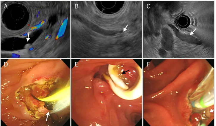
Fig. 1. Endosonographic and endoscopic findings of choledocholithiasis. Endosonography revealed the presence of hyperechoic foci in the common bile duct; posterior shadowing (arrows) indicated choledocholithiasis (A, B), and amorphous materials in the common bile duct without posterior shadowing (arrow) indicated sludge (C). Endoscopy showed the presence of a single dark brown stone, which was extracted after endoscopic sphincterotomy (arrow) (D), and the presence of yellow amorphous material (sludge), which was drained during endoscopic retrograde cholangiopancreatography (arrows) (E, F).
scopic papillary balloon dilatation (EPBD), and the insertion of biliary or pancreatic stents, were analyzed. ERCP-related complications, including aggravated acute pancreatitis, per- foration, bleeding, infection, and cardiorespiratory events, were also analyzed. The materials detected in the CBD were classified as BDS, sludge, or none (Fig. 1).
5. Statistical analysis
Data were analyzed with PASW Statistics for Windows soft- ware version 18.0 (IBM Co., Armonk, NY, USA). Continuous variables were presented as means±standard deviation and compared between groups using t-tests. Categorical varia- bles were compared using 2 tests. Null hypotheses of no dif- ference were rejected if p-values were less than 0.05.
RESULTS
1. Patient characteristics
During the study period, 47 patients were admitted under
the diagnosis of suspected ABP although BDS were un- detectable on MDCT. Six patients were excluded from this study because of previous Billroth II gastrectomy in two cas- es, recurrent acute pancreatitis in one case, or previous sphincterotomy in three cases. Forty-one patients (male:fe- male ratio, 27:14; mean age, 5718 years) were enrolled in the study (Fig. 2). The presenting symptoms of the patients were abdominal pain in 35 (85.4%) cases and fever in five (12.2%) cases. The diagnostic criteria of ABP were satisfied with more than one item on the basis of laboratory data in 33 (80.5%) cases, gallbladder stones or sludge in 18 (43.9%) cases, or dilated CBD in 18 (43.9%) cases. The severity of acute pancreatitis was mild to moderate in all patients. The mean diameter of the CBD was 5.8±2.5 mm (range, 2.0-14.0 mm) (Table 1).
2. Diagnosis of BDS or sludge on EUS
Among the 41 patients evaluated, BDS or sludge was diag- nosed on EUS in 13 (31.7%) and seven (17.1%) patients,
Table 1. Characteristics of Patients with and without Bile Duct Stones (BDS)/Sludge on Endosonography
Characteristic BDS/sludge (+) BDS/sludge (–) p-value
Patient 20 21
Age (yr) 63±17 53±17 0.062
Sex (M/F) 11/9 16/5 0.197
Laboratory finding
Amylase (IU/L) 322±599 539±724 0.458
Lipase (IU/L) 540±1,031 1,105±1,295 0.542
AST (IU/L) 162±187 205±188 0.856
ALT (IU/L) 168±175 171±134 0.562
Bilirubin (mg/dL) 1.6±1.6 2.2±2.6 0.854
ALP (IU/L) 412±289 558±655 0.564
GGT (IU/L) 485±638 834±1,131 0.236
WBC (/mm3) 8,519±3,236 10,510±4,594 0.254 CBD diameter (mm) 6.7±2.9 5.0±1.9 0.043
Cholangitis (+) 2 2 0.865
GB stone/sludge (+) 8 (40.0) 10 (47.6) 0.562 CT severity index 1.4±1.1 1.9±1.3 0.161 Values are presented as n only, mean±SD, or n (%).
M, male; F, female; WBC, white blood cell; CBD, common bile duct;
GB, gallbladder.
Fig. 2. Flow chart of the procedures used.
MDCT, multidetector computed tomo- graphy; CBD, common bile duct.
respectively. The mean diameter of the BDS was 4.2±1.7 mm (range, 1.4-6.9 mm). The other 21 (51.2%) patients showed no evidence of BDS, and six (14.6%) patients presented with dilated CBD (6.2-8.0 mm). Gallbladder stones was evident in 16 (39.0%) and sludge in two (4.9%) patients. The compar- ison between patients with and without BDS/sludge on EUS revealed no differences in liver function tests, pancreatic en-
zymes, or presence of gallbladder stones. However, the mean CBD diameter was larger in patients with BDS or sludge than in those without them (6.7±2.9 mm vs. 5.0±1.9 mm, re- spectively; p<0.043) (Table 1). No EUS-related complica- tions were observed.
3. Diagnosis of BDS or sludge on ERCP
ERCP was performed in 23 (56.1%) patients. The ERCP re- sults revealed the presence of BDS and sludge in 12 (29.3%) and 7 (17.1%) patients, respectively. EUS and ERCP were per- formed in the same session in 22 patients, who were diag- nosed with BDS or sludge on EUS or with coexisting chol- angitis, and within 72 hours in one patient with worsening liv- er enzyme levels. The ERCP procedures consisted of EST in 21 (91.3%) patients, EPBD in two (8.7%) patients, insertion of a plastic biliary stent in 13 (56.5%) patients, and insertion of a plastic pancreatic stent in six (26.1%) patients. Biliary/pan- creatic stents were inserted for prevention of post-ERCP chol- angitis/pancreatic with 7 Fr/3 Fr, 5-cm length, and one pigtail without internal flap. Most of them migrated distally, if not, were removed using grasping forceps. The comparison of pa- tients with or without BDS/sludge on ERCP revealed no differ- ences in the pancreatic enzyme levels, the presence of gall- bladder stones, or CBD diameter. However, the ALT level was higher in patients with BDS or sludge than in those without
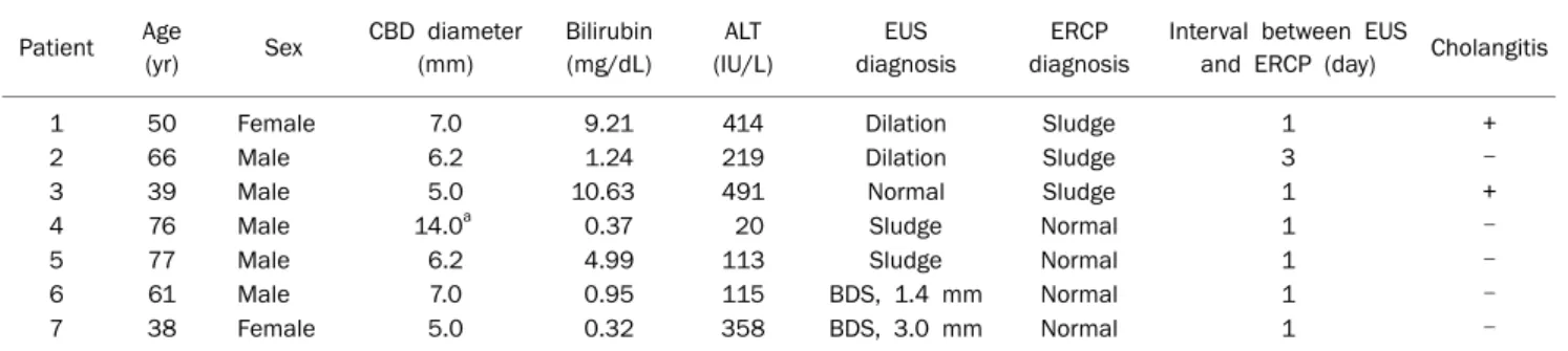
Table 3. Characteristics of Seven Patients with Discordant Diagnoses on EUS vs. ERCP
Patient Age
(yr) Sex CBD diameter (mm)
Bilirubin (mg/dL)
ALT (IU/L)
EUS diagnosis
ERCP diagnosis
Interval between EUS
and ERCP (day) Cholangitis
1 50 Female 7.0 9.21 414 Dilation Sludge 1 +
2 66 Male 6.2 1.24 219 Dilation Sludge 3 –
3 39 Male 5.0 10.63 491 Normal Sludge 1 +
4 76 Male 14.0a 0.37 20 Sludge Normal 1 –
5 77 Male 6.2 4.99 113 Sludge Normal 1 –
6 61 Male 7.0 0.95 115 BDS, 1.4 mm Normal 1 –
7 38 Female 5.0 0.32 358 BDS, 3.0 mm Normal 1 –
CBD, common bile duct; BDS, bile duct stones.
aHistory of cholecystectomy.
Table 2 Diagnosis of Choledocholithiasis in Acute Biliary Pan- creatitis on EUS and ERCP
ERCP
EUS Normal CBD
(n=15)
Stone (n=13)
Sludge (n=7)
CBD dilation, only (n=6) ERCP (+) (n=23)
Stone (n=12) 0 11 1 0
Sludge (n=7) 1a 0 4 2a
None (n=4) 0 2 2 0
ERCP (–) (n=18) 14 0 0 4
CBD, common bile duct.
aThree patients underwent ERCP because of coexisting cholangitis in two and worsening liver function tests in one patient.
them (221±190 IU/L vs. 126±99 IU/L; p=0.047). Major com- plications related to ERCP occurred in two out of 23 (8.7%) patients, including major bleeding and periampullary perforation. Other complications, such as aggravated acute pancreatitis, infection, and cardiorespiratory events, were not observed.
4. Comparison of EUS and ERCP
BDS, sludge, or none were identified on both ERCP and EUS in 16 patients (Table 2). Two patients with BDS and two patients with sludge on EUS were not diagnosed on ERCP, performed during the same session. Three patients with nei- ther BDS nor sludge on EUS but with cholangitis (two pa- tients) and worsening liver enzyme levels (one patient) were diagnosed with sludge on ERCP (Table 3).
5. Record tracking following EUS and ERCP
All patients improved with treatment. Cholecystectomy was performed after the endoscopic procedure in 18 (43.9%) patients. The mean length of hospital stay in patients who un-
derwent ERCP was 9.0±9.9 days, and 7.1±3.3 days for those who did not, but this difference was not significant (p=0.397).
Major complications related to ERCP occurred in 2 out of 23 patients. Bleeding occurred in one patient who under- went hemodialysis for chronic renal insufficiency. In this pa- tient, BDS were found on both EUS and ERCP and were re- moved with EST. Bleeding occurred 72 hours after ERCP at the EST site and was controlled via endoscopic hemostasis.
However, the patient experienced clinical deterioration and improved only after 41 days of conservative management.
Perforation occurred in one patient with gallbladder stones and BDS on EUS. Cannulation was done using a needle-knife papillotome. However, there was no evidence of BDS or sludge on ERCP. Retroperitoneal perforation in the peri- vaterian area was detected immediately after the procedure.
Peritonitis developed and was managed with percutaneous cystic drainage. The patient improved after 37 days of con- servative management.
DISCUSSION
This study demonstrated that EUS is effective as an add-on test before ERCP in patients with suspected ABP. In this study, 44% of the ABP patients who underwent early EUS were treat- ed conservatively without ERCP. Because most ABP cases in- volve small BDS or sludge, which sometimes are not detected on MDCT, the evaluation of biliary tract via EUS was effective in selecting patients for therapeutic ERCP.
The current approach to patients with suspected BDS has been adopted on the basis of the risk factors for BDS.
Accordingly, patients at high risk of BDS (cholangitis, CBD di- ameter >6 mm, bilirubin level >4 mg/dL, or BDS on ultra- sonography) should undergo ERCP directly.8 However, this
approach results in a high number of unnecessary ERC procedures. A recent study reported that only 50% of the pa- tients with high-probability criteria presented with BDS.9 In this study, four patients with dilated CBD (7-8 mm) without BDS/sludge on EUS recovered from ABP without ERCP.
Therefore, EUS is useful for patients with possible ABP and whose BDS are not detected on MDCT.9,15 Fogel and Sherman4 reported that, in patients with suspected ABP, ERCP is indicated for the diagnosis of coexisting cholangitis, persistent biliary obstruction (conjugated bilirubin level >5 mg/dL), BDS on imaging, clinical deterioration, and in- creased liver enzyme levels. The application of these criteria to our data allowed the classification of six patients in the high-risk group and revealed the presence of BDS/sludge on ERCP. Therefore, we suggest that patients with revised cri- teria may undergo ERCP directly. Otherwise, EUS or MRCP should be done before ERCP in those patients.
The comparison between EUS and MRCP indicates that EUS has a higher resolution (0.1 mm vs. 1.5 mm), which ex- plains the lower sensitivity of MRCP for small stones in ABP.
Moreover, the presence of blind spots, including the papillary and peripapillary regions, has been reported in MRCP.16 However, clinical studies revealed that the diagnostic accu- racy of EUS and MRCP were similar, even for small (1-5 mm) BDS.17 Despite this similar accuracy, EUS has advantages in the clinical setting, especially when stones are diagnosed via EUS or ERCP/EST, and stone extraction can be performed in the same session.
The proper timing of EUS in suspected ABP has not been defined. As we expected, a positive EUS was more commonly found during the acute phase of the illness than in the chronic phase.18 The performance of EUS within 24 hours after ad- mission revealed a higher number of cases of BDS/sludge than in other studies.19 Spontaneously excreted BDS/sludge has been recovered in the stool of 80% of the patients with acute pancreatitis,20 and the number of patients with sludge has been reported to decrease over time: 88.2% on day 1, 83.7% on day 2, 69.6% on day 3, and 68.6% on day 4.21 The advantages of early EUS include avoiding the aggravation of ABP by the early removal of BDS/sludge, shortening the hos- pital stay. However, this method cannot effectively select pa- tients with BDS remnants in the biliary tree. The proper timing of EUS in patients with suspected ABP still needs to be determined.
The appropriate timing of EUS can be determined by estab- lishing the appropriate timing of ERCP, because EUS should be followed by ERCP under the same sedation when BDS are detected.22 The timing of ERCP has changed by the under- standing of the natural course of BDS/sludge and the devel- opment of imaging tests. In the 1990s, urgent ERCP (within 24 hours after admission) was recommended by the authors of a randomized controlled trial, who found good outcomes in the ERCP group.23 However, a recent meta-analysis re- vealed that early ERCP (within 72 hours after admission) in patients with acute gallstone pancreatitis did not improve mortality or either local or systemic complications of pan- creatitis, regardless of the severity.24,25 On the other hand, another study reported that early ERCP reduced pan- creatitis-related complications in severe pancreatitis but had no advantage for the patients with mild pancreatitis.26 The determination of which subgroup of patients will benefit from early ERCP remains challenging. Urgent ERCP is usually rec- ommended for a limited number of patients with cholangitis whereas early ERCP is performed in cases of persistent BDS.4
In this study, ERCP treatment of patients with suspected ABP depended on the results of early EUS. All patients im- proved irrespective of ERCP and hospital days were not differ- ent between groups. Therefore, we recommend that patients with suspected ABP and no evidence of BDS on early EUS be treated conservatively without ERCP.
In this study, ERCP-related complications involved one pa- tient of retroperitoneal perforation and one patient of de- layed bleeding. Although the clinical status of these two pa- tients improved after conservative management with percu- taneous cystic drainage or endoscopic hemostasis, the dura- tion of hospitalization was very long. No EUS-related morbid- ity was observed. Therefore, early EUS may be useful to select patients for therapeutic ERCP in cases of suspected ABP with undetectable BDS on MDCT.
The criteria used for the differentiation between sludge and small BDS are not entirely clear; however, it has been sug- gested that a BDS has a diameter greater than 2 mm and can- not be crushed by digital compression.27 BDS or sludge are observed within the CBD as hyperechoic foci on EUS with an acoustic shadow (stones) or without an acoustic shadow (sludge). The identification of sludge on ERCP is defined on the basis of endoscopic visualization of the flow of sand-like bile without evidence of stones. Gallbladder sludge is treated
in the same manner as gallstones. However, biliary sludge passes spontaneously to the intestine in a higher number of cases.20 Our results indicated that four patients diagnosed with BDS or sludge on EUS had none on ERCP; this may be caused by the passage of small stones in the interval be- tween the two procedures, or the stones may not have been visible on fluoroscopy.28
In this study, biliary pancreatitis was diagnosed when the values of one or more biochemical tests were greater than or equal to the cutoff values for ALP, ALT, and bilirubin.13 These cutoff values can adequately separate the biliary from the non-biliary groups with a sensitivity of 73%, specificity of 94%, positive predictive value of 97%, and negative pre- dictive value of 57%.13 Moreover, our results indicated that patients with and without BDS/sludge presented different serum ALT levels on ERCP and different CBD diameters on EUS. These results are consistent with those of other studies that reported the suspected biliary origin of ABP in cases of jaundice, elevated ALT (three times greater than normal), or dilated CBD.29,30
The present study has several limitations. First, the retro- spective and cross-sectional nature of the study limited the number of patients with ABP who underwent EUS and whose BDS were undetectable on MDCT. Second, our study did not include a control group who did not undergo EUS or under- went late EUS.
In conclusion, EUS is useful as an add-on test before ERCP in patients with suspected ABP because BDS associated with acute pancreatitis are too small to be detected on MDCT. EUS is an accurate diagnostic tool for the diagnosis of BDS/
sludge and is safe; therefore, all patients with suspected ABP should undergo EUS before ERCP. However, further studies are needed to determine the optimal timing and methods to as- sess which patients require closer observation, EUS, or ERCP.
REFERENCES
1. Attasaranya S, Fogel EL, Lehman GA. Choledocholithiasis, as- cending cholangitis, and gallstone pancreatitis. Med Clin North Am 2008;92:925-960.
2. Anderson SW, Lucey BC, Varghese JC, Soto JA. Accuracy of MDCT in the diagnosis of choledocholithiasis. AJR Am J Roentgenol 2006;187:174-180.
3. Sgouros SN, Bergele C. Endoscopic ultrasonography versus oth- er diagnostic modalities in the diagnosis of choledocholithiasis.
Dig Dis Sci 2006;51:2280-2286.
4. Fogel EL, Sherman S. ERCP for gallstone pancreatitis. N Engl J Med 2014;370:150-157.
5. Liu CL, Lo CM, Chan JK, et al. Detection of choledocholithiasis by EUS in acute pancreatitis: a prospective evaluation in 100 con- secutive patients. Gastrointest Endosc 2001;54:325-330.
6. Chak A, Hawes RH, Cooper GS, et al. Prospective assessment of the utility of EUS in the evaluation of gallstone pancreatitis.
Gastrointest Endosc 1999;49:599-604.
7. Prat F, Edery J, Meduri B, et al. Early EUS of the bile duct before endoscopic sphincterotomy for acute biliary pancreatitis.
Gastrointest Endosc 2001;54:724-729.
8. Arguedas MR, Dupont AW, Wilcox CM. Where do ERCP, endo- scopic ultrasound, magnetic resonance cholangiopancreatog- raphy, and intraoperative cholangiography fit in the manage- ment of acute biliary pancreatitis? A decision analysis model.
Am J Gastroenterol 2001;96:2892-2899.
9. Adams MA, Hosmer AE, Wamsteker EJ, et al. Predicting the like- lihood of a persistent bile duct stone in patients with suspected choledocholithiasis: accuracy of existing guidelines and the im- pact of laboratory trends. Gastrointest Endosc 2015;82:88-93.
10. Anderloni A, Galeazzi M, Ballarè M, et al. Early endoscopic ultra- sonography in acute biliary pancreatitis: a prospective pilot study. World J Gastroenterol 2015;21:10427-10434.
11. Liu CL, Fan ST, Lo CM, et al. Comparison of early endoscopic ultra- sonography and endoscopic retrograde cholangiopancreatog- raphy in the management of acute biliary pancreatitis: a pro- spective randomized study. Clin Gastroenterol Hepatol 2005;3:1238-1244.
12. Cavdar F, Yildar M, Tellioğlu G, Kara M, Tilki M, Titiz Mİ. Controver- sial issues in biliary pancreatitis: when should we perform MRCP and ERCP? Pancreatology 2014;14:411-414.
13. Goodman AJ, Neoptolemos JP, Carr-Locke DL, Finlay DB, Fossard DP. Detection of gall stones after acute pancreatitis. Gut 1985;26:125-132.
14. Leung TK, Lee CM, Lin SY, et al. Balthazar computed tomography severity index is superior to Ranson criteria and APACHE II scor- ing system in predicting acute pancreatitis outcome. World J Gastroenterol 2005;11:6049-6052.
15. Andari R, Modiri A, Makipour K. Endoscopic ultrasound should be performed before endoscopic retrograde cholangiopan- creatography in all patients with mild acute gallstone pancrea- titis. Pancreas 2014;43:147-148.
16. Bergele C, Giovannini M. EUS and common bile duct stones. Ann Gastroenterol 2004;17:246-252.
17. Giljaca V, Gurusamy KS, Takwoingi Y, et al. Endoscopic ultra- sound versus magnetic resonance cholangiopancreatography for common bile duct stones. Cochrane Database Syst Rev 2015;(2):CD011549.
18. Shapiro T, Melzer E, Binder Y, et al. Selective utilization of pre-op- erative endoscopic ultrasound to exclude choledocholithiasis prior to laparoscopic cholecystectomy: a retrospective study.
Hepatogastroenterology 2013;60:456-460.
19. De Lisi S, Leandro G, Buscarini E. Endoscopic ultrasonography versus endoscopic retrograde cholangiopancreatography in acute biliary pancreatitis: a systematic review. Eur J Gastroenterol Hepatol 2011;23:367-374.
20. Tranter SE, Thompson MH. Spontaneous passage of bile duct stones: frequency of occurrence and relation to clinical presentation. Ann R Coll Surg Engl 2003;85:174-177.
21. Kohut M, Nowak A, Nowakowska-Duiawa E, Marek T. Presence and density of common bile duct microlithiasis in acute biliary pancreatitis. World J Gastroenterol 2002;8:558-561.
22. Fabbri C, Polifemo AM, Luigiano C, et al. Single session versus separate session endoscopic ultrasonography plus endoscopic retrograde cholangiography in patients with low to moderate risk for choledocholithiasis. J Gastroenterol Hepatol 2009;24:1107- 1112.
23. Fan ST, Lai EC, Mok FP, Lo CM, Zheng SS, Wong J. Early treatment of acute biliary pancreatitis by endoscopic papillotomy. N Engl J Med 1993;328:228-232.
24. Tse F, Yuan Y. Early routine endoscopic retrograde cholangiopan- creatography strategy versus early conservative management strategy in acute gallstone pancreatitis. Cochrane Database Syst Rev 2012;(5):CD009779.
25. Petrov MS, van Santvoort HC, Besselink MG, van der Heijden GJ, van Erpecum KJ, Gooszen HG. Early endoscopic retrograde chol- angiopancreatography versus conservative management in
acute biliary pancreatitis without cholangitis: a meta-analysis of randomized trials. Ann Surg 2008;247:250-257.
26. Moretti A, Papi C, Aratari A, et al. Is early endoscopic retrograde cholangiopancreatography useful in the management of acute biliary pancreatitis? A meta-analysis of randomized controlled trials. Dig Liver Dis 2008;40:379-385.
27. Keizman D, Ish-Shalom M, Konikoff FM. The clinical significance of bile duct sludge: is it different from bile duct stones? Surg Endosc 2007;21:769-773.
28. Ney MV, Maluf-Filho F, Sakai P, Zilberstein B, Gama-Rodrigues J, Rosa H. Echo-endoscopy versus endoscopic retrograde chol- angiography for the diagnosis of choledocholithiasis: the influ- ence of the size of the stone and diameter of the common bile duct. Arq Gastroenterol 2005;42:239-243.
29. Tenner S, Dubner H, Steinberg W. Predicting gallstone pan- creatitis with laboratory parameters: a meta-analysis. Am J Gastroenterol 1994;89:1863-1836.
30. Şurlin V, Săftoiu A, Dumitrescu D. Imaging tests for accurate di- agnosis of acute biliary pancreatitis. World J Gastroenterol 2014;20:16544-16549.