I. 서론
치주조직은 육안적으로 백악질, 치조골이라는 경 조직과 치은과 치주인대라는 연조직으로 이루어져 있는 복잡하고 고도로 특정화된 기관이다. 두 연조 직인 치주인대와 치은을 이루고 있는 주된 세포는 섬유아세포로 조직의 발달, 재생, 정상기능, 병리적 변화 등에서 중요한 역할을 하고 있다1). 치주인대세 포와 치은섬유아세포는 치주조직내 위치의 독특함 때문에 기능에 있어서 차이점을 나타내는데, 치은섬 유아세포는 치은연조직의 형성과 유지를 담당하고 치주인대세포는 치주인대의 형성과 유지를 담당할 뿐만 아니라 치조골과 백악질의 치유, 개조, 재생 및 치근면에 새로운 결합조직의 부착에도 기여한다고 알려져 있다2,3).1)
전자 현미경상에서 관찰한 치주인대세포와 치은섬 유아세포의 형태는 서로 비슷하게 나타나며 섬유아 세포 유사형태인 방추형을 나타내었고4,5) 치은섬유아 세포는 치주인대세포보다 초기에 밀생상태에 더 빨
리 도달하는데 세포의 크기가 더 크기 때문이며6) 세 포가 성긴 상태에서는 형태차이를 보이지 않지만 밀 생 상태가 되면 치은섬유아세포는 평행하게 한 층으 로 자라는 반면, 치주인대세포는 임의로 배열된 세 포들이 여러 층을 형성하는 것으로 알려져 있다7). 성장률은 비슷하거나4,5) 치주인대세포가 치은섬유아 세포보다 더 빠르다는 보고7)가 있는가 하면, 계대 배양에 있어서 세대수가 거듭될수록 치주인대세포가 치은섬유아세포보다 더 빨리 노쇠하여 증식속도가 감소된다는 보고8)도 있다.
세포외기질의 합성에 있어서는 치주인대세포가 치 은섬유아세포보다 교원질 제I, III형을 더 활발히 합 성하며5), Gianopoulou 등9)은 세포외기질 가운데 교 원질 제I, III형은 치은섬유아세포의 부착을 증진시 키고 gelatin, laminin, vitronectin은 치주인대세포 의 부착을 증진시킨다고 보고하였다. 세포골격단백 질의 발현이나 수축성에 대해서는 많이 알려진 바가 없으며 단지 치은섬유아세포에 비해 치주인대세포가 총 actin을 3배정도 많이 함유한다고 알려져 있다10,11).
본 연구는 한국학술진흥재단 (R04-2003-000-10165-0) 연구비 지원에 의해 수행되었음.
*교신저자:서조영, 대구광역시 중구 삼덕2가 50번지, 경북대학교 치과대학 치주과학교실, 우편번호 700-422 E-mail:[email protected]
대한치주과학회지 : Vol. 36, No. 3, 2006
배양된 치주인대세포와 치은섬유아세포에서 상이하게 발현된 유전자들의 검토 양상
윤혜정1, 최미혜1, 여신일1, 박진우1, 최병주2, 김문규3, 김정철3, 서조영1*
1. 경북대학교 치과대학 치주과학교실, 2. 치과약리학교실
3. 경북대학교 의과대학 면역학교실3
골아세포 유사 특징과 관련된 치주인대세포와 치 은섬유아세포의 성상을 살펴보면 알칼리인산효소의 생성이나 부갑상선 호르몬에 대한 cAMP의 생성은 치주인대세포에서 더 활발하였고12), Arceo 등13)은 치주인대세포가 배양과정에서 광물화된 결절을 형성 함을 보고하였다. Nobutcu 등14)과 Kapila 등15)은 세 포실험에서 다른 연구들과는 달리 dexamethasone 없이 단지 ascorbic acid와 β-glycerophosphate 만으로 치주인대세포의 광물화가 나타남을 보고하면 서 치주인대세포가 골아세포와 유사한 세포를 포함 하고 있거나 그 자체가 골아세포 계열이거나 혹은 다기능적인 세포라고 제시하였다. 골관련 단백질의 합성에 있어서는 Ivanovsky 등16)은 치주인대세포에 서는 BMP-2가 치은섬유아세포에서는 osteopontin 이 더 많이 발현된다고 보고하였다.
한편, Rose 등17)과 Cho 등18)은 적당한 환경 조건 이 주어질 때 치은섬유아세포도 골상을 형성할 수 있다고 제시하였으며 Carnes 등19)은 치은섬유아세포가 알칼리인산효소를 합성하고 1,25-dihydroxyvitamin D3에 반응하여 osteocalcin을 분비하며 광물화된 기 질를 형성하는 것을 관찰함으로 치은조직에서 기원 한 세포가 섬유아세포 혹은 골아세포로 분화할 수 있을 것이라고 보고하였다. 또한 치주인대세포와 치 은섬유아세포의 성상에 대하여 여러 개체로부터 배 양된 실험군을 설정하였을 때 동일 세포 내에서도 실험군 간의 차이가 있었음을 보고하였다19,15,4).
이상의 연구결과에서 보듯이 치주인대세포와 치은 섬유아세포는 섬유아세포라는 점과 관련하여 유사성 을 지님에도 불구하고 생물학, 생화학적 성상에서와 개체 간에서의 성상 발현 정도에서의 차이들을 볼 수 있었다. 이러한 치주인대세포와 치은섬유아세포 의 차이점에 관한 연구들이 단편적이었으므로 cDNA microarray를 이용한 동시 유전자 분석을 시행하여 치주인대세포와 치은섬유아세포의 전반적 유전자 발 현 양상의 차이점과 개체간의 차이점들을 알아보고 자 본 실험을 시행하였다.
II. 연구재료 및 방법
1. 세포 배양
교정치료를 목적으로 경북대학교병원에 내원한 환 자 중 14세 남자 환자 두 명과 18세 여자 환자 한 명 에게서 각각 소구치를 발치하고 그 치아의 치은유두 의 결합 조직 부위를 채취하여 서 등20)의 방법으로 치주인대세포와 치은섬유아세포를 분리, 배양하였 다. 배양된 세포가 밀생상태에 이르면 1 : 3으로 계 대배양한 후 3-5세대 사이의 세포를 사용하여 100mm 배양접시당 1×10⁶개의 세포를 접종하였다.
단, 동일 환자, 동일 부위에서 얻은 치주인대세포와 치은섬유아세포는 같은 세대의 것을 사용하였다.
2. 총 RNA의 추출
Chomczynski와 Sacchi21)의 방법을 기본으로 하 여 RNA를 다음과 같이 분리하였다. 배양접시에서 배양액을 제거하고 세포층을 찬 PBS 용액으로 씻은 후 TRI REGENTⓇ로 세포를 용해시켰다. 용해물을 긁어모아 미량원침관에 담고 chlorform을 넣고 10초 간 강하게 교반하여 얼음에서 15분간 방치한 후 원 심분리(12000rpm, 20분, 4℃)하여 상층액을 다른 미량원침관에 옮기고 동량의 isopropanol을 섞어 70% 알코올로 씻고, 상온에서 15분 정도 방치하여 말린 후 물을 각각 50㎕씩 넣어 녹였다. RNA 양은 UV 분광광도계로 260/280nm에서 흡광도를 잰 후 양과 순도를 계산하였다.
3. cDNA microarray
본 실험에서는 경북대학교 기질세포 유전자 은행 에서 모발 간엽세포로부터 제작한 KNU Human 3K 유전자칩을 이용하였다. cDNA microarray는 1.8 cm2 면적의 amino-silane으로 코팅된 slide(CMT -GAPSTM coated slide) (Corning Inc., 미국)에 전 체 3,063개의 cDNA가 배열, 인쇄되어지고 후처리
과정을 거쳐 제작되었다. 이 중 1,290개는 expressed sequences tags(ESTs)와 novel genes이고 1,741개는 named genes이었다. β-actin과 glutaraldehyde -3-phosphate dehydrogenase(GAPDH)를 유전자 발현의 보정 목적으로 사용하였다.
4. 총 RNA로부터 poly A+ mRNA의 분리 및 형광 cDNA probe 제작
총 RNA로부터 oligotex mRNA midi kit를 이용하 여 mRNA를 분리하고 UV spectrophotometer (DU530, Beckman사, 미국)로 정량하고 cDNA probe 제작은 DeRisi 등의 방법22)을 변형하여 이용하였다.
mRNA 3㎍에 oligo-dT(16), Superscript II re- verse transcriptase (Gibco사, 미국), fluorescent nucleotide(Cy3-dUTP 또는 Cy5-dUTP) (Amersham 사, 미국)등을 가하여 역전사시키고 1.5M sodium hydroxide 1㎕와 30mM EDTA 1㎕를 넣어 65℃에서 10분간 처리한 후 468㎕의 TE buffer(pH 7.4)를 첨 가하여 반응을 중지시킨 다음, Microcon30 (Millipore사, 미국)을 사용해서 정제 및 농축시켰다.
3×SSC, 0.5㎎/㎖ poly A blocker (Amersham사) 을 함유하는 0.1% SDS, 0.5㎎/㎖ yeast tRNA (Life Technologies사)가 최종 25㎕로 되어있는 용액에서 Cy3와 Cy5의 형광 probe을 혼합하였다.
5. cDNA microarray hybridization
cDNA microarray를 10㎎/㎖ BSA, 3.5×SSC, 0.1% SDS(sodium dodecyl sulfate)용액에 넣어 5 0℃에서 20분간 prehybridization 시행한 후 증류수 와 isopropanol에 담궜다가 원심분리를 이용하여 건 조시켰다. 준비된 probe를 95-100℃에서 2분간 가 열하여 변성시키고 cDNA microarray slide에 점적 하여 cover glass를 덮은 후 CMT-Hybridization chamber (Corning사)에 넣어 50℃의 수조에서 20 시간 hybridization하였다. 그리고 나서 cDNA mi- croarray를 상온에서 0.1×SSC/0.1% SDS에 5분간
한 번, 0.1×SSC에 두 번 수세하고 원심분리를 이용 하여 건조시켰다.
6. Scanning과 분석
Hybridization 결과인 형광 세기는 confocal la- ser scanner인 Scanarray 4000 (GSI Lumonics)을 사용하여 두 가지 형광 상(Cy3와 Cy5)의 경쟁적 결 합에 의한 상대적 발현 수치를 측정하고 Quantarray software (GSI Lumonics)를 이용하여 각 이미지를 합성하고 분석하여 치주인대세포와 치은섬유아세포 의 유전자별 발현 비율 (Cy3/Cy5비, red와 green의 intensity 비)을 결정하였다. 결과는 각각의 cDNA microarray에 있는 housekeeping gene인 β-actin 과 GAPDH를 이용하여 보정하고 Cy3와 Cy5의 발현 비가 2.0 이상인 것, 혹은 0.5 이하인 것을 발현에 유의한 차이가 있는 것으로 결정하였다.
7. Northern blot
총 RNA중 10㎍을 65℃의 50% formamide, 0.02% formaldehyde, 40mM의 MOPS(3-[-N-mor- pholino]propanesulfonic acid), 10mM sodium acetate, 1mM EDTA용액에서 15분간 데운 후 1%
agarose, 55% formaldehyde, 40mM MOPS, 10mM sodium acetate와 1mM EDTA에서 전기영동하였다.
그 후 RNA를 20×SSPE에서 Hybond-N+ mem- brane (Amersham사)에 옮기고 나서 RNA를 UV에 노출시켜 cross-linking한 후 공기에서 건조시켰다.
probe를 Ramdomprime labelling system kit (Amersham pharmacia biotech사)를 사용하여 [α -32p]-dCTP (Amersham사)로 label하고 prehy- bridization과 hybridization을 Express Hyb sol- ution (Clontech사)를 사용하여 시행하였고 hy- bridization후 membrane을 2×SSC/0.1% SDS용액 으로 실온에서 씻고 나서 55℃의 0.1×SSC/0.1%
SDS용액으로 씻고 -70℃에서 intensifying screen 으로 Agfa X-ray film에 노출시켰다.
III. 결과
세 명의 환자로부터 치주인대세포와 치은섬유아세 포를 배양하여 실험하였으며 실험군 간의 변이가 커 서 공통적으로 강하게 발현되는 유전자의 수가 적었 다. 전체 3,063개의 유전자들 가운데에서, 61개의 유전자들이 세 번의 실험에서 일관성 있게 특정세포 에서 더 강하게 발현되었다. 그 중 12개의 유전자들 은 치은섬유아세포에서, 49개의 유전자들은 치주인 대세포에서 더 강하게 발현되었다. 이렇게 특정세포 에서 더 강하게 발현된 유전자들을 유전자의 기능에 따라 세포의 신호전달에 관여하는 유전자(Table 1), 세포분열과 DNA 합성에 관여하는 유전자(Table 2), 세포골격 및 유주에 관여하는 유전자(Table 3), 세포 방어 및 항상성 유지에 관여하는 유전자(Table 4), 유전자 및 단백질 발현에 관계하는 유전자(Table 5), 대사에 관여하는 유전자(Table 6), 그리고 작용이 알 려지지 않은 유전자(Table 7)로 나누었다.
치주인대세포에서 더 강하게 발현된 유전자들 중 intensity 차이가 가장 큰 것은 insulin-like growth factor binding protein(IGFBP)3이었고 치
은섬유아세포에서 더 강하게 발현된 유전자 가운데 intensity 차이가 가장 큰 것은 osteoprotegerin이 었다.
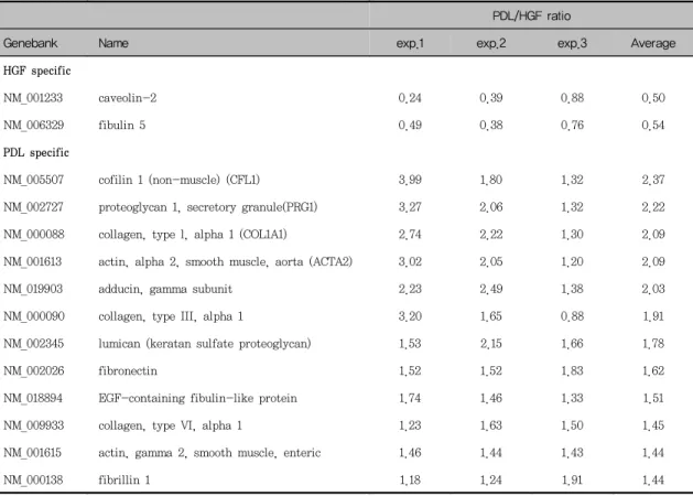
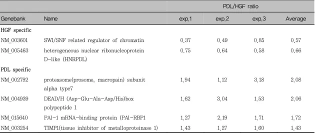
세포골격 및 유주에 관여하는 유전자들과 유전자 및 단백질 발현에 관계하는 유전자들 중에는 치주인 대세포에서 더 강하게 발현된 유전자들이 많았으며 (Table 3, 5) 그 중 cofilin 1, proteoglycan 1 sec- retary granule, 교원질 제I형(α1), α-smooth muscle actin(aorta), adducin γ subunit, 교원질 제III형(α1), lumican(keratan sulfate proteogly- can), fibronectin은 세포골격 및 유주에 관여하는 유전자들이었고, proteasome subunit α type 7, DEAD/H box polypeptide 1, PAl-1 mRNA- binding protein, tissue inhibitor of metal- loproteinase(TIMP)1은 유전자 및 단백질발현에 관 계하는 유전자들이었다.
세포방어 및 항상성 유지에 관여하는 유전자인 heat shock 70kda protein 4와 heat shock pro- tein 27은 치은섬유아세포에서 강하게 발현되었고, 대사에 관여하는 유전자인 ATP synthase β와 phosphoglycerate dehydrogenase는 치주인대세포
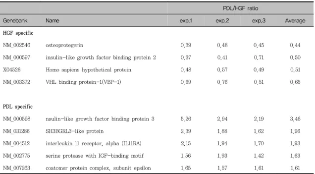
Table 1. Differentially expressed genes related with cell signaling and communication
PDL/HGF ratioGenebank Name exp.1 exp.2 exp.3 Average
HGF specific
NM_002546 osteoprotegerin 0.39 0.48 0.45 0.44
NM_000597 insulin-like growth factor binding protein 2 0.37 0.41 0.71 0.50
X04526 Homo sapiens hypothetical protein 0.48 0.57 0.49 0.51
NM_003372 VHL binding protein-1(VBP-1) 0.69 0.76 0.51 0.65
PDL specific
NM_000598 nsulin-like growth factor binding protein 3 5.26 2.94 2.19 3.46
NM_031286 SH3BGRL3-like protein 2.39 1.88 1.62 1.96
NM_004512 interleukin 11 receptor, alpha (IL11RA) 2.15 1.94 1.70 1.93
NM_002775 serine protease with IGF-binding motif 1.56 1.93 1.42 1.63
NM_007263 coatomer protein complex, subunit epsilon 1.65 1.57 1.61 1.61
Table 2. Differentially expressed genes related with cell division
PDL/HGF ratioGenebank Name exp.1 exp.2 exp.3 Average
HGF specific
NM_001758 cyclin D1 0.73 0.76 0.74 0.74
PDL specific
NM_022662 meiotic checkpoint regulator(MCPR) 2.34 4.27 1.95 2.85
Table 3. Differentially expressed genes related with cell structure and motility
PDL/HGF ratioGenebank Name exp.1 exp.2 exp.3 Average
HGF specific
NM_001233 caveolin-2 0.24 0.39 0.88 0.50
NM_006329 fibulin 5 0.49 0.38 0.76 0.54
PDL specific
NM_005507 cofilin 1 (non-muscle) (CFL1) 3.99 1.80 1.32 2.37
NM_002727 proteoglycan 1, secretory granule(PRG1) 3.27 2.06 1.32 2.22
NM_000088 collagen, type l, alpha 1 (COL1A1) 2.74 2.22 1.30 2.09
NM_001613 actin, alpha 2, smooth muscle, aorta (ACTA2) 3.02 2.05 1.20 2.09
NM_019903 adducin, gamma subunit 2.23 2.49 1.38 2.03
NM_000090 collagen, type III, alpha 1 3.20 1.65 0.88 1.91
NM_002345 lumican (keratan sulfate proteoglycan) 1.53 2.15 1.66 1.78
NM_002026 fibronectin 1.52 1.52 1.83 1.62
NM_018894 EGF-containing fibulin-like protein 1.74 1.46 1.33 1.51
NM_009933 collagen, type VI, alpha 1 1.23 1.63 1.50 1.45
NM_001615 actin, gamma 2, smooth muscle, enteric 1.46 1.44 1.43 1.44
NM_000138 fibrillin 1 1.18 1.24 1.91 1.44
Table 4. Differentially expressed genes related with Cell and organism defence and homeostasis
PDL/HGF ratioGenebank Name exp.1 exp.2 exp.3 Average
HGF specific
AB023420 heat shock 70 kda protein 4 0.36 0.63 0.79 0.59
X54079 heat shock protein27 0.40 1.02 0.44 0.62
PDL specific
Table 5. Differentially expressed genes related with expression of genes and proteins
PDL/HGF ratioGenebank Name exp.1 exp.2 exp.3 Average
HGF specific
NM_003601 SWI/SNF related regulator of chromatin 0.37 0.49 0.85 0.57
NM_005463 heterogeneous nuclear ribonucleoprotein D-like (HNRPDL)
0.75 0.64 0.58 0.66
PDL specific
NM_002792 proteasome(prosome, macropain) subunit alpha type7
1.94 1.12 3.18 2.08
NM_004939 DEAD/H (Asp-Glu-Ala-Asp/His)box polypeptide 1
1.62 3.04 1.53 2.06
NM_015640 PAl-1 mRNA-binding protein (PAl-RBP1 1.27 2.19 1.71 1.72
NM_003254 TIMP1(tissue inhibitor of metalloproteinase 1) 1.43 1.27 1.60 1.43
Table 6. Differentially expressed genes related with metabolism
PDL/HGF ratioGenebank Name exp.1 exp.2 exp.3 Average
HGF specific
PDL specific
NM_001686 ATP synthase beta 2.80 2.12 1.77 2.23
NM_005469 peroxisomal acyl-CoA thioesterase 2.68 1.57 1.39 1.88
NM_032692 phosphoglycerate dehydrogenase 2.51 1.10 1.58 1.73
NM_001084 procollagen-lysine, 2-oxoglutarate 5-dioxygenase 3
2.28 1.32 1.33 1.64
Table 7. Differentially expressed genes related with unclassifed genes
PDL/HGF ratioGenebank Name exp.1 exp.2 exp.3 Average
HGF specific
NM_004859 clathrin, heavy polypeptide (Hc) 0.44 0.66 0.53 0.54
PDL specific
NM_022543 Rattus norvegicus steroid sensitive gene-1 protein
1.70 2.23 2.00 1.98
NM_004791 osteoblastic specific cytein-rich protein 2.88 1.54 1.23 1.88
NM_016081 palladin (KIAA0992) 1.62 1.93 1.56 1.70
NM_007278 GABA(A) receptor-associated protein 2.13 1.54 1.25 1.64
NM_021034 interferon-induced transmembrane protein 3 (1-8U)
2.01 1.35 1.33 1.56
NM_003311 Tumor suppressing subtransferable cadidate 3 1.56 1.46 1.44 1.49
에서 강하게 발현되었다.
세포의 신호전달에 관여하는 유전자 가운데에서 IGFBP2는 치주인대세포에서, IGFBP3는 치은섬유아 세포에서 더 강하게 발현되었다.
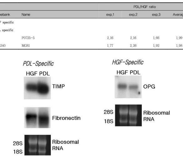
cDNA microarray의 결과를 바탕으로 특정세포에 서 더 강하게 발현된 유전자들 중에서 치은섬유아세 포에서 강하게 발현된 유전자인 osteopreotegerin, 치주인대세포에서 강하게 발현된 유전자들인 TIMP1 과 fibronectin을 선택하여 northern blot analysis 를 시행한 결과는 각각의 유전자들이 우세하게 나타 났던 세포에서 더 강하게 발현되었다(Figure 2).
IV. 고안
최근 유전자 발현에 관한 연구에서 많이 이용되고 있는 cDNA microarray는 한번의 hybridization 과 정으로 동시에 수천 가지의 유전자 발현 정도를 측 정할 수 있다. 치주인대세포와 치은섬유아세포의 유 전자 발현 정도의 차이를 살펴보고자 시행한 본 실 험에서는 경북대학교 기질세포 유전자 은행에서 모 발 간엽세포로부터 제작한 유전자 칩을 사용하였다.
동일 부위의 치주인대세포와 치은섬유아세포를 계 대 배양하여 동일 환자, 동일 세대의 세포에서 mRNA 를 추출하여 cDNA microarry와 hybridization하는
Table 8. Differentially expressed genes related with unknown/novel genes
PDL/HGF ratio
Genebank Name exp.1 exp.2 exp.3 Average
HGF specific PDL specific
P0725-5 2.16 2.16 1.66 1.99
L08240 MG81 1.77 2.26 1.92 1.98
Figure 1. Northern blot analysis of gene expression of PDL cells and HGFs. To verify mi- croarray results on selected genes, we performed northern blot.
PDL : periodontal ligament, HGF : human gingival fibroblast, TIMP : Tissue inhibitor of
metalloproteinase, OPG : osteoprotegerin
과정을 세 명의 환자에게 시행한 결과 세 실험군 간 의 변이가 커서 공통적으로 한 세포에서 우세하게 발현되는 유전자의 수는 적었다. 전체 3,063개의 유 전자들 중에서, 61개의 유전자들이 세 명의 환자에 게 시행한 실험에서 일관성 있게 특정세포에서 더 강하게 발현되었다. 그 중 12개의 유전자들은 치은 섬유아세포에서, 49개의 유전자들은 치주인대세포에 서 더 강하게 발현되었다.
치주인대세포에서 강하게 발현된 유전자들이 가장 많았던 것은 세포골격 및 유주에 관한 유전자들이었 으며 섬유아세포의 주요 교원질 형태인 교원질 제I 형(α1), 교원질 제III형(α1), 그리고 교원질 제IV형 (α1), 섬유아세포의 세포외기질인 fibronectin과 lumican(keratan sulfate proteoglycan), 세포의 수축성의 표지인 α-smooth muscle actin(aorta, enteric) 등이 있었다.
Fibronectin은 고분자량(440,000)의 결합조직 내 에 존재하는 주요 당단백질이며 주된 세포 부착 부 위인 Arg-Gly-Asp(RGD) 배열을 지니고 있는 세포 외기질로서 세포와 기질의 접착에 있어서 단백질 부 착을 제공하고 세포를 교원질쪽으로 이주하게 하는 것으로 알려져 있다23). Hou와 Yaeger 등5)은 같은 배양 조건 하에서 치은섬유아세포보다 치주인대세포 에서 교원질 제I형과 fibronectin의 발현이 더 강하 게 나타남을 관찰함으로써 치주인대세포의 생합성적 활성도가 더 활발함을 보고하였으며 이들의 결과는 본 실험과 일치하였다. 치주인대세포가 fibronectin 의 부분 조각보다는 전체 fibronectin에 부착하며
5,15), 치주인대세포내에서 fibronectin과 상호작용하
는 αv와 α5가 관찰됨으로 integrin 수용체를 통해 서 치주인대세포와 fibronectin이 상호 작용하는 것 으로 알려져 있다24).
본 실험에서 lumican은 치주인대세포에서 더 강 하게 발현되었으며, lumican에 관한 연구는 아직 미 미하였으나 Cheng 등25)은 백악질에서 조직의 광물 화와 관계있는 proteoglycan에 관한 연구에서 ker- atan sulfate proteoglycan으로 lumican과 fi- bromodulin의 존재를 밝혔고 이들은 precementum
과 pericementocyte와 같은 비광물성 부위에서 넓 게 존재함으로 백악질의 광물화 조절에 관여하고 있 음을 보고한 바 있다.
Arora와 McCulloch26)는 collagen gel contraction 을 이용한 연구에서 collagen gel contraction 정도 는 α-smooth muscle actin의 발현 정도에 의존적 이었음을 관찰함으로써 α-smooth muscle actin이 세포가 세포외기질을 빠르게 개조하고 있다는 기능 적 표지가 됨을 보고하였으며, 또한 α-smooth muscle actin은 근섬유아세포를 나타내는 특징중의 하나라고도 알려져 있다27,28). 이러한 α-smooth muscle actin이 본 실험에서는 치주인대세포에서 더 강하게 발현되었으며 이는 교원질, fibronectin과 같 은 세포외기질이 치주인대세포에서 더 강하게 발현 된 결과와 함께 미루어볼 때 치주인대세포가 세포외 기질의 생성과 개조에 관여하고 있음을 고려해 볼 수 있다.
치주인대세포에서 더 우세하게 발현되었던 유전자 들 중에서 교원질 제I형(α1), 제IV형(α1), fi- bronectin, lumican, α-smooth muscle actin은 다른 연구28)에서는 근섬유아세포의 표지로 이용되었 으며, 근섬유아세포는 상처 치유, 세포외기질의 항 상성 유지, 조직의 재생에 관여하는 것으로 알려져 있다29). 육아조직내의 섬유아세포는 형태학적으로, 생물학적으로 섬유아세포와 평활근 세포의 중간 형 태의 성질을 나타내며30), 이러한 세포들은 근섬유아 세포와 유사한 특징을 지니면서 상처 수축에 필요한 힘을 제공한다고 보고하였다31). 본 실험의 결과에서 나타난 바와 같이 치주인대세포가 α-smooth muscle actin을 포함한 세포골격 및 유주에 관한 유 전자를 강하게 발현하고 배양과정에서 형태학적, 생 물학적으로 몇 가지의 특수 형태로 분화가능하다고 알려져 있으나 치주인대세포가 근섬유아세포 유사 특징을 나타낼 수 있는지에 대한 근거는 아직 미약 하다.
치주인대세포에서 강하게 발현되었던 TIMP는 유 전자 및 단백질 발현에 관여하는 유전자로서 섬유아 세포에서 생성되고 지금까지 네 가지 형태가 보고
32-34)되었으며 TIMP-1과 TIMP-2는 모든 matrix metalloproteinase(MMP)를 억제하며 gelatinase와 는 특수한 결합을 보이는 것으로 알려져 있다32). MMP는 단백질 분해 효소의 한 구성원으로서 결합 조직, 교원질, proteoglycan의 분해, 태아형성기와 성숙한 조직에서의 항상성 유지, 만성 염증에서 중 요한 역할을 하는 것으로 알려져 있다35). 이 두 종류 의 단백질 사이의 균형이 교원질의 형성과 유지를 조절하는 기전이며31) MMP와 TIMP사이의 불균형이 초래되면 결합조직의 파괴가 나타나는 것으로 여겨 진다.
Osteoprotegerin/osteoclastogenesis inhibitory factor(OPG/OCIF)는 tumor necrosis factor(TNF) receptor superfamily의 새로운 구성원으로서 파골 세포의 활성도를 억제함으로써 골 흡수를 억제하는 기능을 가지고 있다37,38). 폐, 심장, 신장, 태반과 같 은 다양한 조직들이 OPG/OCIF transcripts를 가지 고 있으며37,39) 세포수준에서는, bone marrow stromal cells, osteoblastic cells, osteosarcoma cell, IMR-90등이 OPG/OCIF mRNA를 발현한다고 알려져 있다39,41,42). Sakata 등43)은 OPG/OCIF가 치 은섬유아세포, 치주인대세포, 치수세포등의 치아간 엽세포들에서 존재함을 발견하고 이들 세포들이 OPG/OCIF를 분비하여 파골세포와 파치세포의 활성 도를 억제함으로써 치조골, 백악질, 상아질과 같은 치아 경조직의 흡수를 조절하는 역할을 한다고 보고 하였다. 본 실험에서는 osteoprotegerin이 치은섬유 아세포에서 더 강하게 발현되었으며 이러한 결과로 유추할 수 있는 치은섬유아세포의 특성은 적당한 조 건이 주어졌을 때 치은섬유아세포가 골상을 형성한 다고 보고한 연구17,18)와 섬유아세포 혹은 골아세포로 의 분화 가능성을 제시한 보고19)에서 나타나는 치은 섬유아세포의 특성과 관련있을 것으로 여겨진다.
cDNA microarray의 결과를 바탕으로 특정세포에 서 더 강하게 발현된 유전자들 중에서 치은섬유아세 포에서 강하게 발현된 유전자인 osteopreotegerin, 치주인대세포에서 강하게 발현된 유전자들인 TIMP1 과 fibronectin을 선택하여 northern blot analysis
를 시행한 결과, 각각의 유전자들이 우세하게 나타 났던 세포에서 더 강하게 발현되었으며 이러한 결과 를 통해서 cDNA microarray를 이용한 유전자 발현 정도의 측정이 신뢰할 만한 방법으로 여겨졌다.
본 실험에서는 세 명의 다른 환자에게서 치아와 치은유두를 채취하여 치주인대세포와 치은섬유아세 포를 배양하고, cDNA microarray를 시행하였다. 세 번의 실험에서 각 유전자들의 발현 정도의 차이가 심하여 세 번 모두 한 세포에서 우세하게 발현된 유 전자들의 수가 적었다. 이와 같이 치주인대세포와 치은섬유아세포의 성상에 관한 실험에서 개체간에 나타나는 차이에 대해 직접적으로 연구 보고한 자료 는 거의 없었지만 Somerman 등4), Kapila 등17), 과 Carnes 등19)의 연구들의 자료를 살펴보면 실험 결과 에서 여러 실험군간의 차이가 크게 나타남을 통해서 개체간의 차이가 있음을 알 수 있었다. 이러한 개체 간의 차이에 대해 몇 가지 사항을 고려해 보면, 먼저 치아와 치은조직을 제공했던 개개인의 특성에 의한 영향을 고려해 볼 수 있다. 치주 조직의 건강에 따른 세포의 생활력 정도, 나이에 따른 세포의 분화 정도 등이 영향을 끼칠 수 있을 것으로 여겨지며 무엇보 다도 배양된 치주인대세포와 치은섬유아세포들이 불 균질한 세포 집단이므로44), 환경에 따라 다양하게 분화할 수 있는 가능성을 가지고 있기 때문에 유전 자들의 발현정도가 개체마다 다르게 나타난 것으로 고려해 볼 수 있다.
치주인대세포와 치은섬유아세포는 같은 섬유아세 포로서의 유사성을 지니고 있지만 서로 다른 특성을 나타내는 것으로 여겨지며 개체간의 변이가 크게 나 타났던 것에 대해서는 앞으로의 연구가 더 진행되어 야 할 것으로 사료된다.
V. 결론
세 명의 환자로부터 배양한 치주인대세포와 치은 섬유아세포로부터 mRNA를 분리하여 제작한 cDNA probe와 3,063개의 유전자를 지닌 cDNA micro- array를 하여 다음과 같은 결론을 얻었다.
세 명의 환자에게 시행한 실험에서 일관성 있게 특정 세포에서 더 강하게 발현된 유전자들의 수가 적었으며 전체 3,063개의 유전자 가운데, 61개의 유 전자들(12개의 유전자들은 치은섬유아세포, 49개의 유전자들은 치주인대세포)이 일관성 있게 특정세포 에서 더 강하게 발현되었다. 세포골격 및 유주에 관 한 유전자들은 치주인대세포에서 더 강하게 발현되 었고, 주요 교원질 형태인 교원질 제I, III. IV형, fibronectin, lumican, 그리고 α-smooth muscle actin 등이 있었다. MMP와 함께 세포외기질을 조절 하는 효소로 알려져 있는 TIMP가 치주인대세포에서 강하게 발현되었고 osteoclastogenesis inhibitory factor인 osteoprotegerin은 치은섬유아세포에서 강 하게 발현되었다.
cDNA microarray의 결과를 바탕으로 시행한 northern blot의 결과, 각각의 유전자들이 우세하게 나타났던 세포에서 그 발현이 강하게 나타난 것으로 보아 cDNA microarray를 이용한 유전자 발현정도 측정이 신뢰할 만한 방법으로 여겨진다.
VI. 참고문헌
1. Lekic P and McCulloch CAG. Periodontal ligament cell populations: the centralrole of fibroblasts in creating a unique tissue.
Anat Res 1996;245:327-341
2. Boyko GA, Mecher AH, and Brunette DM.
Formation of new periodontal ligament by periodontal ligament cells implanted in vivo after culture in vitro. J Periodont Res 1981;16:73-88
3. Gottlow J, Nyman S, Karring T, and Lsindhe J. New attachment formation as the result of controlled tissue regeneration.
J Clin Periodontol 1984;11:494-503
4. Somerman MJ, Archer SY, Imm GR, and Foster RA. A Comparative Study of Human Periodontal Ligament Cells and Gingival
Fibroblasts in vitro. J Dent Res 1988;67:
66-70
5. Hou LT and Yager A. Cloning and Characterization of Human Gingival and Periodontal Ligament Fibroblasts. J Periodontol 1993;64:1209-1218
6. Mariotti A and Cochran DL. Characterization of Fibroblasts Derived From Human Periodontal Ligament and Gingiva. J Periodontol 1990;61:103-111
7. Ogata Y, Niisato N, Sakurai T, Furuyama S, and Suglya H. Comparison of the Characteristics of Human Gingival Fibroblsts and Periodontal Ligament cells.
J Periodontol 1995;66:1025-1031
8. Somerman MJ, Young MF, Foster RA, Moehring JM, Imm G, and Sauk JJ.
Characteristics of Human Periodontal Ligament Cells in vitro. Archs Oral Biol 1990;35:241-247
9. Giannopoulou C and Cimasoni G. Functional Characteristics of Gingival and Periodontal Ligament Fibroblasts. J Dent Res 1996;75:
895-902
10. Beertsen W, Everts V, and Van den Hooff A. Fine structure of fibroblasts in the pe- riodontal ligament of the rat incisor and their possible role in tooth eruption. Archs Oral Biol 1974;19:1087-1098
11. Pender N, McCulloch CAG. Quantitation of actin polymerisation in two human fibro- blast subtypoes responding to mechanical stretching. J Cell Sci 1991;100:187-193 12. Nojima N, Kobayashi M, Shinonome M,
Takahashi N, Suda T, Hasegawa K.
Fibroblastic cells derived from bovine perio- dontal ligaments have the phenotypes of osteoblasts. J Periodont Res 1990;25:179-185 13. Arceo N, Sauk JJ, Moehring J, Foster RA,
Somerman MJ. Human periodontal cells in- itiate mineral-like nodules in vitro. J Periodontol 1991;62:499-503
14. Nohutcu RM, McCauley LK, Koh AJ, Somermana MJ. Expression of extracellular matrix proteins in human periodontal liga- ment cells during mineralization in vitro. J Periodontol 1997;68:320-327
15. Kapila YL, Lancero H, Johnson PW. The Response of Periodontal Ligament Cells to Fibronectin. J Periodontol 1998;69:1008-1019 16. Ivanovski S, Li H, Haase R, Bartold PM.
Expression of bone associated macro- molecules by gingival and periodontal liga- ment fibroblasts. J Periodont Res 2001;36:
131-141
17. Rose GC, Yamaski A, Mahan CJ. Bone in- duction in vitro I. Human gingival fibro- blast cell lines versus tooth matrix. J Periodont Res 1981;16:344 -357
18. Cho MI, Lin WL, Genco RJ. Platelet-de- rived growth factor-modulated guided tis- sue regenerative therapy. J Periodontol 1995;66:522-530
19. Carnes DL, Maeder CL, Graves DT. Cells with osteoblastic phenotypes can be ex- planted from human gingiva and periodontal ligament. J Periodontol 1997;68:701-707 20. 서조영, 최제용, 유현모, 박준봉, 조준승: 치주
인대세포와 치은섬유아세포의 성상에 관한 비 교, 대한구강생물학회지 1991;15:14-28
21. Chomczynski P, Sacchi N. Single-step method of RNA isolation by acid guanidinium thiocyanate-phenol-chloroform extraction.
Anal Biochem 1987;162:156-159
22. DeRisi J, Penland L, Brown PO, Bittner ML, Meltzer PS, Ray M, Chen Y, Su YA, Trent JM. Use of a cDNA microarray to analyse gene expression patterns in human
cancer. Nat Genet 1996;14:457-460
23. Hynes RO. Fibronectins. Sci Am 1986;254:
42-51
24. Steffensen B, Duong AH, Milam SB.
Immunohistological localization of cell ad- hesion proteins and integrins in the periodontium. J Periodontol 1992;63:584-592 25. Cheng H, Caterson B, Neame PJ, Lester
GE, Yamauchi M. Differential distribution of lumican and fibromodulin in tooth cementum. Con Tissue Res 1996;34:87-96 26. Arora PD, McCulloch CAG. Dependence of
Collagen Remodelling on α-smooth muscle actin expression by fibroblasts. J Cell Physiol 1994;159:161-175
27. Valentich JD, Popov V, Saada JI, Powell DW. Phenotypic characterization of an in- testinal subepithelial myofibroblast cell line. Am J Physiol 1997;272:1513-1524 28. Weill FX, Blazejewski S, Blanc JF, Huet S,
Gauthier JM, Neaud V, Olaso E, Dubuisson L, Azais-Braesco V, Vidal-Vanaclocha F, Balabaud C, Bioulac-Sage P, Rosenbaum J.
Characterization of a new human liver- myofibroblast cell line : transcriptional regulation of plasminogen activator in- hibitor type l by transforming growth fac- tor beta 1. Lab lnvest 1997;77: 63-70, 1997 29. Mermelstein CS, Guma FC, Mello TG,
Fortuna VA, Guaragna RM, Costa ML, Borojevic R. Induction of the lipocyte phe- notype in murine hepatic stellate cells : reorganisation of the actin cytoskeleton.
Cell Tissue Res 2001;306:75 -83
30. Hakkinen L, Larjava H. Characterization of fibroblast clones from periodontal gran- ulation tissue in vitro. J Dent Res 1992;71 :1901-1907
31. Darby I, Skalli O, Gabbiani G. α-smooth
muscle actin is transiently expressed by myofibroblasts during experimental wound healing. Lab Invest 1990;63:21-29
32. Apte SS, Mattei MG, Olsen BR. Cloning of the cDNA encoding human tissue inhibitor of metalloproteinase-3(TIMP-3) and map- ping of the TIMP3 gene to chromosome 22.
Genoomics 1984;19:86-90
33. Reynolds JJ. Collagenases and tissue in- hibitors of metalloproteinases : A func- tional balance in tissue degradation. Oral Dis 1996;2:70-76
34. Greene J, Wang M, Liu YE, Raymond LA, Rosen C, Shi YE. Molecular cloning and characterization of human tissue ingibitor of metaloproteinase-4. J Biol Chem 1996;
271:30375-30380
35. Woessner FJ. Matrix metalloproteinases and their inhibitors in connective tissue remodelling. FASEB J 1991;5:2145-2154 36. Khokha R, Denhardt DT. Matrix metal-
loproteinases and tissue inhibitor of metal- loproteinases : A review of their role in tumorigenesis and tissue invasion. Invasion Metastasis 1989;9:391-405
37. Simonet WS, Lacey DL, Dunstan CR et al.
Osteoprotegerin : A novel secreted
protein involved in the regulation of bone density. Cell 1997;89:309-310
38. Lacey DL, Timms E, Tan HL et al.
Osteoprotegerin ligand is a cytokine that regulates osteoclast differentiation and activation. Cell 1998;93:165-176
39. Yasuda H, Sgima N, Nakagaua N, Mochizuki S, Higashio K. Identity of OCIF and OPG : A mechanism by which OPG/OCIF inhibits osteoclastogenesis in vitro. Endocrinology 1998;139:1329-1337 40. Brandstrom H, Jonsson KB, Ohlsson C,
Vidal O, Ljundghall S, Ljunggren O.
Regulation of osteoprotegerin mRNA levels by prostaglandin E2 in human bone mar- row stroma cells, Biochem. Biophys Res Commun 1998;247:338-341
41. Brandstrom H, Jonsson KB, Ohlsson C, Vidal O, Ljundghall S, Ljunggren O. Tumor necrosis factor-α and -β upregulate the levels of osteoprotegerin mRNA in human osteosarcoma MG-63 cells. Biochem Biophys Res Commun 1998;248:454-457 42. Hofbauer LC, Dunstan CR, Spelsberg TC,
Riggs BL, Khosla S. Osteoprotegerin pro- duction by human osteoblast lineage cells is stimulated by vitamin D, bone morpho- genetic protein-2, and cytokines. Biochem Biophys Res Commun 1998;250:776-781 43. Sakata M, Shiba H, Komatsuzawa H, Fujita
T, Ohta K, Sugai M, Suginaka H, Kurihara H. Expression of osteoproteger- in(osteoclatogensis inhibitory factor) in cultures of human dental mesenchymal cells and epithelial cel. J Bone Miner Res 1999;14:1486-1492
44. McCulloch CAG. Progenitor cell populations in the periodontal ligament of mice. Anat Rec 1985;258:262
-Abstract-
Screening of genes differentially expressed in cultured human periodontal ligament cells and human gingival
fibroblasts
Hye-Jeong Yoon1, Mi Hye Choi1, Shin-Il Yeo1, Jin-Woo Park1, Byung-Ju Choi2, Moon-Kyu Kim3, Jung-Chul Kim3, Jo-Young Suh1
1. Department of Periodontology
2. Department of Dental Pharmacology, College of Dentisrtry, 3Department of Immunology, School of Medicine, Kyungpook National University
Periodontal ligament(PDL) cells and human gingival fibroblasts(HGFs) play important roles in de- velopment, regeneration, normal function, and pathologic alteration. PDL cells and HGFs have the similarity related with general characteristics of fibroblast such as spindle shaped morphology, the presence of vimentin intermediate filament and the synthesis of interstitial collagens and fibronectin.
There were many studies about the differences between PDL cells and HGFs, but they were not about whole gene level. In this study, we tried to explain the differences of gene expression profiles be- tween PDL cells and HGFs, and the differences among three individuals by screening gene expression patterns of PDL cells and HGFs, using cDNA microarray.
Although there were some variants among three experiments, a set of genes were consistentely and differentially expressed in one cell type. Among 3,063 genes, 49 genes were more highly ex- pressed in PDL cells and 12 genes were more highly expressed in HGFs.
The genes related with cell structure and motility were expressed more highly in PDL cells. These are cofilin 1, proteoglycan 1 secretory granule, collagen type I(α1), adducin gamma subunit, collagen type III(α1), fibronectin, lumican(keratan sulfate proteoglycan), and α-smooth muscle actin. Tissue inhibitor of metalloproteinase known as the enzyme controlling extracellular matrix with matrix metalloproteinase is more highly expressed in PDL cells, osteoprotegerin known as osteoclastogenesis inhibitory factor is more highly expressed in HGFs.
We performed northern blot to verify cDNA microarray results on selected genes such as tissue in- hibitor of metalloproteinase, fibronectin, osteoprogeterin. The result of northern blot analysis showed that each cell expressed the genes in similar pattern with cDNA microarray result. This result in- dicates that cDNA microarray is a reliable method in screening of gene expression profiles.2)
Key words : cDNA microarray, human gingival fibroblast, periodontal ligament cell