서론
난소종양은 그 기원에 따라 상피성 난소종양, 생식세포종 양, 성기삭-기질종양으로 분류하는데, 흔히 난소암이라 하 면 상피성 난소암을 일컫는다. 상피성 난소암은 전체 난소 암 중 90% 이상을 차지하며, 50-60대 폐경 여성에 주로 발 생한다. 전세계적으로 난소암은 매년 약 22만 명 이상의 환 자들이 새롭게 진단되고 있으며, 14만 명 이상의 환자들이 사망하는 것으로 보고된다[1]. 국내에서는 여성에게 발생하
는 암 중 10위를 차지하고 있는데, 여성 생식기관에 발생하 는 암 중에서는 유방암과 자궁경부암에 이어 세 번째로 흔 히 발생하며, 2012년 한 해 동안 2,167명의 환자가 새롭게 난소암 진단을 받았고, 910명이 사망한 것으로 보고되었다 [2]. 이처럼 높은 사망률을 보이는 이유는 난소암 환자의 약 3분의 2가 암이 골반을 벗어나 진행된 상태인 International Federation of Gynecology and Obstetrics (FIGO) 병기 3기 혹은 4기 상태로 발견이 되고 조기진단을 위한 효과적인 방법이 없기 때문이다[3-5].
수술은 항암화학요법과 더불어 난소암 치료에 근간을 이 루는 가장 중요한 요소인데, 수술을 통해 난소암의 진단과 병기 설정, 치료가 이루어지기 때문이다[6]. 따라서 모든 난 소암 환자는 일부 예외적인 경우를 제외하고는 다 수술을 받 게 된다. 조기 난소암 환자의 경우 철저한 병기설정수술을 통해 병소를 제거하고 정확한 병기를 파악하여 그에 따른 적 절한 항암화학요법을 제공함으로써 예후를 향상시킬 수 있
난소암의 수술적 치료
장 석 준 | 아주대학교 의과대학 산부인과학교실
Surgical management of ovarian cancer
Suk-Joon Chang, MD
Department of Obstetrics and Gynecology, Ajou University School of Medicine, Suwon, Korea
Ovarian cancer is the most lethal of the gynecologic cancers worldwide because most patients present with advanced stage disease at the time of diagnosis. Although multiple therapeutic modalities are employed in the management of ovarian cancer, and despite advances in chemotherapeutic and biologic agents, primary surgery followed by adjuvant chemotherapy remains the cornerstone treatment of this disease. Adequate, comprehensive surgical staging in women with early stage ovarian cancer has been shown to improve oncologic outcomes. Complete surgical cytoreduction leaving no gross residual disease is known to be the only physician-driven prognostic factor for patients with advanced disease. This review describes the rationale and surgical steps for full surgical staging for women with early ovarian cancer, and outlines the cytoreductive surgical procedures required to achieve optimal cytoreduction in patients with advanced ovarian cancer. In addition, the impact of radical surgery (as part of maximal tumor debulking) on the amount of residual tumor and on survival rates will be discussed.
Key Words: Ovarian neoplasms; Surgery; Neoplasm staging; Cytoreduction
Received: December 12, 2015 Accepted: December 29, 2015 Corresponding author: Suk-Joon Chang
E-mail: drchang@ajou.ac.kr
© Korean Medical Association
This is an Open Access article distributed under the terms of the Creative Commons Attribution Non-Commercial License (http://creativecommons.
org/licenses/by-nc/3.0) which permits unrestricted non-commercial use, distribution, and reproduction in any medium, provided the original work is properly cited.
다[7]. 진행된 난소암 환자에서는 적극적인 종양감축수술을 통해 잔류종양을 최소화하는 것이 생존율을 향상시키는 중 요한 예후인자임이 많은 연구들을 통해 밝혀져 있다[8-11].
본 글에서는 원발성 난소암의 수술적 치료에 대한 이론적 근 거와 수술 술식에 대한 고찰, 그리고 최근의 연구동향에 대 한 내용을 다루고자 한다.
난소암의 전파양식과 병기설정
난소암은 인접 골반 조직(자궁, 난관, 골반 복막, 방광, 요 관, 직결장, 후맹낭)으로의 직접적인 침윤, 림프 배출을 통한
골반 및 대동맥주위 림프절 전이, 그리 고 혈행성 전파를 통한 간, 비장, 폐 등 원격 장기로의 전이를 통해 종양이 퍼 지게 된다. 그러나 난소암의 가장 주된 전파경로는 복막을 따라 복강 내 여러 장기에 전이를 일으키는 transcoelomic spread이다[12]. 난소암 세포가 종괴로 부터 탈락되어 골반강 내 떨어지고, 이 세포들이 복강 내 체액의 흐름에 따라 복부와 골반 내 여러 곳으로 이동을 하 게 되는데, 결국 복막표면에 착상하여 자라게 됨으로써 복막의 전이성 결절들 이 나타나게 된다. 복강 내 체액은 호흡 운동에 따라 골반에서 우측 대장주위 홈 을 거쳐 우측 횡격막으로 시계방향의 순 환을 하게 된다. 또한 장의 정상적인 연 동운동을 통해 암세포는 대망, 장 표면 과 장간막, 그리고 전체 복강 내로 퍼지 게 된다.
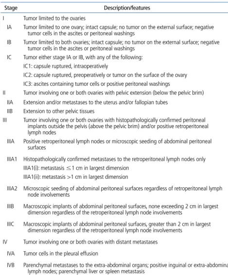
FIGO는 이러한 난소암의 독특한 전파 방식을 반영한 수술적 병기체계를 확립 하였고, 1980년대를 거치며 몇 차례에 걸친 개정을 거쳐 사용해왔다. 그러나 2000년대 들어 난소암이 homogeneous 한 질환이 아니라 다른 형태학적, 생물학적 특성을 보이는 질환군이라는 질병 자체의 개념에 대한 변화, 세계보건기구 의 난소암 조직학적 분류의 개정, 그리고 난소암의 병인으로 써 high-grade serous tubal intraepithelial carcinoma의 발 견 등 큰 과학적인 성취가 있었고, 이들을 반영하여 2014년 에 새로운 병기설정체계가 개정, 수립되었다[13] (Table 1).
진단 당시 병기에 따른 환자 분포를 보면 1기가 24%, 2기가 6%, 3기가 55%, 4기가 15%로, 3기 이상이 70%를 차지한다.
과거에 미국 Gynecologic Oncology Group은 난소암 임상 시험을 위한 분류에서 1기와 2기를 조기 난소암으로, 3기와 4기를 진행성 난소암으로 정의하였는데, 최근에는 높은 재 발률을 고려하여 2기 이상을 진행성으로 분류하고 있다[14].
Table 1. International Federation of Gynecology and Obstetrics staging system for ovarian cancer
Stage Description/features
I Tumor limited to the ovaries
IA Tumor limited to one ovary; intact capsule; no tumor on the external surface; negative tumor cells in the ascites or peritoneal washings
IB Tumor limited to both ovaries; intact capsule; no tumor on the external surface; negative tumor cells in the ascites or peritoneal washings
IC Tumor either stage IA or IB, with any of the following:
IC1: capsule ruptured, intraoperatively
IC2: capsule ruptured, preoperatively or tumor on the surface of the ovary IC3: ascites containing tumor cells or positive peritoneal washings
II Tumor involving one or both ovaries with pelvic extension (below the pelvic brim) IIA Extension and/or metastases to the uterus and/or fallopian tubes
IIB Extension to other pelvic tissues
III Tumor involving one or both ovaries with histopathologically confirmed peritoneal implants outside the pelvis (above the pelvic brim) and/or positive retroperitoneal
lymph nodes
IIIA Positive retroperitoneal lymph nodes or microscopic seeding of abdominal peritoneal surfaces
IIIA1 Histopathologically confirmed metastases to the retroperitoneal lymph nodes only IIIA1(i): metastasis ≤1 cm in largest dimension
IIIA1(ii): metastasis >1 cm in largest dimension
IIIA2 Microscopic seeding of abdominal peritoneal surfaces regardless of retroperitoneal lymph node involvements
IIIB Macroscopic implants of abdominal peritoneal surfaces, none exceeding 2 cm in largest dimension regardless of the retroperitoneal lymph node involvements
IIIC Macroscopic implants of abdominal peritoneal surfaces, greater than 2 cm in largest dimension regardless of the retroperitoneal lymph node involvements
IV Tumor involving one or both ovaries with distant metastases IVA Tumor cells in the pleural effusion
IVB Parenchymal metastases to the extra-abdominal organs; positive inguinal or extra-abdominal lymph nodes; parenchymal liver or spleen metastasis
Modified from Prat J; FIGO Committee on Gynecologic Oncology. J Gynecol Oncol 2015;26:87-89 [13].
난소암 수술적 치료의 역사
문헌상에 보고된 최초의 난소종양 수술은 19세기 초반으 로 거슬러 올라간다. 1809년 미국 켄터키의 외과의사였던 McDowell은 큰 난소종양을 성공적으로 제거하였고, 환자는 이후 33년 동안 생존하였다. McDowell은 총 13명의 환자에 게 ovariotomy를 시행하였고 그 중 8명이 수술 후 생존하였 다[15]. 이후 병이 있는 난소는 제거해야 한다는 수술적 원칙 이 받아들여지게 되었고, 1879년 Tate는 난소종양의 감별을 위한 시험적 개복술의 개념을 도입하였으며, 1900년대 초까 지 난소암이 의심되는 여성의 난소를 제거하는 수술이 시행 되었다[16]. 1930년대 독일의 Peham과 미국의 Meigs는 난 소암 환자에서 수술 후 방사선치료의 효과를 극대화하기 위 해 가능한 한 많은 종양을 최대한 절제하는 종양감축수술의 개념을 처음 제안하였고[10], 1940년 Pemberton[17]은 난 소암 종양감축수술의 일환으로 대망절제술의 시행을 주장 하였으며, 1968년 Munnell[18]은 전자궁절제술, 자궁부속 기절제술, 대망절제술 외에 S자결장까지 절제하는 적극적인 수술 시행 경험을 보고하였다.
1975년 Griffiths [19]는 난소암 수술 후 남는 잔류종양 과 생존율과의 역상관관계를 보고한 기념비적인 논문을 발 표하였다. 그는 진행성 난소암 환자에서 수술 후 복강 내 남 는 잔류종양의 최대 직경이 1.5 cm 이상이면 대부분의 환 자들이 2년 내 사망하지만, 잔류종양의 최대직경이 1.5 cm 이하이면 5년 생존율이 20%까지 연장되며, 다변량 분석 결 과 종양의 조직학적 등급과 수술 후 남는 잔류종양의 최대 직경이 생존율에 영향을 미치는 독립적인 예후인자라고 보 고하였다. 1990년대 중반 Hoskins 등[20]은 난소암 환자들 의 5년 생존율을 잔류종양의 크기에 따라 분석하였는데, 육 안적 잔류종양이 보이지 않는 환자들의 생존율이 60%, 잔 류종양의 크기가 2 cm 이하인 경우 35%, 2 cm 이상인 경 우 20%로 각각 보고함으로써 잔류종양이 작으면 작을수록 생존율이 향상된다는 것을 보여주었다. 이후 많은 후향적 연구들이 잔류종양의 크기와 생존율과의 유의한 역상관성 을 보여 Griffiths의 주장이 타당함을 입증하였고, 2002년 Bristow 등[21]은 메타분석을 통해 최대 종양감축이 이루어
진 정도가 클수록 생존율이 향상되며, 최대 종양감축수술의 시행이 가장 강력한 clinician-driven prognostic factor임 을 보여줌으로써, 결국 적극적인 종양감축수술과 수술 후 항암치료라는 치료전략이 진행성 난소암 치료의 표준으로 자리잡게 되었다.
조기 난소암의 병기설정수술
앞서 언급한대로 난소암은 복막으로의 transcoelomic spread, 인접 장기로의 직접 침윤, 림프절 전이, 혈행성 전이 를 통해 병이 진행되므로, 정확한 병기설정을 위해서는 골반 과 복부를 포함한 전체 복강 내 평가와 후복막 평가가 다 이 루어져야 한다. 조기 난소암에서는 특히 병기설정수술이 중 요한데, 철저한 수술로 확인된 병기에 따라서 이후의 치료 방향이 결정되고, 환자에게 예후와 관련된 정확한 정보를 제 공할 수 있기 때문이다.
병기설정을 위한 수술로는 복막세척검사, 전자궁절제술,
양측자궁부속기절제술, 후맹낭, 골반벽, 대장주위홈, 횡경
막 및 기타 전이 의심 및 가능 부위에 대한 다발성 복막
생검, 대망절제술, 골반 및 대동맥주위 림프절절제술을 시
행한다. 육안적으로 난소에 국한된 것으로 보이는 조기 난
소암의 경우라도 이러한 철저한 병기설정수술을 통해 약
18-30%의 환자에서 육안으로 발견되지 않는 숨겨진 골반
외 병소를 찾아낼 수 있다[7,22-24]. 수술 중 육안적으로,
또 수술 전 영상검사에서 난소에 국한된 조기 난소암으로
추정되는 경우라도 많게는 25%의 환자에서는 림프절전이
가 있는 것으로 보고가 되고 있기 때문에 후복막 림프절에
대한 철저한 평가가 이루어져야 한다[23,24]. 초기 병기설
정이 불충분하게 이루어지면 아무리 수술 후 항암치료를 시
행한다 하더라도 재발의 위험성이 증가하므로, 이런 경우에
는 항암치료 시행 전 부인종양의사에 의한 재병기설정이 필
요하다[22-24]. 또한 수술의사의 전문성 역시 중요한데, 여
러 연구들을 통해 일반부인과의사나 일반외과의사에 의해
수술을 받은 난소암 환자들을 부인종양의사가 다시 수술하
였을 때 30% 이상의 환자들에서 병기가 상향되었다는 보고
가 있어서[7], 난소암 수술은 부인종양의사에 의해 시행되 어야 한다[25].
병기설정수술 술식
난소암 병기설정수술은 복부 정중절개를 통한 개복수술 이 원칙이다. 최근 일부 환자에서 선택적으로 복강경을 이 용한 최소침습수술이 사용이 시도되기도 하지만 종양학적 인 안전성에 대한 근거는 아직 미약하다. 절개는 통상적으 로 치골에서부터 배꼽까지 이루어지는데, 종양이 있는 난 소를 우선적으로 확인하게 되며, 종양 피막 파열과 피막 침 범 여부 등을 확인해야 한다. 피막이 수술 중 파열될 수 있 는데, 수술 중 파열이 예후에 영향을 미치는지 여부는 아직 확실히 밝혀져 있지 않고, 최근의 메타분석 결과는 무병생 존율에 영향을 주지 않는 것처럼 보인다고 밝히고 있으나 [26], 가능한 한 터트리지 않고 제거하도록 해야 한다. 동 결절편검사를 통해 난소암이 확진 되면 절개를 배꼽부터 칼 돌기까지 연장한다. 견인기를 이용하여 전체 복강과 골반강 을 충분히 노출시킨 후 surgical exploration을 시행한다.
복수가 있다면 세포학적 검사를 위해 50-100 mL의 복수 를 채취하고, 만약 복수가 없다면 골반과 복부의 복막을 물 로 세척한 뒤 세포검사를 위해 채집한다. 1기의 난소암 환 자의 약 30%가 세포검사에서 양성으로 나온다고 알려져 있 다[27]. 전자궁절제술 및 반대쪽의 난소난관절제술을 시행 하고 난소 외의 병변이 있는지 확인하기 위해 체계적인 시 진과 촉진을 시행한다. 정해진 것은 없으나 일반적으로 우 측 대장주위홈에서 시작하여 우측 신장, 간하부, 우측 횡격 막, 간우엽, 담낭, Morrison’s pouch, 좌측 횡격막, 간좌엽, 비장, 위, 대망, 횡행결장, 좌측 신장, 좌측 대장주위홈의 순 으로 시계방향으로 확인한다. 작은복막주머니는 위주름창 자인대의 좌측이나 작은 대망을 열어 확인한다. 소장과 대 장, 그리고 장간막 표면을 확인하고 후복막의 혈관 부위 역 시 촉진하여 림프절이 커져 있는지 여부를 확인한다. 논란 의 여지는 있으나 육안적으로 병변이 없는 조기 난소암으 로 생각되더라도 무작위 복막 생검과 대망절제술은 시행한
다. 일부 연구자들은 무작위 복막 생검과 대망절제술을 통 해 2.4-4.7%의 환자에서 병기가 상향되었음을 보고한 바 있다[28,29]. 점액성 종양인 경우엔 충수돌기절제술을 시 행한다. 암이 난소에 국한된 경우에 림프절절제술의 시행 여부도 그 치료적 효과에 대해서는 분명하게 입증되어 있지 는 않지만 10-25%의 환자에서는 림프절 전이가 보고되고 있으므로[23,24], 골반 및 대동맥주위 림프절 생검 혹은 절 제술의 시행이 권장된다. 대동맥주위림프절절제술을 시행 할 때의 범위 또한 논란이 있는 부분인데, 하장간막동맥 상 방의 림프절 전이가 4.3-8.6% 환자에서 보고 되고 있어서 [24], 대동맥 분기부터 신장정맥 하방까지 철저한 대동맥 주 위 림프절에 대한 평가가 이루어져야 한다.
진행성 난소암의 종양감축수술
종양감축수술의 목적은 가능한 많은 종양을 제거하여 육 안적으로 보이는 잔류 종양을 최소화 하는 것인데, 진행성 난소암에서 종양감축수술을 시행하는 이론적 근거는 다음 과 같다. 첫째, 항암치료 전 tumor burden을 감소시켜 항 암치료의 효과를 극대화 할 수 있다. 항암치료에 의해서 종 양세포는 각 주기 당 일정 분율 만큼 제거 되는데, 수술로 종양세포의 절대 수를 줄이게 되면 필요한 항암치료의 주기 가 줄어들게 되고, 보다 효과적으로 종양세포를 제거할 수 있게 된다[30]. 또한 종양의 크기가 클수록 세포의 자연변 이가 증가하여 항암제에 내성을 보이는 세포가 증가하게 되 고[31], 또 항암제에 노출이 되면 내성 세포의 발현이 증가 한다[32]. 따라서 종양감축수술을 통해 종양세포의 절대 수 를 줄임으로써 내성을 극복할 수 있는 기회를 높이게 된다.
둘째, 효과적으로 항암제가 종양에 전달될 수 있다. 크기가 큰 종양은 혈관 분포가 미약한 hypoperfuzed area가 있어 서 항암제의 전달이 효과적이지 않을 수 있는데, 수술로 이 를 제거함으로써 항암제 diffusion이 개선될 수 있다[33].
셋째, 증식하는 종양세포는 항암제에 민감하고 증식하지 않
는 종양세포(세포주기의 G
0phase에 해당하는)는 본질적으
로 항암에 내성을 보이는데, 크기가 큰 종양은 대부분의 세
포들이 분열을 하지 않는 G
0세포들로 구성된다. 종양감축 수술로 종양을 제거하면 남아 있는 G
0세포들이 분열을 시 작하여 자라게 됨으로써 항암제에 민감한 종양세포의 비율 이 증가하게 된다[34]. 넷째, 큰 종양을 제거함으로써 복수 를 줄이고 장폐색 등을 유발할 수 있는 특정 위치의 종양 을 제거함으로써 환자의 면역학적, 영양학적 상태를 개선 할 수 있다[35].
부인종양의사는 진행성 난소암 종양감축수술 시 가능한 한 모든 종양을 제거하기 위해 노력한다. 그러나 병변의 위 치나 환자의 상태에 따라 잔류종양이 생길 수 밖에 없는 상 황에 처하게 되는데, 이때 어느 정도의 잔류종양이 남게 되 었을 때 적절한 수술이 이루어졌다고 할 수 있는지에 대한 기준이 필요하다. 1980년대 후반 Gynecologic Oncology Group에서 난소암 임상시험에 참여한 환자들의 수술 결과 를 기술함에 있어 잔류 종양의 최대 직경이 1 cm 이하인 경 우를 최적의 종양감축이 이루어진 것으로 정의한 이후[36], 최근까지도 이 기준이 사용되어 왔다. 그러나 1990년대 중 반 이후 후향적 연구이기는 하지만 종양감축수술로 모든 종 양이 완전히 제거되어 잔류종양이 없도록 된 환자들이 유의 하게 긴 생존기간을 가진다는 결과들이 보고됨으로써 적절
한 수술의 기준이 바뀌기 시작했다[10,37-42]. 최근의 메 타분석은 육안적으로 보이는 잔류종양이 없도록 완전 종양 감축 되었을 때 생존기간의 변화를 정량적으로 분석한 결 과, 완전 종양감축이 시행된 환자들의 분율이 10%씩 증가 할 때마다 생존기간이 2.3개월 증가함을 보여주었으며, 완 전 종양감축의 달성이 독립적인 예후인자라고 하였다[43].
따라서 현재는 진행성 난소암 환자의 종양감축수술 시 완전 한 종양 절제 시행을 수술의 궁극적 목표로 하고 있고, 적 절한 수술의 기준은 육안적 잔류종양이 없는 상태로 정의하 고 있다[25,44].
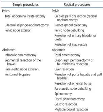
적극적인 종양감축수술
진행성 난소암에서는 잔류종양을 최소화하고 가능한 한 완전한 종양절제를 달성하기 위해 다음과 같은 수술을 시 행한다(Table 2). Simple procedures만으로 적절한 종양감 축을 달성할 수도 있겠지만, 대부분의 진행성 난소암 환자 는 복강 내 여러 곳에 전이를 동반하고 있으므로 수술의 목 표를 달성하기 위해 자궁과 자궁부속기 외에 복막, 횡격막, 간, 위, 비장, 대장, 소장, 장간막 등 전이가 있는 조직을 최 대한 제거하는 다장기 절제를 포함한 radical procedures를 시행하기도 한다. 그러나 이러한 적극적인 수술의 시행에 비 판적인 의견이 있어왔는데, radical surgery가 잔류종양을 최소화하는 데는 효과적일 수 있으나, 상당한 합병증을 동 반할 수 있고 또한 생존율을 결정짓는 것이 수술자체라기 보다는 종양자체가 가진 생물학적인 특성에 좌우된다는 의 견이 바로 그것이다. 최근 영국과 미국에서 발표된 두 연구 는 수술 비판론자들의 의견을 뒷받침하는 근거로 종종 이용 되는데, 이들은 난소암 항암관련 임상연구에 참여한 환자들 의 수술 데이터를 후향적으로 분석한 결과 수술 당시 비교 적 양호한 tumor biology를 보이는(병기가 낮고, 종양의 전 이 정도가 심하지 않고, 복수가 적은) 환자군에서 optimal debulking이 이루어졌을 때 생존율의 향상이 있었고, 나쁜 biology를 보이는 종양에서는 optimal debulking이 이루어 지더라도 생존율의 향상을 기대하기 어렵다고 하였다. 특히,
Table 2. Cytoreductive surgical procedures for advanced ovarian cancer Simple procedures Radical procedures
Pelvis Pelvis
Total abdominal hysterectomy En bloc pelvic resection (radical oophorectomy)
Bilateral salpingo-oophorectomy Rectosigmoid colectomy Pelvic node excision Pelvic node debulking
Resection of urinary bladder or ureter
Resection of iliac vessels
Abdomen Abdomen
Infracolic omentectomy Total omentectomy Segmental resection of the
bowel Diaphragm peritonectomy or full-thickness resection Para-aortic node excision Liver resection
Peritoneal biopsies Resection of porta hepatis and gall bladder
Resection of omental bursa Para-aortic node debulking Splenectomy
Distal pancreatectomy Gastric resection Multiple bowel resection
높은 정도의 radical procedures를 시행 받은 환자라도 수술 전 암의 전이 정도가 심했던 경우에는 생존율의 향상이 없었 다고 보고하여 적극적인 수술이 tumor biology를 극복할 수 는 없다고 주장하고 있다[45,46].
그러나 대부분의 experienced centers에서 나온 연구들 은 FIGO 병기 3기말에서 4기의 진행성 난소암에서 적극적 인 수술을 통해 잔류종양을 최소화할 경우 유의한 생존율 의 향상을 가져올 수 있음을 보여주고 있다[37-39,42]. 최 근 Chang과 Bristow [10]는 진행성 난소암환자에 pacli- taxel-platinum 기반의 복합 항암요법이 표준 항암치료 로 사용되기 시작한 2003년 이후 발표된 논문을 대상으 로 하여 총 14개 연구에서 13,949명의 환자들을 확인하였 고, 해당 환자들의 생존율을 잔류종양의 크기에 따라 분석 하였다. 그 결과, 육안적으로 보이는 잔류종양이 없도록 완 전 종양감축이 달성된 환자가 77.8개월, 1 cm 이하의 잔류 종양을 보이는 환자가 39개월, 1 cm 이상 잔류종양을 가 지는 환자가 31개월의 평균 생존기간을 가지는 것으로 나 타나서, FIGO 병기 3기 이상의 난소암 환자라도 완전 종 양감축수술이 이루어지면 5년 이상의 생존기간을 보일 수 있는 것을 확인할 수 있었다. 또한 진단 당시 FIGO 병기 4기의 난소암이나 같은 3기말이더라도 복막 파종을 동반한 난소암은 나쁜 tumor biology를 시사한다고 할 수 있는데, 적극적인 수술을 통해 잔류종양을 최소화 할 경우 생존율을 향상을 가져올 수 있으며[39,42,47,48], 진행성 난소암의 생 존율은 수술의사와 기관의 expertise에 따라 달라질 수 있음 이 알려져 있다[39,49,50]. 이상의 연구들은 비록 후향적 연 구이긴 하지만, 적극적인 종양감축수술이 진행성 난소암환 자의 생존기간을 향상시킬 수 있음을 일관되게 보여주고 있 어서, 수술이 종양의 생물학적인 특성을 부분적으로는 극복 할 수 있음을 보여주는 증거라 할 수 있을 것이다.
결론
난소암의 치료에 있어서 수술은 가장 핵심적인 요소이다.
수술은 진단과 병기설정, 그리고 치료의 목적으로 사용되며,
조기 난소암의 경우 철저한 병기설정수술을 통해 정확한 병 기, 그에 따른 예후와 관련된 정보, 그리고 적절한 수술후 항암 치료를 환자에게 제공할 수 있다. 진행성 난소암의 경우 종양 감축수술을 통해 잔류종양을 최소화 하는 것이 중요하며, 수 술의 목표는 완전한 종양 절제에 두어야 한다. 난소암은 “수술 의사가 차이를 만든다”는 말을 끝으로 본 글을 마치고자 한다.
찾아보기말: 난소암; 수술; 병기; 종양감축수술
ORCID
Suk-Joon Chang, http://orcid.org/0000-0002-0558-0038
REFERENCES
1. Trimbos B, Timmers Jemal A, Bray F, Center MM, Ferlay J, Ward E, Forman D. Global cancer statistics. CA Cancer J Clin 2011;61:69-90.
2. Jung KW, Won YJ, Kong HJ, Oh CM, Cho H, Lee DH, Lee KH.
Cancer statistics in Korea: incidence, mortality, survival, and prevalence in 2012. Cancer Res Treat 2015;47:127-141.
3. Kim WY, Choi JS, Park CS, Kim BG, Lee JH, Bae DS. An analysis of clinicopathologic prognostic factors affecting sur- vival in patients with epithelial ovarian cancer. Korean J Obstet Gynecol 2002;45:1800-1807.
4. Park HM, Kim YS, Shin EK, Kim MK, Jung KA, Park MH, Chun SH, Ahn JJ, Kim CI, Kim SC. The ability of preoperative serum CA-125 level to predict the outcome of primary cytore- ductive surgery of epithelial ovarian carcinoma. Korean J Gyne- col Oncol Colposc 2003;14:158-166.
5. Kim SC. Consolidation and maintenance therapy for patients with advanced epithelial ovarian cancer. Korean J Obstet Gyne- col 2005;48:2261-2284.
6. No JH, Park YS, Kim SM, Chung HH, Kim JW, Park NH, Song YS, Kang SB, Lee HP. Pattern of care: epithelial ovarian cancer in case of incomplete previous surgery. Korean J Gynecol Oncol 2007;18:195-200.
7. Le T, Adolph A, Krepart GV, Lotocki R, Heywood MS. The benefits of comprehensive surgical staging in the management of early-stage epithelial ovarian carcinoma. Gynecol Oncol 2002;85:351-355.
8. Yun JH, Lee HY, Park HW, Shin JW, Lee JM, Park CY. The analysis of prognostic factors in patients with epithelial ovarian cancer. Korean J Obstet Gynecol 2006;49:566-571.
9. Shih KK, Chi DS. Maximal cytoreductive effort in epithelial ovarian cancer surgery. J Gynecol Oncol 2010;21:75-80.
10. Chang SJ, Bristow RE. Evolution of surgical treatment para-
digms for advanced-stage ovarian cancer: redefining ‘optimal’
residual disease. Gynecol Oncol 2012;125:483-492.
11. Chang SJ, Bristow RE, Chi DS, Cliby WA. Role of aggressive surgical cytoreduction in advanced ovarian cancer. J Gynecol Oncol 2015;26:336-342.
12. Tan DS, Agarwal R, Kaye SB. Mechanisms of transcoelomic metastasis in ovarian cancer. Lancet Oncol 2006;7:925-934.
13. Prat J; FIGO Committee on Gynecologic Oncology. FIGO’s staging classification for cancer of the ovary, fallopian tube, and peritoneum: abridged republication. J Gynecol Oncol 2015;
26:87-89.
14. Jelovac D, Armstrong DK. Recent progress in the diagnosis and treatment of ovarian cancer. CA Cancer J Clin 2011;61:183-203.
15. Othersen HB Jr. Ephraim McDowell: the qualities of a good surgeon. Ann Surg 2004;239:648-650.
16. Tait L. The pathology and treatment of disease of the ovaries.
New York: William Wood & Co; 1883.
17. Pemberton FA. Carcinoma of the ovary. Am J Obstet Gynecol 1940;40:751-763.
18. Munnell EW. The changing prognosis and treatment in cancer of the ovary: a report of 235 patients with primary ovarian carcinoma 1952-1961. Am J Obstet Gynecol 1968;100:790-805.
19. Griffiths CT. Surgical resection of tumor bulk in the primary treatment of ovarian carcinoma. Natl Cancer Inst Monogr 1975;42:101-104.
20. Hoskins WJ, McGuire WP, Brady MF, Homesley HD, Creasman WT, Berman M, Ball H, Berek JS. The effect of diameter of largest residual disease on survival after primary cytoreductive surgery in patients with suboptimal residual epithelial ovarian carcinoma. Am J Obstet Gynecol 1994;170:
974-979.
21. Bristow RE, Tomacruz RS, Armstrong DK, Trimble EL, Montz FJ. Survival effect of maximal cytoreductive surgery for advanced ovarian carcinoma during the platinum era: a meta- analysis. J Clin Oncol 2002;20:1248-1259.
22. Young RC, Decker DG, Wharton JT, Piver MS, Sindelar WF, Edwards BK, Smith JP. Staging laparotomy in early ovarian cancer. JAMA 1983;250:3072-3076.
23. Grabowski JP, Harter P, Buhrmann C, Lorenz D, Hils R, Kommoss S, Traut A, du Bois A. Re-operation outcome in patients referred to a gynecologic oncology center with pre- sumed ovarian cancer FIGO I-IIIA after sub-standard initial surgery. Surg Oncol 2012;21:31-35.
24. Chang SJ, Bristow RE, Ryu HS. Analysis of para-aortic lympha- denectomy up to the level of the renal vessels in apparent early- stage ovarian cancer. J Gynecol Oncol 2013;24:29-36.
25. Stuart GC, Kitchener H, Bacon M, duBois A, Friedlander M, Ledermann J, Marth C, Thigpen T, Trimble E; participants of 4th Ovarian Cancer Consensus Conference (OCCC); Gyne- cologic Cancer Intergroup. 2010 Gynecologic Cancer Inter- Group (GCIG) consensus statement on clinical trials in ovarian cancer: report from the Fourth Ovarian Cancer Consensus Conference. Int J Gynecol Cancer 2011;21:750-755.
26. Kim HS, Ahn JH, Chung HH, Kim JW, Park NH, Song YS, Lee HP, Kim YB. Impact of intraoperative rupture of the ovarian
capsule on prognosis in patients with early-stage epithelial ovarian cancer: a meta-analysis. Eur J Surg Oncol 2013;39:279- 289.
27. Zuna RE, Behrens A. Peritoneal washing cytology in gyne- cologic cancers: long-term follow-up of 355 patients. J Natl Cancer Inst 1996;88:980-987.
28. Powless CA, Bakkum-Gamez JN, Aletti GD, Cliby WA. Ran- dom peritoneal biopsies have limited value in staging of appa- rent early stage epithelial ovarian cancer after thorough explora- tion. Gynecol Oncol 2009;115:86-89.
29. Lee JY, Kim HS, Chung HH, Kim JW, Park NH, Song YS.
The role of omentectomy and random peritoneal biopsies as part of comprehensive surgical staging in apparent early-stage epithelial ovarian cancer. Ann Surg Oncol 2014;21:2762-2766.
30. Skipper HE. Adjuvant chemotherapy. Cancer 1978;41:936-940.
31. Goldie JH, Coldman AJ. A mathematic model for relating the drug sensitivity of tumors to their spontaneous mutation rate.
Cancer Treat Rep 1979;63:1727-1733.
32. Curt GA, Clendeninn NJ, Chabner BA. Drug resistance in cancer. Cancer Treat Rep 1984;68:87-99.
33. Skipper HE. Thoughts on cancer chemotherapy and combi- nation modality therapy (1974). JAMA 1974;230:1033-1035.
34. Gunduz N, Fisher B, Saffer EA. Effect of surgical removal on the growth and kinetics of residual tumor. Cancer Res 1979;
39:3861-3865.
35. Hacker NF. Controversial aspects of cytoreductive surgery in epithelial ovarian cancer. Baillieres Clin Obstet Gynaecol 1989;3:49-57.
36. Omura GA, Bundy BN, Berek JS, Curry S, Delgado G, Mortel R. Randomized trial of cyclophosphamide plus cisplatin with or without doxorubicin in ovarian carcinoma: a Gynecologic Oncology Group Study. J Clin Oncol 1989;7:457-465.
37. Eisenkop SM, Friedman RL, Wang HJ. Complete cytoreductive surgery is feasible and maximizes survival in patients with advanced epithelial ovarian cancer: a prospective study. Gyne- col Oncol 1998;69:103-108.
38. Chi DS, Eisenhauer EL, Lang J, Huh J, Haddad L, Abu-Rustum NR, Sonoda Y, Levine DA, Hensley M, Barakat RR. What is the optimal goal of primary cytoreductive surgery for bulky stage IIIC epithelial ovarian carcinoma (EOC)? Gynecol Oncol 2006;103:559-564.
39. Aletti GD, Dowdy SC, Gostout BS, Jones MB, Stanhope CR, Wilson TO, Podratz KC, Cliby WA. Aggressive surgical effort and improved survival in advanced-stage ovarian cancer. Obstet Gynecol 2006;107:77-85.
40. Winter WE 3rd, Maxwell GL, Tian C, Carlson JW, Ozols RF, Rose PG, Markman M, Armstrong DK, Muggia F, McGuire WP; Gynecologic Oncology Group Study. Prognostic factors for stage III epithelial ovarian cancer: a Gynecologic Oncology Group Study. J Clin Oncol 2007;25:3621-3627.
41. du Bois A, Reuss A, Pujade-Lauraine E, Harter P, Ray-Coquard I, Pfisterer J. Role of surgical outcome as prognostic factor in advanced epithelial ovarian cancer: a combined exploratory analysis of 3 prospectively randomized phase 3 multicenter trials: by the Arbeitsgemeinschaft Gynaekologische Onkologie
Studiengruppe Ovarialkarzinom (AGO-OVAR) and the Groupe dʼInvestigateurs Nationaux Pour les Etudes des Cancers de l’Ovaire (GINECO). Cancer 2009;115:1234-1244.
42. Chang SJ, Bristow RE, Ryu HS. Impact of complete cytore- duction leaving no gross residual disease associated with radical cytoreductive surgical procedures on survival in advanced ova- rian cancer. Ann Surg Oncol 2012;19:4059-4067.
43. Chang SJ, Hodeib M, Chang J, Bristow RE. Survival impact of complete cytoreduction to no gross residual disease for ad- vanced-stage ovarian cancer: a meta-analysis. Gynecol Oncol 2013;130:493-498.
44. Elattar A, Bryant A, Winter-Roach BA, Hatem M, Naik R.
Optimal primary surgical treatment for advanced epithe- lial ovarian cancer. Cochrane Database Syst Rev 2011;(8):
CD007565.
45. Crawford SC, Vasey PA, Paul J, Hay A, Davis JA, Kaye SB. Does aggressive surgery only benefit patients with less advanced ovarian cancer? Results from an international comparison within the SCOTROC-1 Trial. J Clin Oncol 2005;23:8802-8811.
46. Horowitz NS, Miller A, Rungruang B, Richard SD, Rodriguez N, Bookman MA, Hamilton CA, Krivak TC, Maxwell GL. Does aggressive surgery improve outcomes? Interaction between preoperative disease burden and complex surgery in patients with advanced-stage ovarian cancer: an analysis of GOG 182. J Clin Oncol 2015;33:937-943.
47. Winter WE 3rd, Maxwell GL, Tian C, Sundborg MJ, Rose GS, Rose PG, Rubin SC, Muggia F, McGuire WP; Gynecologic Oncology Group. Tumor residual after surgical cytoreduction in prediction of clinical outcome in stage IV epithelial ovarian cancer: a Gynecologic Oncology Group Study. J Clin Oncol 2008;26:83-89.
48. Wimberger P, Wehling M, Lehmann N, Kimmig R, Schmalfeldt B, Burges A, Harter P, Pfisterer J, du Bois A. Influence of residual tumor on outcome in ovarian cancer patients with FIGO stage IV disease: an exploratory analysis of the AGO- OVAR (Arbeitsgemeinschaft Gynaekologische Onkologie Ova- rian Cancer Study Group). Ann Surg Oncol 2010;17:1642-1648.
49. Wimberger P, Lehmann N, Kimmig R, Burges A, Meier W, Du Bois A; Arbeitsgemeinschaft Gynaekologische Onkologie Ovarian Cancer Study Group. Prognostic factors for complete debulking in advanced ovarian cancer and its impact on sur- vival: an exploratory analysis of a prospectively randomized phase III study of the Arbeitsgemeinschaft Gynaekologische Onkologie Ovarian Cancer Study Group (AGO-OVAR). Gyne- col Oncol 2007;106:69-74.
50. Bristow RE, Chang J, Ziogas A, Campos B, Chavez LR, Anton- Culver H. Impact of National Cancer Institute Comprehensive Cancer Centers on ovarian cancer treatment and survival. J Am Coll Surg 2015;220:940-950.
Peer Reviewers’ Commentary
부인암 중에 가장 사망률이 높은 난소암의 중요한 치료방법인 수 술적 치료에 대한 논문으로, 특히 진행된 난소암에서 환자의 예 후에 가장 중요한 영향을 미치는 적극적인 종양 감축술에 대한 자세한 기술은 젊은 부인종양 의사들에게 많은 도움이 될 것으 로 사료된다. 초기 암일지라도 반드시 완벽한 병기 설정 수술이 향후 적절한 치료와 예후를 위해 필요한데 이는 아무리 강조하여 도 지나치지 않을 정도로 중요한 내용이다. 또 이러한 수술은 전 문적인 지식과 경험을 갖고 있는 부인 종양 전문 의사에 의해서 행해져야 하며 이 경우 환자의 치료 결과도 가장 좋은 것임을 본 논문은 잘 지적하고 있다. 근간에 우리나라의 난소암 환자는 완 만한 증가 추세를 보이고 있는데, 그 조기 진단이나 예방법이 아 직은 마땅치 않은 점이 문제이다. 또 항암화학요법에 대한 내성 으로 치료 후 생존율도 높지 않아 가장 무서운 부인암인 난소암 환자의 수술적 치료에 대한 좋은 지침이 될 수 있는 논문으로 이 용될 수 있기를 기대한다.
[정리: 편집위원회]