Effect of Treadmill Exercise Training on the Expression of PGC-1α, GLUT-1, Tfam Proteins and Antioxydent Ezymes in Brain of STZ-Induced Diabetic Rats
Noh-Hwan Park1, Jin Lee2, Kook-Hyun Jung1, Bong-Am Choi3, Hyung-Chae Jang1, Suk-In Lee4, Dong-Soo Lee4 and Joon-Yong Cho1*
1Exercise Biochemistry Laboratory, Korea National Sport University, Seoul 138-763, Korea
2Department of Anatomy and Cell Biology, Collage of Medicine, Han-Yang University, Seoul 133-791, Korea
3Collage of Golf, Dae-Gu University, Dae-gu 712-714, Korea
4College of Physical Education, Chung-Ang University, Seoul 156-756, Korea Received January 10, 2011 /Accepted March 7, 2011
The purpose of this study is to identify the effects of exercise training [ET, 10~18 m/min (speed), 20~30 min (exercise duration)/a day for 5 day/wk, 6 wk) on PGC-1α, GLUT-1, Tfam, Cu,Zn-SOD and Mn-SOD proteins in brain of STZ-induced diabetic rats. The male Sprague-Dawley (SD) rats were sin- gle-injected intraperitoneally with 50mg/kg of streptozotocin (STZ) to produce STZ-induced diabetic rats. Rats were divided into 3 experimental groups with 8 rats in each group, as follows: (1) non-STZ group (n=8), (2) STZ-CON group (n=8), (3) STZ-EXE group (n=8). The results of this study suggest that i) serum glucose level was significantly reduced in STZ-EXE group compared with STZ-CON group (p<0.05), ii) PGC-1α (p<0.001), mtPGC-1α (p<0.001), GLUT-1 (p<0.001), and mtTfam (p<0.001) proteins in brain of STZ-induced diabetic rats were significantly increased in STZ-EXE group com- pared with STZ-CON group, iii) Cu,Zn-SOD (p<0.001) and Mn-SOD (p<0.01) proteins in the STZ-in- duced diabetic rats were significantly increased in STZ-EXE group compared with STZ-CON group.
In conclusion, the findings of the present study reveal that treadmill exercise training increases brain GLUT-1 protein level possibly through up-regulation of PGC-1α and Tfam proteins which represent key regulatory components of stimulation of brain mitochondrial biogenesis. In addition, treadmill ex- ercise training may prevent oxidative stress by up-regulation of Cu,Zn-SOD and Mn-SOD proteins in the STZ-induced diabetic rats.
Key words: Treadmill exercise training, STZ-induced diabetic rat, PGC-1α, GLUT-1, Tfam, Cu,Zn-SOD, Mn-SOD
*Corresponding author
*Tel:+82-2-410-6867, Fax:+82-2-410-6945
*E-mail : [email protected]
서 론
당뇨병은 고혈당, 인슐린 분비 부족 및 결핍과 인슐린 수용체 의 민감도 저하 등의 원인으로 생기는 질환이며, 장기간 지속되 면 합병증이 나타나며, 대표적인 합병증은 당뇨성 망막증 (retinopathy), 신장병(nephropathy), 신경병증(neuropathy) 등을 유발한다[2,7].
당뇨에 대한 병태·생화학적 기전들을 보면 인체 내 항산화 방어기전이 손상되어 활성산소(free radical)의 생성을 높여 세 포 내 단백질과 세포막 지질의 변형, 공간 배열의 손상, 조직 내 미토콘드리아 DNA (mtDNA)의 손상[2,7]과 고혈당증 (hyperglycemia)으로 생성된 반응성 산소종(reactive oxygen species)인한 저밀도 지단백(low-density lipoprotein)의 지질 과산화 증가[18,40] 등이 있다.
최근 당뇨로 인한 고혈당은 뇌 미토콘드리아의 기능을 손상
시켜 학습능력과 기억력을 저하시키고 알츠하이머와 파킨슨 질환과 같은 신경퇴행성질환의 발병률을 높게 만든다고 보고 [4,5,6]되고 있다. 즉 뇌에서 미토콘드리아 기능 이상은 mtDNA복제 수와 양을 크게 감소시켜 학습능력과 기억력을 손상시킨다[1,20,24]. 한편 미토콘드리아에서는 미토콘드리아 생성 및 활성을 유지하기 위해 peroxisome proliferators- activated receptor gamma coactivator-1α (PGC-1α)와 mitochondrial transcription factor A (Tfam) 등의 단백질을 발현시킨다[10]. PGC-1α는 미토콘드리아 DNA에 의해 합성 되어 미토콘드리아의 생성과 활성을 조절하여 미토콘드리아 의 기능과 복제[9,23,30,31]를 하며, Tfam은 주로 DNA를 합성 시켜 미토콘드리아를 복제하는데 주요한 인자로 보고되어 있 다[12]. 따라서 당뇨와 관련된 신경퇴행성질환에 대응하기 위 해서는 뇌 미토콘드리아 수 생성관련인자들에 대한 활성 방안 이 절실히 필요하다.
뇌의 중추신경계에 필요한 주 에너지원인 글루코스를 말초 혈액에서 뇌까지 공급하고 이동시키는데 관여하는 단백질인 GLUT-1 (glucose transporter-1)은 글루코스가 뇌혈관장벽
(blood-brain barrier)의 내피세포를 지나가도록 도와주기 때 문에 혈당조절에 중요한 인자[12]로써 활동을 하며, 뇌의 피질 부분과 시상 및 해마를 포함한 거의 모든 신경세포체에서 광 범위하게 분포되어 뇌의 기억력활동을 조절해 준다[32]. 그러 나 당뇨병 환자들의 뇌에서는 GLUT-1의 발현이 억제 되는 것으로 알려져 있기 때문[11]에 GLUT-1 단백질을 활성화시키 는 방안이 시급하다.
당뇨 치료와 관련된 연구들은 주로 의·약학 분야에서 약물치 료로 혈당 감소에 초점을 두고 수행되어 왔으며 최근에는 당뇨 병이 신경퇴행성질환인 알츠하이머 질환과 파킨슨 질환의 발병 과 관계가 높다고 보고되면서 당뇨 상태에서 뇌 조직의 글루코 스 대사 및 산화적 스트레스에 대한 연구가 상당한 관심을 받고 있다. 이러한 측면에서 최근 약물이외에 규칙적인 신체활 동 혹은 운동이 당뇨병 개선 및 뇌 기능 향상에 긍정적인 영향을 미치며 신경퇴행성질환인 알츠하이머질환의 예방과 치료에 있 어서 긍정적인 효과가 있다고 보고되면서 운동에 대한 관심이 증가하고 있다.
운동과 뇌 기능 관련 선행연구들을 보면 운동은 조직 내 항산화효소의 발현을 증가시키고[36] 대뇌피질과 시상하부 및 다양한 조직에서 인슐린 신호전달을 향상시킬{28]뿐만 아니라 심장과 골격근 세포 내 에너지 요구에 의해 PGC-1α 의 발현을 유도시킨다[34]고 알려져 있다. 또한 Cotaman 등 [8]은 운동수행으로 뇌신경의 가소성을 조절하는 유전자들 과 학습능력과 기억력을 자극하는 BDNF (brain derived neurotrophic facor)와 같은 특정 분자들을 활성화시켜 뇌의 인지기능에 긍정적인 영향을 미친다고 보고하였으며, Park 등[28]과 Sander 등[34]도 운동이 대뇌피질과 시상하부에서 인슐린 신호전달 능력 향상 등 뇌기능 향상에 긍정적인 영 향을 미친다고 보고하였다.
이와 같이 운동이 혈당 강하와 뇌 기능 향상(글루코스, 대 사, 미토콘드리아 생합성)에 효과가 있음에도 불구하고 당뇨 상태에서 운동이 뇌 기능에 어떠한 영향을 미치는가에 관한 연구는 부족한 실정이다. 더욱이 당뇨 상태에서 운동이 뇌조 직의 산화적 손상을 예방하는데 효과가 있는지에 관한 기초자 료도 부족한 실정이다. 따라서 이 연구는 streptozotocin (STZ) 투여로 당뇨병을 유발시킨 Sprague-Dawley (SD)계 흰쥐를 대 상으로 트레드밀 지구성 운동이 글루코스 수준, 인슐린 수준, HOMA-IR 및 PGC-1α, GLUT-1, Tfam 단백질과 항산화 효소 (Cu,Zn-SOD, Mn-SOD)의 발현에 미치는 효과를 규명하는데 목적이 있다.
재료 및 방법
실험동물
본 실험에 사용된 실험동물은 생후 6주령 된 Sprague- Dawley (SD)계 수컷 흰쥐를 실험 동물센터에서 분양 받은 후
1주간의 적응기간과 1주간의 적응훈련을 거친 후 6주간의 훈 련을 수행하였다. 이들은 실험 8주 동안 동일한 조건, 즉 온도 20±2℃, 습도 50%로 조절된, 조명은 주간(07:00-19:00)에 소등 하고, 야간(19:00-07:00)에는 자동적으로 전등하도록 한 실험동 물실에서 사육하였다. 모든 쥐들은 케이지 당 1마리씩 넣어 사육하였으며 실험기간 동안 미국영양학회에서 발표된 AIN-76A 실험동물용 식이조정에 따라 제조된 식이를 제공하 였고, 식이와 수분섭취는 제한 없이 공급하였다. 이들 실험동 물들은 무작위로 정상 SD 비교집단(n=8, NON STZ-CON), 당뇨 비교집단(n=8, STZ-CON), 당뇨 운동집단(n=8, STZ- EXE)으로 구분하였다.
당뇨유도
환경적응훈련을 1주 실시하는 동안 실험에 들어가기 3일 전에 흰쥐 췌장의 베타(β)세포만을 특이적으로 파괴시키는 streptozotocin을 0.1M citric acid buffer (pH 4.5) 용액에 용 해시킨 50 mg/kg을 복강에 1회 주사하여 당뇨병을 유도하 였다. 당뇨병의 유발 확인은 streptozotocin 주사 48시간에서 72시간 사이에 공복상태에서 꼬리 정맥으로부터 채혈하여 공복 시 혈당 수준이 300 mg/dl 이상일 때 당뇨가 유발되 었음을 확인하였다. 정상 SD 비교집단은 동일한 양의 saline 만을 주사하였다[17].
훈련방법
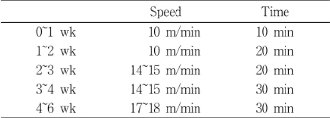
Rodent 트레드밀(8 Lanes, Dae-myung Scientific Co, Ltd, Korea)을 이용하여 지구성 운동을 시키기 위해 1주일 정도 사전적응 훈련(10분 동안 10 m/min, 주 5일)을 실시하였으 며, 본 훈련은 실온(22~24℃)에서 주 5일 총 6주간 실시하였 다[17]. 훈련 프로토콜은 Table 1과 같다.
혈중 생화학 성분 분석
NON STZ-CON 집단, STZ-CON 집단, STZ-EXE 집단을 대상으로 혈액을 채혈하기 위해 pentobarbital sodium (50 mg/kg)으로 마취 시켜 개복하여 심장에서 8 ml의 혈액을 채 혈한 후 혈청 글루코스와 인슐린 수준을 분석하기 위해 혈청 분리 튜브를 이용하여 혈액을 약 30분 동안 방치한 후 15분 간 원심분리(3,000 rpm)하여 혈청을 분리하고, 혈중 생화학적 검사를 실시하였다. 글루코스와 인슐린 수준은 각각 Glucose Hexokinase kit (Bayer, Pittsburg, USA)와 enzyme immuno- assay ELISA 분석용 kit (Mercodia AB, Uppsala, Sweden)를 이용하여 분석하였으며 인슐린 저항성의 지표로 사용되고 있 는 HOMA-IR (Homeostasis model assessment of insulin re- sistance=[Fasting Insulin (μU/ml)×Fasting Plasma Glucosea (mmol/l)/22.5])를 산출하였다.
Table 1. Treadmill program
Speed Time
0~1 wk 10 m/min 10 min
1~2 wk 10 m/min 20 min
2~3 wk 14~15 m/min 20 min
3~4 wk 14~15 m/min 30 min
4~6 wk 17~18 m/min 30 min
뇌 적출
6주간 트레드밀 지구성 운동을 수행한 후, pentobarbital (50 mg/kg)을 복강 내 주입시켜 마취 시킨 후, 뇌 조직을 적출하 여 분석 때까지 -80℃에서 냉동 보관하였다.
미토콘드리아 분리방법
미토콘드리아의 분리는 Mitochondria Extraction Kit (IMGENEX Corporation, San Diego, CA)을 이용하여 분리하 였다. 분석방법은 조직 1 g 당 5 ml의 homogenizing buffer를 넣어 잘게 균질화 시킨 후, 균질화된 샘플은 4℃에서 10분간 3,000 rpm으로 원심 분리하였다. 분리된 상층액을 다시 4℃에 서 30분간 12,000 rpm으로 원심 분리한 후, 상층액을 제거하고 남은 pellet을 5 ml의 Suspension buffer를 넣어 잘 섞어 준 다음 다시 4℃에서 10분간 12,000 rpm으로 원심 분리하였다.
이후 상층액을 제거 후 1회 더 Suspension buffer 5 ml를 넣고 잘 섞어 준 후 다시 4℃에서 10분간 12,000 rpm으로 원심 분리 하였다. 상층액을 제거한 후 남은 pellet은 1 ml의 Complete Mitochondrial lysis buffer를 넣어 4℃에서 30분간 녹인 후 분리된 mitochondrial extract를 4℃에서 5분간 12,000 rpm으 로 원심 분리하여 획득한 상층액(mitochondria fraction)을 얻 었다. 총 단백질량은 BSA (Bovine Serum Albumin, 570 nm)를 이용하여 Bradford [6]의 방법으로 정량화하였다.
SDS-PAGE방법
10% separating gel (30% acrylamide, 1.5M tris pH8.8, 10%
SDS, 10% Ammonium persulfate, TEMED)와 5% stacking gel (30% acrylamide, 1M tris pH 6.8, 10% SDS, 10% Ammonium persulfate, TEMED)을 만들어 사용하였으며, 원심분리(15,000 rpm, 20분) 한 후, 상층액(total cytosol fraction)과 SDS load- ing buffer (60mM tris pH6.8, 25% glycerol, 2% SDS, 14.4mM 2-mercaptoethanol, 0.1% Bromophennol Blue)을 잘 혼합한 후 100℃에 10분간 끓여 단백질을 변성시킨 다음, 얼음에 10분 식힌 후, 다시 원심분리(15,000 rpm, 20분간, 4℃)하였다. 스탠 다드 마커(SDS-PAGE Molecular Weight Strandards, BioRad) 와 함께 각 샘플을 Mini-protein Ⅱ dual-slab apparat us (Bio-Rad, CA, USA)에 준비된 stacking gel well에 총 단백질 량이 100 μg이 되도록 분주하고 80 V에서 2시간 정도 샘플이
바닥에 닿을 때까지 전기영동 하였다.
Western blot 분석방법
Transfer buffer (190 mM glycine, 50 mM Tris-base, 0.05 SDS 20% methanol)에 적신 nitrocellulose membrane (Amer- sham), Whatman 3M paper을 차례로 겹친 후, Mini trans- blot module (Bio-Rad)에 장치하고 40 V로 2시간 전사하였다.
Membrane으로 증착이 끝나면 rocker platform 위에서 1시간 동안 membrane을 3% skim milk 용액(in PBS-T: 10mM Tris-base pH 8.0, 150 mM NaCl, 0.05% Tween-20)으로 block- ing시켰다. 1차 항체인 anti-GLUT-1 (sc-7903, Santa Cruz, CA, USA), anti-PGC-1α (sc-13067, Santa Cruz, CA, USA), anti-Tfam (sc-23588, Santa Cruz, CA, USA), anti-SOD- 1(sc-11407, Santa Cruz, CA, USA) and anti-SOD-2 (sc-30080, Santa Cruz, CA, USA), anti-GAPDH (sc-0357, Santa Cruz, CA, USA)를 1:1,000 으로 bolcking (3% skim milk) 용액으로 각각 희석시켜 12시간 동안 흔들어 준 다음 TBS-T 용액으로 10분씩 3차례 세척한 후 2차 항체(horseradish peroxidase-conjugated goat anti-rabbit 65-6120, ZYMED, CA, USA; horseradish peroxidase-con- jugated rabbit anti-goat 81-1620, ZYMED, CA, USA)를 Blocking 용액으로 1:5,000으로 희석시켜 90분 동안 흔들어 주고 난 후 TBS-T 용액으로 10분씩 3차례 세척하였다. 마지막 단계로 ECL substrate 용액 Santa cruz Biotechnology, CA, USA)에 membrane을 넣고 5분간 발색하고 얻어진 membrane을 이미지 분석 시스템(Molecular Imager ChemiDoc XRS System, Bio-Rad, USA)을 이용하여 스캔한 후 Quantity 1-D Analysis Soft ware Bio-Rad, USA)를 이용하여 단백질량을 산출하였다.
자료처리방법
이 연구에서 얻어진 모든 결과는 SPSS/PC 11.0 통계 프로 그램을 이용하여, 각 변인에 대한 기술 통계치(mean±SD)를 산출하고 집단 간 변인의 차이를 확인하기 위해 일원변량분석 (one-way ANOVA)을 실시하였으며 집단 간 유의한 차이가 있을 경우 LSD (least significant difference)를 이용하여 사후 검증을 실시하였다. 이때 가설 유의 기준은 α=0.05 수준으로 설정하였다.
결 과
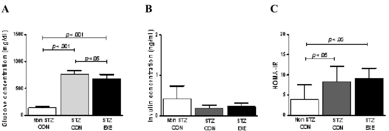
집단 간 혈청 글루코스, 인슐린 수준 및 HOMA-IR STZ를 투여한 SD 쥐에서 6주간의 트레드밀 지구성 운동 후 집단 간 혈청 글루코스, 인슐린 수준 및 HOMA-IR를 분석 한 결과, 혈청 글루코스 수준의 경우, 집단 간에 p<0.014로 유 의한 차이가 있는 것으로 나타나 사후 검증한 결과 NON STZ- CON 집단(100%)과 STZ-CON 집단 간에 p<0.001 수준에서
A B C
Fig. 1. Glucose concentration, Insulin concentration and HOMA-IR at the end of 6week of exercise training. Values are expressed as mean±SD of 8 animals/groups.
차이가 있는 것으로 나타났으며, NON STZ-CON 집단과 STZ- EXE 집단 간에 p<0.001수준에서 차이가 있는 것으로 나 타났다. 마지막으로 STZ-CON 집단과 STZ-EXE 집단 간에도 p<0.05 수준에서 차이가 있는 것으로 나타났다(Fig. 1A). 혈청 인슐린의 경우, 집단 간에 p>0.05 수준에서 통계적으로 차이가 없는 것으로 나타났다(Fig. 1B). 인슐린 저항성 지표로 알려진 HOMA- IR 경우, 집단 간에 p<0.05로 유의한 차이가 있는 것 으로 나타나 사후 검증한 결과 NON STZ-CON 집단과 STZ-CON 집단 간에 p<0.05 수준에서 차이가 있는 것으로 나 타났으며, NON STZ- CON 집단과 STZ-EXE 집단 간에 p<0.05 수준에서 차이가 있는 것으로 나타났다(Fig. 1C).
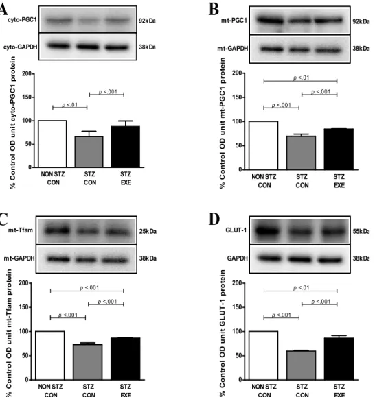
집단 간 PGC-1α, TGLUT-1, Tfam 단백질 발현 수준 차이 6주간 트레드밀 지구성 운동 후 뇌 미토콘드리아의 생성과 기능에 중요한 조절 단백질인 PGC-1α에 미치는 영향을 분석 한 결과, 세포질 내 PGC-1α 단백질의 경우 집단 간에 p<0.013 으로 유의한 차이가 있는 것으로 나타나 사후 검증한 결과 NON STZ-CON 집단과 STZ-CON 집단 간에 p<0.01 수준에서 차이가 있는 것으로 나타났으며, STZ-CON 집단과 STZ-EXE 집단 간에 p<0.001수준에서 차이가 있는 것으로 나타났다(Fig.
2A). 미토콘드리아 PGC-1α 단백질의 경우, 집단 간에 p<0.001 로 유의한 차이가 있는 것으로 나타나 사후 검증한 결과, NON STZ-CON 집단과 STZ-CON 집단 간에 p<0.001 수준에서 차 이가 있는 것으로 나타났으며, STZ-CON 집단과 STZ-EXE 집 단 간에도 p<0.001 수준에서 차이가 있는 것으로 나타났으며, NON STZ-CON 집단과 STZ-EXE 집단 간에도 p<0.001 수준 에서 차이가 있는 것으로 나타났다(Fig. 2B). 또한, 6주간 트레 드밀 지구성 운동 후 뇌 미토콘드리아 내 전사인자인 Tfam에 미치는 영향을 분석한 결과, 집단 간에 p<0.01로 유의한 차이 가 있는 것으로 나타나 사후 검증한 결과, NON STZ-CON 집단(100%)과 STZ-CON 집단 간에 p<0.001 수준에서 차이가
있는 것으로 나타났으며, STZ-CON 집단과 STZ-EXE 집단 간 에 p<0.001 수준에서 차이가 있는 것으로 나타났다. NON STZ-CON 집단과 STZ-EXE 집단 간에도 p<0.001 수준에서 차 이가 있는 것으로 나타났다(Fig. 2C). 6주간 트레드밀 지구성 운동 후 뇌에서 글루코스를 세포 내로 유입시키는 역할을 담 당하는 GLUT-1에 미치는 영향을 분석한 결과, 집단 간에 p<0.001 수준에서 차이가 있는 것으로 나타나 사후 검증한 결 과, NON STZ-CON 집단과 STZ-CON 집단 간에 p<0.001 수준 에서 차이가 있는 것으로 나타났으며 STZ-CON 집단과 STZ-EXE 집단 간에 p<0.001수준에서 차이가 있는 것으로 나 타났다. 마지막으로 NON STZ-CON 집단과 STZ-EXE 집단 간에도 p<0.001 수준에서 차이가 있는 것으로 나타났다(Fig.
2D).
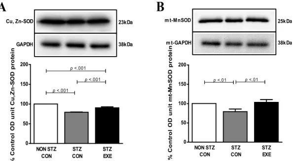
집단 간 Cu,Zn-SOD와 Mn-SOD 단백질 발현 수준 차이 STZ를 투여한 SD 쥐에서 6주간 트레드밀 지구성 운동 후 뇌에서 활성산소 제거에 관여하는 항산화제인 Cu, Zn-SOD가 미치는 영향을 뇌 조직에서 분석한 결과(Fig. 3A), NON STZ- CON 집단과 STZ-CON 집단 간에 p<0.001 수준에서 유의한 차이가 있는 것으로 나타났다. STZ-CON 집단과 STZ-EXE 집 단 간에 p<0.001 수준에서 차이가 있는 것으로 나타났다. STZ 를 투여한 SD 쥐에서 6주간 트레드밀 지구성 운동 후 뇌에서 활성산소제거에 관여하는 항산화제인 Mn-SOD가 미치는 영 향을 분리한 미토콘드리아에서 분석한 결과(Fig. 3B) NON STZ- CON 집단과 STZ-CON 집단 간에 p<0.01 수준에서 차이 가 있는 것으로 나타났으며 STZ-CON 집단과 STZ-EXE 집단 간에 p<0.01 수준에서 차이가 있는 것으로 나타났다.
고 찰
당뇨병은 심각한 대사성 질환으로서 고혈압, 뇌졸중과 같은
0 50 100 150 200
p <.001 cyto-PGC1
cyto-GAPDH
92kDa
38kDa
p <.01
NON STZ CON
STZ CON
STZ
% Control OD unit cyto-PGC1 protein EXE
0 50 100 150
200 p <.01
p <.001 m t-PGC1
m t-GAPDH
92kDa
38kDa
p <.001
NON STZ CON
STZ CON
STZ
% Control OD unit mt-PGC1 protein EXE
0 50 100 150
200 p <.001
p <.001 m t-Tfam
m t-GAPDH
25kDa
38kDa
p <.001
NON STZ CON
STZ CON
STZ
% Control OD unit mt-Tfam protein EXE
0 50 100 150
200 p <.01
p <.001 GLUT-1
GAPDH
55kDa
38kDa
p <.001
NON STZ CON
STZ CON
STZ
% Control OD unit GLUT-1 protein EXE
A B
C D
Fig. 2. Effect of exercise training on PGC-1α (A), mito PGC-1α (B), mito Tfam (C), GLUT-1 (D) protein in the brain of STZ-induced diabetic rats and untreated diabetic rats. The expression of PGC-1α, mito PGC-1α, mito Tfam and GLUT-1 in the brain was analyzed by western blot analysis. Values are expressed means±SD of 8 animals/groups.
합병증을 초래할 뿐만 아니라 뇌에서 글루코스 대사의 장애는 학습, 기억력과 인지능력의 감소와도 관련 있다[15,21]. 최근에 는 당뇨로 인한 신경퇴행성 뇌질환의 발병 기전 중 미토콘드 리아의 기능 이상이 중요하게 관여하는 것으로 보고되고 있다 [4,5,16], 특히 제 I 형 당뇨의 경우 고혈당과 함께 산화적 스트 레스 수준이 높게 나타남으로써 뇌 허혈, 미세혈관병증, 뇌 위축 현상이 유발된다고 Rosen 등[33], Nishikawa 등[26], West 등[42]이 제시하였다. Muranyi 등[25]은 미토콘드리아 기능 이상과 미토콘드리아 의존성 세포사멸 경로가 대부분 제 I형 당뇨병성 허혈에 의한 뇌 손상으로 작용되고 있음을 보고하였다.
또한, 당뇨가 미토콘드리아의 산화적 인산화에 관여하는 유 전자의 발현을 감소[24]시켜 미토콘드리아 생성의 변화[1]와
근육에서 PGC-1α의 발현이 감소[29]됨을 주장하였다. 따라서 이 연구는 STZ로 유발된 당뇨 흰쥐를 대상으로 6주간의 트레 드밀 지구성 운동이 미토콘드리아 생합성과 글루코스 대사 관련 단백질인 PGC-1α, GLUT-1, Tfam 및 항산화효소인 Cu, Zn-SOD와 Mn-SOD의 발현 양상을 분석하였다.
그 결과 혈중 인슐린 수준(Fig. 1B)은 STZ-CON 집단과 STZ-EXE 집단 간에 유의한 차이는 나타나진 않았지만, 혈중 글루코스 수준(Fig. 1A)은 STZ-CON 집단에 비해 STZ-EXE 집단에서 유의하게 감소한 것으로 나타났다. 이러한 결과는 인슐린 수준과는 관계없이 글루코스 수준이 유의하게 감소된 것이며, Luis 등[22]의 연구와 일치된 결과로써 인슐린 신호 전달이 아닌 지구성 운동으로 인한 또 다른 보상기전에 의해 나타난 결과로 생각된다. 또한 뇌에서의 글루코스 수송을 담
0 50 100 150
200 p <.001
p <.001 Cu, Zn-SOD
GAPDH
23kDa
38kDa
p <.001
NON STZ CON
STZ CON
STZ EXE
% Control OD unit Cu,Zn-SOD protein
0 50 100 150 200
p <.01 m t-M nSOD
m t-GAPDH
25k Da
38k Da
p <.01
NON STZ CON
STZ CON
STZ
% Control OD unit mt-MnSOD protein EXE
A B
Fig. 3. Effect of exercise training on Cu,Zn-SOD (A), mito Mn-SOD (B) protein in the brain of STZ-induced diabetic rats and untreated diabetic rats. The expression of Cu,Zn-SOD and mito Mn-SOD in the brain was analyzed by western blot analysis. Values are expressed means±SD of 8 animals/groups.
당하는 GLUT-1 단백질(Fig. 2D)의 경우, STZ-EXE 집단이 STZ-CON 집단에 비해 현저하게 증가한 것으로 나타났다. 이 러한 결과는 4주간의 지구성 운동이 당뇨 조건에서 글루코스 유입과 이용의 감소를 보상하기 위해 GLUT-1 단백질 발현을 증가시켰다는 결과[17]와 일치하는 것으로 운동이 당뇨 조건 에서 뇌의 글루코스 이용률을 촉진시켜 신경세포의 대사적 요구량을 효율적으로 보상해주어서 나타난 현상으로 판단되 며11] 뇌에서 신경세포의 활성을 향상시킬 수 있음을 알 수 있다.
한편 PGC-1α는 심장, 신장, 뇌, 골격근, 갈색지방 등 여러 조직의 핵 DNA에 의해 합성되어 미토콘드리아 생성과 활성 을 조절하는 기능을 가지고 있으며[30], 노화와 당뇨 조건하에 서 PGC-1α의 발현이 감소되는 것[24,29]처럼 이 연구에서도 PGC-1α를 세포질과 미토콘드리아를 분리하여 실험한 결과 (Fig. 2A, 2B), STZ-CON 집단에서 가장 낮게 나타났지만 STZ-EXE 집단은 STZ-CON 집단에 비해 유의하게 증가한 것 으로 나타났다. 이는 장기적인 운동 적응에서 PGC-1α의 발현 수준이 증가되었다는 여러 선행연구[4,27,39]들의 결과들과 일 치하는 것으로 나타났다.
운동은 세포 내 cAMP와 칼슘(calcium) 수준을 증가시킬 뿐만 아니라 신호전달 물질인 AMPK의 활성을 일으키고, 결 국 PGC-1α의 발현을 유도한다[15]고 알려져 있다. 따라서 운 동으로 증가된 PGC1-α의 발현은 세포 내 미토콘드리아의 생 성과 활성에 관여하는 단백질들의 발현을 증가시킬 뿐만 아니 라 Tfam의 발현을 돕는 인자들을 전사시키고[44], 이들 인자 들은 미토콘드리아 전자전달복합체의 구성 성분인 β-ATP
synthase, cytochrome c, cytochrome c oxidase Ⅳ 등의 발현 을 증가시킨다[35]. 더욱이 Tfam이 mitochondria 속으로 이동 (translocation)하게 되면 mitochondria의 DNA 복제와 유전 자 발현증가를 촉진시킨다[14,19]. 더욱이 이 연구는 STZ으로 유도된 당뇨 쥐의 뇌 미토콘드리아 PGC-1α와 Tfam의 발현은 억제되었지만, 트레드밀 지구성 운동이 PGC-1α와 Tfam 단백 질 발현을 증가시킴으로 뇌 조직 내 mtDNA 수를 증가시킬 수 있는 가능성을 보여준 것으로 간주된다.
일반적으로 STZ 투여는 췌장의 β세포에 치명적인 손상을 주기 때문에 당뇨가 유발된다고 알려져 있다. 즉 β세포에서 H2O2을 생성을 자극시켜 반응성 산소종 생성을 높이기 때문 에 산화적 손상의 결과인 당뇨가 유발[38]된 것으로 설명할 수 있으며, 당뇨로 인한 산화적 손상에 대한 방어기전으로 여 러 연구자들은 항산화 효소의 발현 증가에 초점을 두고 있다. Superoxide dismutase (SOD)는 세포 내 호흡작용의 부산물로 나타나는 superoxidie radical을 제거함으로써 과산화수소가 세포 내에 잔존되는 것을 막아주기 때문에 DNA 손상으로부 터 보호할 수 있다[20].
이 연구에서도 지구성 운동이 당뇨 조건에서 뇌 조직의 항 산화 방어기전을 알아보기 위해 Cu,Zn-SOD와 미토콘드리아 Mn-SOD를 분석하였다. 그 결과 STZ-EXE 집단은 STZ-CON 집단에 비해 Cu,Zn-SOD와 미토콘드리아 Mn-SOD 단백질 발 현량이 현저하게 증가한 것으로 나타났다. 이와 같이 당뇨 조 건에서 지구성 운동에 의해 항산화 효소의 활성도가 증가한 이유는 장기간 지구성 운동이 세포 내 발전소인 미토콘드리아 내 호흡비율을 높여 ATP 대사 및 산소 소비량이 증가시키고
이를 통해 체내 항산화 효소(Cu,Zn-SOD, Mn-SOD, catalase) 들을 활성화시켜 체내의 산소 독성을 물과 산소로 변환시키는 과정을 촉진시켰기 때문인 것으로 볼 수 있다. 즉 지구성 운동 이 세포 내 항상성 유지[36]를 위해 항산화 방어기전을 촉진시 킨 것으로 볼 수 있다[10]. 따라서 지구성 운동은 산화적 스트 레스가 높은 당뇨 조건에서 뇌의 항산화 효소(Cu,Zn-SOD, Mn-SOD)의 발현량을 증가시켜 당뇨로 인한 mtDNA 손상을 어느 정도 억제하는데 도움을 주는 것으로 판단된다.
결론적으로 트레드밀 지구성 운동은 당뇨 조건에서 뇌 조직 의 PGC-1α, GLUT-1, Tfam 단백질의 발현량과 항산화 효소 (Cu,Zn-SOD, Mn-SOD)의 활성도를 증가시켜 뇌 조직의 글루 코스 이용능력과 미토콘드리아의 생합성에 어느 정도 긍정적 인 영향을 미치고 당뇨병으로 인한 산화적 손상으로부터 뇌 조직을 보호하는데 긍정적인 효과를 가져올 것으로 생각된다.
감사의 글
이 연구는 2007년도 한국체육대학교 자체학술연구과제의 부분적인 지원에 의해 수행되었음.
References
1. Anabela, P. R. and M. P. Carlos. 2006. Diabetes and mi- tochondrial function: Role of hyperglycemia and oxidative stress. Toxicol. Appl. Pharmacol.212, 167-178.
2. Atonetti, D. A., C. Reynet, and C. R. Kahn. 1995. Increased expression of mitochondrial-encoded genes in skeletal mus- cle of humans with diabetes millitus. J. Clin. Invest. 95, 1383-1388.
3. Baar, K., A. R. Wende, T. E. Jones, M. Marison, L. A. Nolte, M. Chen, D. P. Kelly, and J. O. Holloszy. 2002. Adaptations of skeletal muscle to exercise: rapid increase in the transcrip- tional coactivator PGC-1. FASEB. J. 16, 1879-1886.
4. Beauquis, J., P. Roig, F. Homo-Delarche, A. De Nicola, and F. Saravia. 2006. Reduced hippocampal neurogenesis and number of hilar neurones in streptozotocin-induced diabetic mice: reversion by antidepressant treatment.Eur. J. Neurosci.
23, 1539-1546.
5. Bossy-Wetzel, E., M. J. Barsoum, A. Godzik, R.
Schwarzenbacher, and S. A. Lipton. 2003. Mitochondrial fis- sion in apoptosis, neurodegeneration and aging.Curr. Opin.
Cell Biol. 15, 706-716.
6. Bradford, M. M. 1976. A rapid and sensitive method for the quantitation of microgram quantities of protein utilizing the principle of protein-dye binding.Analytical Biochem.72, 248-254.
7. Ceriello, A. 2000. Oxidative stress and glycemic regulation.
Metabolism 49, 27-29.
8. Cotman, C. W. and C. Engesser-Cesar. 2002. Exercise enhan- ces and protects brain function.Exerc. Sport Sci. Rev. 30, 75-79.
9. Daniel, P. K. and C. S. Richard. 2004. Transcriptional regu- latory circuits controlling mitochondrial biogenesis and function. Genes Dev. 18, 357-368.
10. De Moraes, C., A. P. Davel, L. V. Rossoni, E. Antunes, and A. Zanesco. 2008. Exercise training improves relaxation re- sponse and SOD-1 expression in aortic and mesenteric rings from high caloric diet-fed rats. BMC Physiol.8, 12.
11. Duelli, R. and W. Kuschinsky. 2001. Brain glucose trans- porters: relationship to local energy demand.News. Physiol.
Sci. 16, 71-76.
12. Endo, N., C. Emilio, M. Salvador, and O. C. Michele. 2004.
Mitochondrial biogenesis as a cellular signaling framework.
Biochem Pharmacol. 67, 1-15.
13. Garesse, R. and C. G. Vallejo. 2001. Animal mitochondrial biogenesis and function: a regulatory cross-talk between two genomes. Gene 263, 1-16.
14. Hardie, D. G. 2004. AMP-activated protein kinase: a key sys- tem mediating metabolic responses to exercise. Med. Sci.
Sports Exerc. 36, 28-34.
15. Hou, W. K., Y. X. Xian, L. Zhang, H. Lai, X. G. Hou, Y.
X. Xu, T. Yu, F. Y. Xu, J. Song, C. L. Fu, W. W. Zhang, and L. Chen. 2007. Influence of blood glucose on the ex- pression of glucose trans-porter proteins 1 and 3 in the brain of diabetic rats. Chin. Med. J. 120, 1704-1709.
16. Jacobs, H. T. 2003. The mitochondrial theory of aging: dead or alive? Aging Cell 2, 9-10.
17. Jeong, L. G., J. H. Yoon, H. H. Lee, J. O. Kim, T. B. Sel, and M. J. Oh. 2007. Effect of exercise training on expression of GLUT 1 and GLUT 3 protein in the hippocampus of streptozotocin-induced diabetic rats.J. Korean Physical Edu. 46, 359-367.
18. Kawamura, M., J. W. Heinecke, and A. Chait. 1994.
Pathophysiological concentrations of glucose promote oxi- dative modification of low density lipoprotein by a super- oxide dependent pathway. J. Clin. Invest. 94, 771-778.
19. Larsson, N. G., J. Wang, H. Wilhelmsson, A. Oldfors, P.
Rustin, M. Lewandoski, G. S. Barsh, and V. Clayton. 1998.
Mitochondrial transcription factor A is necessary for mtDNA maintenance and embryogenesis in mice. Nat.
Genet. 18, 231-236.
20. Lee, S. Z., S. H. Park, and H. S. Lee. 2001. Chainges in vivo lipid peroxidation and antioxidant defense system in strep- tozotocin induced diabetic rats: a time course study. J.
Korean Nutr. Soc. 34, 253-264.
21. Li, Z. G. and A. A. Sima. 2004. C-peptide and central nervous system complications in diabetes.Exp. Diabesity Res. 5, 79-90.
22. Luis, D. M., B. Lamvert, N. Sash, R. R. Ghazala, P. Norman, and A. F. Paul. 2001. Effect of streptozotocin-induced dia- betes on glycogen resynthesis in fasted rats post-high-in- tensity exercise.Am. J. Physiol. Endocrinol. Metab.280, E83-91.
23. Monsalve, M., Z. Wu, G. Adelmant, P. Puigserver, M. Fan, and B. M. Spiegelman. 2000. Direct coupling of transcription and mRNA processing through the thermogenic coactivator PGC-1. Mol. Cell 6, 307-316.
24. Mootha, V. K., C. M. Lindgren, K. F. Eriksson, A.
Subramanian, S. Sihag, J. Lehar, P. Pulgserver, E. Carlsson,
M. Ridderstrale, E. Laurlla, N. Houstls, M. J. Daly, N.
Patterson, J. P. Mesirov, T. R. Golub, P. Tamayo, B.
Spiegelman, E. S. Lander, J. N. Hirschhorn, D. Altshuler, and L. C. Grouup. 2003. PGC-1, Lpha-responsive genes in- volved in oxidative phosphorylation are coordinately down- regulated in human diabetes. Nat. Genet.34, 267-273.
25. Muranyi, M., M. Fujioka, Q. He, A. Han, G. Yong, K. Csiszar, and P. A. Li. 2003. Diabetes activates cell death pathway after transient focal cerebral ischemia.Diabetes52, 481-486.
26. Nishikawa, T., D. Edelstein, and M. Brownlee. 2000. The missing link: a single unifying mechanism for diabetic complications. Kidney Int. Suppl. 77, S26-30.
27. Norrbom, J., C. J. Sundberg, H. Ameln, W. E. Kraus, E.
Jansson, and T. Gustafsson. 2004. PGC-1alpha mRNA ex- pression is influenced by metabolic perturbation in exercis- ing human skeletal muscle. J. Appl. Physiol.96, 189-194.
28. Park, S., J. S. Jang, D. W. Jun, and S. M. Hong. 2005. Exercise enhances insulin and leptin signaling in the cerebral cortex and hypothalamus during dexamethasone-induced stress in diabetic rats. Neuroendocrinol. 82, 282-293.
29. Patti, M. E., A. J. Butte, S. Crunkhorn, K. Cusi, R. Berria, S. Kashyap, Y. Miyazaki, I. Kohane, M. Costello, R. Saccone, E. J. Landarker, A. B. Goldfine, E. Mun, R. DeFronzo, J.
Finlayson, C. R. Kahn, and L. J. Mandarino. 2003.
Coordinated reduction of genes of oxidative metabolism in human with insulin resistance and diabetes: potential role of PGC1 and NRF1. Proc. Natl. Acad. Sci. USA 100, 8466-8471.
30. Puigserver, P., Z. Wu, C. W. Park, R. Graves, M. Wright, and B. Spiegelman. 1998. A cold-inducible coactivator of nu- clear receptors linked to adaptive thermogenesis. Cell92, 829-839.
31. Puigserver, P. and B. M. Spiegelman. 2003. Peroxisome pro- liferator-activated receptor-gamma coactivator 1 alpha (PGC-1 alpha): transcriptional coactivator and metabolic regulator. Endocr. Rev. 24, 78-90.
32. Reagan, L. P., N. Gorovits, E. K. Hoskin, S. E. Alves, E.
B. Katz, C. A. Grillo, G. G. Piroli, B. S. McEwen, and M.
J. Charron. 2001. Localization and regulation of GLUT×1 glucose transporter in the hippocampus of streptozotocin diabetic rats. Proc. Natl. Acad. Sci. USA 98, 2820-2825.
33. Rösen, P., P. P. Nawroth, G. King, W. Möller, H. J. Tritschler, and L. Packer. 2001. The role of oxidative stress in the onset and progression of diabetes and its complications: a sum- mary of a Congress Series sponsored by UNESCO-MCBN,
the American Diabetes Association and the German Diabetes Society. Diabetes Metab. Res. Rev.17, 189-212.
34. Sander, M. H. and A. Johan.2004. PGC-1α: Turbocharging Mitochondria. Cell 119, 5-7.
35. Scarpulla, R. C. 2002. Transcriptional activators and co- activators in the nuclear control of mitochondrial function in mammalian cells. Gene286, 81-89.
36. Sen, C. K. 1995. Oxidants and antioxidants in exercise.J.
Appl. Physiol. 79, 675-686.
37. Serradas, P., M. H. Giroix, C. Saulnier, M. N. Gangnerau, L. A. Borg, M. Welsh, B. Portha, and N. Welsh. 1995.
Mitochondrial deoxyribonucleic acid content is specifically decreased in adult, but not fetal, pancreatic islets of the Goto-Kakizaki rat, a genetic model of noninsulin-dependent diabetes. Endocrinol. 136, 5623-5631.
38. Takasu, N., I. Komiya, T. Asasa, Y. Nagasawa, and T, Yamada. 1991. Streptozotocin- and alloxan-induced H2O2
generation and DNA fragmentation in pancreatic islets.
H2O2 as mediator for DNA fragmentation. Diabetes 40, 1141-1145.
39. Terada, S. and I. Tabata. 2004. Effects of acute bouts of run- ning and swimming exercise on PGC-1alpha protein ex- pression in rat epitrochlearis and soleus muscle. Am. J.
Physiol. Endocrinol. Metab. 286, E208-216.
40. Tsai, E. C., I. B. Hirsch, J. D. Brunzell, and A. Chait.
1994. Reduced plasma peroxyl radical trapping capacity and increased susceptibility of LDL to oxidation in poorly con- trolled IDDM. Diabetes 43, 1010-1014.
41. Yang, W., J. Li, and S. Hekimi. 2007. A Measurable increase in oxidative damage due to reduction in superoxide detox- ification fails to shorten the life span of long-lived mitochon- drial mutants of Caenorhabditis elegans. Genetics 177, 2063-2074.
42. West, I. C. Radicals and oxidative stress in diabetes.Diabet.
Med. 17, 171-180.
43. Winder, W. W., E. B. Taylor, and D. M. Thomson. 2006.
Role of AMP-activated protein kinase in the molecular adaptation to endurance exercise.Med. Sci. Sports Exerc.38, 1945-1949.
44. Wu, Z., P. Puigserve, U. Andersson, C. Zhang, G. Adelmant, V. Mootha, A. Troy, S. Cinti, B. Lowell, R. C. Scarpulla, and B. M. Spiegelman. 1999. Mechanisms controlling mitochon- drial biogenesis and respiration through the thermogenic coactivator PGC-1. Cell98, 115-124.
초록:트레드밀 지구성 운동이 streptozotocin으로 유발된 당뇨 흰쥐의 뇌에서 PGC-1α, GLUT-1, Tfam 단백질 및 항산화 효소(Cu, Zn-SOD, Mn-SOD)의 발현량에 미치는 영향
박노환1․이진2․정국현1․최봉암3․장형채1․이석인4․이동수4․조준용1*
(1한국체육대학교 운동생화학실,2한양대학교 해부세포생물학실,3대구대학교 골프학과,4중앙대학교 체육교 육학과)
이 연구는 지구성 운동이 streptozotocin (STZ)으로 유발된 제 1형 당뇨 특징을 가진 쥐 뇌의 글루코스 운반, 미토콘드리아 기능 및 항산화효소 단백질 발현에 미치는 영향을 규명하는데 목적이 있다. 제 1형 당뇨 모델 쥐는 50 mg/kg의 streptozotocin을 수컷 Sprague-Dawley (SD) 흰쥐의 복강에 1회 주입하여 생산하였으며 본 실험 시 집단은 NON-STZ 집단(n=8), STZ-CON 집단(n=8) 및 STZ-EXE 집단(n=8) 등 3집단으로 구분하여 실시하였다.
트레드밀 지구성 운동은 총 6주, 주 5일, 2주 간격으로 속도를 약 3∼4 m/min으로 점증적으로 증가시켰으며 운 동시간은 1주와 3주차에 10분씩 증가시켰다. 분석 결과 혈청 글루코스 수준은 STZ-EXE 집단은 STZ-CON 집단 에 비해 현저하게 감소(p<0.05)하였으며 PGC-1α (p<0.001), mtPGC-1α (p<0.001), GLUT-1 (p<0.001), Tfam (p<0.001), Cu,Zn- SOD (p<0.001), Mn-SOD (p<0.01) 경우도 STZ-EXE 집단이 STZ-CON 집단에 비해 현저하게 증가하였다. 이러한 결과는 장기간 지구성 운동이 뇌의 글루코스 이용능력과 관련된 단백질인 GLUT-1과 미토콘 드리아 기능 향상과 관련된 단백질인 PGC-1α과 Tfam을 증가시키고 산화적 스트레스의 방어 기전으로서 역할을 수행하는 항산화 효소인 Cu,Zn-SOD와 Mn-SOD를 활성화시키는데 긍정적인 역할을 수행한 것으로 나타났다.