Expression of Tbr2 in the Hippocampus Following Pilocarpine-induced Status Epilepticus
Yun-Sik Choi*
Department of Pharmaceutical Science and Technology, Catholic University of Daegu, Gyeongsan, Gyeongsangbuk-do, 712-702, Korea
Received November 7, 2013 /Revised December 4, 2013 /Accepted December 11, 2013T-box transcription factor 2 (Tbr2) is a member of the T-box family of transcription factors and it plays an important role in brain development, progenitor cell proliferation, and the modulation of differ- entiation and function in immune cells, such as CD8+ T cells and natural killer cells. This study aims to elucidate the involvement of Tbr2 in the pathophysiological events following pilocarpine-induced status epilepticus in mice. Status epilepticus resulted in prominent neuronal cell death in discrete brain regions, such as CA3, the hilus, and the piriform cortex. Interestingly, when the immunor- eactivity of Tbr2 was examined two days after status epilepticus, it was transiently increased in CA3 and in the piriform cortex. Tbr2-positive cells in CA3 and the piriform cortex were double-labeled with CD11b, a marker of microglia and a subset of white blood cells, such as monocytes, CD8+ T cells, and natural killer cells. Moreover, the double-labeled cells with Tbr2 and CD11b showed amoe- boid morphology, and this data indicates that Tbr2-expressing cells may be reactive microglia or in- filtrating white blood cells. Furthermore, clustered Tbr2-positive cells were observed in the platelet en- dothelial cell adhesion molecule-1 (PECAM-1)-positive blood vessels near the CA3 area, which sug- gests that Tbr2-positive cells may be infiltrating the white blood cells. Based on this data, this study is the first to indicate the involvement of Tbr2 in neuropathophysiology in status epilepticus.
Key words : CD11b, hippocampus, pilocarpine, status epilepticus, T-box transcription factor 2 (Tbr2)
*Corresponding author
*Tel : +82-53-850-2561, Fax : +82-53-359-6820
*E-mail : [email protected]
This is an Open-Access article distributed under the terms of the Creative Commons Attribution Non-Commercial License (http://creativecommons.org/licenses/by-nc/3.0) which permits unrestricted non-commercial use, distribution, and reproduction in any medium, provided the original work is properly cited.
Journal of Life Science 2013 Vol. 23. No. 12. 1532~1540 DOI : http://dx.doi.org/10.5352/JLS.2013.23.12.1532
Introduction
The T-box family of transcription factor genes play an im- portant role in early embryonic cell fate decisions, develop- ment of extraembryonic structures, embryonic patterning, and organogenesis [21]. Among these genes, T-box tran- scription factor 2 (Tbr2), also known as eomesodermin (Eomes), comprises the Tbr2 subfamily with Tbr1 and Tbx21 (T-bet), and regulates the differentiation of excitatory projec- tion neurons and retinal ganglion cells [11, 17]. In addition, Tbr2 is required for the maintenance of excitatory-inhibitory balance in the olfactory bulb circuitry [20, 26] and condi- tional ablation of Tbr2 during development results in the loss of intermediate progenitor cells, decrease in cortical sur- face expansion, and thickness with neuronal reduction [27].
Furthermore, in adult mouse hippocampus, Tbr2 protein is specifically expressed by intermediate-stage progenitor cells,
a type of transit amplifying cells, which respond to neuro- genic stimuli, such as voluntary wheel running [12].
Tbr2 is also expressed in a subset of white blood cells, such as CD8+ T cells and natural killer cells. In CD8+ T cells, Tbr2 with T-bet induces the expression of granzyme B, perforin, and interferon-γ (IFN-γ), and both transcription factors are thought to be involved in differentiation of effec- tor and memory T cells [9, 13, 16, 22]. Interestingly, the amount of memory CD8+ T cells decreased when Tbr2 gene was deleted whereas it increased when T-bet was absent [3].
Additionally, Tbr2 expression was promoted by inter- leukin-2 (IL-2) and Tbr2 expression with T-bet facilitated tu- mor infiltration by CD8+ T cells [24, 29]. In case of mature natural killer cells, the deletion of Tbr2 causeed reversion to a more immature state and deficiency in both T-bet and Tbr2 resulted in the loss of classical natural killer cell anti- gens [10]. All these observations indicate that Tbr2 is a crit- ical transcription factor modulating not only early brain de- velopment and hippocampal neurogenesis but also the dif- ferentiation and function of white blood cells.
Although the role of Tbr2 in the peripheral tissues and
during brain development has been well characterized, its
role in the central nervous system under pathological con-
ditions, such as status epilepticus (SE) or stroke is not
known. SE is a devastating disease, resulting in selective neuronal cell death, microglial reaction, and recurrent seiz- ure [6, 7, 14]. Moreover, blood-brain barrier (BBB) leakage has been reported following pilocarpine-induced SE, which may provide an environment for peripheral leukocyte infiltration. On the other hand, when examined 2 days after SE, pilocarpine-induced SE increased progenitor cell pro- liferation in the subgranular zone through insulin-like growth factor-1 (IGF-1)-ERK-MSK signaling pathways [4, 5].
Considering the distinct roles of Tbr2 in proliferation and differentiation of progenitor cells as well as in the infiltration of CD8+ T cells and natural killer cells, it may be interesting to investigate the intervention of Tbr2 in progenitor cell pro- liferation and/or immune reaction following SE. Thus, this study was designed to elucidate the involvement of Tbr2 in the pathophysiological events following pilocarpine-in- duced SE in mice.
Materials and Methods Animals
Male C57BL/6 mice (8 weeks old, Harlan, Indianapolis, IN) were housed at standard temperature (22±1°C) and hu- midity (50±1%) levels, and in a light-controlled environment (lights on from 8:00 a.m. to 8:00 p.m.) with ad libitum access to food and water. The animal experiments were conducted in accordance with the National Institutes of Health Guide for the Care and Use of Laboratory Animals (NIH Publication No. 80-23, revised 1996). All efforts were made to minimize animal suffering and to use the minimum num- ber of animals possible.
Pilocarpine-induced status epilepticus and BrdU injection
The mice were initially given an intraperitoneal injection (i.p.) of atropine methyl nitrate (1 mg/kg in saline, Sigma, St. Louis, MO) and 30 min later, SE was elicited by an intra- peritoneal injection (i.p.) of pilocarpine (325 mg/kg in saline, Sigma). SE was defined as a continuous motor seizure per- sisting for over 3 hr. The control animals were initially in- jected with atropine methyl nitrate (1 mg/kg) and sub- sequently with saline, instead of pilocarpine. To label the proliferating cells, 5-bromo-2´-deoxyuridine (100 mg/kg in saline, Sigma) was injected (i.p.) 2 hr before sacrifice.
Tissue processing
The mice were anesthetized with 15% chloral hydrate and
transcardially perfused with saline, followed by 4% paraf- ormaldehyde in 0.1 M phosphate buffer (PB), pH 7.4. The brains of the mice were postfixed for 4 hr and subsequently cryoprotected in 30% sucrose in 0.1 M PB. A cryotome was used to prepare sequential coronal sections (30 μm thick) through the hippocampus.
Immunofluorescence
For immunofluorescence labeling, sections were blocked with 10% normal goat serum, followed by overnight in- cubation with rabbit polyclonal anti-Tbr2 antibody (1:500;
Abcam, Cambridge, MA). Next, sections were incubated (2 hr at room temperature) with secondary antibodies con- jugated with Alexa 488 and Alexa 594 (1:1,000; Invitrogen, San Diego, CA) and subsequently mounted with Cytoseal (Richard-Allan Scientific, Kalamazoo, MI). For double label- ing with BrdU, sections were incubated in 2XSSC/50% for- mamide for 2 hr at 65°C, followed by incubation in 2 N HCl at 37°C for 1 hr. After washing with 0.05 M borate buf- fer (pH 8.5) for 10 min and then washing with PBS, sections were blocked with 10% normal goat serum, followed by overnight incubation with rabbit polyclonal anti-Tbr2 anti- body and rat monoclonal anti-BrdU antibody (1:400;
Accurate Chemical, Westbury, NY). Sections were incubated (2 hr at room temperature) with secondary antibodies con- jugated with Alexa 488 and Alexa 594 (1:1,000). For the visu- alization of the nucleus, 80 μM Hoechst 33342 (Fluka, Switzerland) was added for 20 min. Fluorescence images were captured using a Zeiss (Oberkochen, Germany) 510 Meta confocal microscope (2-μm-thick optical section).
Cresyl violet staining
In brief, sections were mounted on gelatin-coated slides and dried. After dehydrating in a graded alcohol series, sec- tions were stained (20 min) with 0.1% cresyl violet solution.
After destaining with a solution of 95% ethanol and 0.1%
glacial acetic acid, sections were dehydrated and mounted with Permount.
Results
Selective cell death following pilocarpine-induced status epilepticus
The selective neuronal damage in the hippocampus fol- lowing pilocarpine-induced SE has been well studied.
Previous study shows that neuronal damage can be ob-
served as early as 6 hr post SE in the hilus and later on
A
B
Hilus CA3
Hilus CA3
Fig. 1. Status epilepticus by pilocarpine injection induces neuronal cell death in the hippocampus. (A) Cresyl violet histology was performed to identify cell viability. In saline-injected control animals, cresyl violet-stained neurons were observed throughout the hippocampus, including the hilus and CA3 regions. (B) Marked cell damage was observed in the CA3 and hilus regions of the hippocampus when examined at 3 days post SE. Boxed areas in the hilus and CA3 are magnified on the right panel.
Hil: hilus. Scale bar=200 μm (low magnification image), 100 μm (high magnification image).
in CA3 and CA1 [6]. In accordance with these findings, com- pared to saline-injected control prominent neuronal damage was observed in the hippocampal CA3 and hilus examined at 3 days post SE (Fig. 1). In this study, cresyl violet, which stains viable cells, was used.
Increased expression of Tbr2 in the CA3 area following pilocarpine-induced status epilepticus
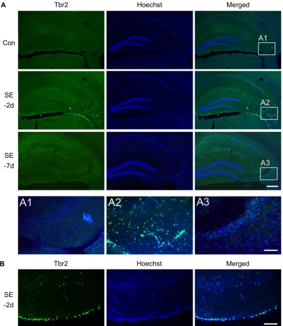
Tbr2 expression was examined by immunofluorescence with anti-Tbr2 antibody. As shown in Fig. 2A, under normal conditions, the Tbr2 expression level was minimal in the hippocampus. However, at 2 days after SE, a high density of Tbr2-expressing cells was observed in the CA3 region where prominent neuronal cell death was also observed, and Tbr2 signaling was almost disappeared at 7 days after SE.
Pilocarpine-induced SE also caused neuronal loss in the piri- form cortex and an emergence of Tbr2-expressing cells at 2 days post SE (Fig. 2B). These spatial and temporal ex- pression profiles may indicate that Tbr2-expressing cells ap- pear in regions that are vulnerable to pilocarpine-induced SE.
Induction of Tbr2 in CD11b-immunoreactive cells
As mentioned earlier, Tbr2 is a marker of intermediate-
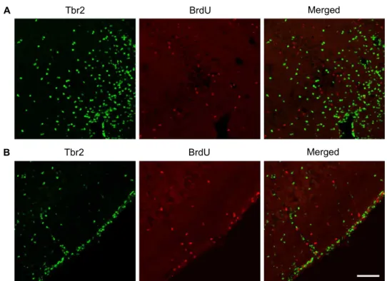
stage progenitor cells. Thus, BrdU, a thymidine analogue, was administered to mice to label actively proliferating pro- genitor cells and the animals were sacrificed 2 hr later.
However, as shown in Fig. 3, there were no double-labeled cells with Tbr2 and BrdU in both CA3 and piriform cortex.
This indicates that Tbr2-positive cells in CA3 and piriform cortex may not be intermediate-stage progenitor cells.
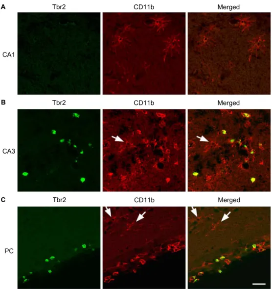
Next, to elucidate the cells that express Tbr2, Tbr2 was double labeled with CD11b, a marker of microglia, mono- cytes, natural killer cells, and a subset of CD8+ T cells [25].
Surprisingly, as shown in Fig. 4, CD11b-positive cells in CA1
area were not double labeled with Tbr2. On the other hand,
a large population of CD11b-positive cells in CA3 and piri-
form cortex was double labeled with Tbr2. Moreover, the
morphology of Tbr2-positive cells was different from that
of the Tbr2-negative cells. In CA1 area, CD11b-positive cells
have a ramified shape, which is a typical morphological
characteristic of inactive microglia. However, double-labeled
cells with Tbr2 and CD11b in CA3 and piriform cortex have
an amoeboid or round shape, which can be observed in acti-
vated microglia and migrating immune cells, such as mono-
cyte, CD8+ T cells, and natural killer cells [15, 28]. Further,
even in CA3 and piriform cortex, CD11b-positive cells with
ramified morphology do not express Tbr2 (arrows). This in-
B
Tbr2 Hoechst Merged
Tbr2 Hoechst Merged
Con
SE -2d
SE -7d
SE -2d A
Fig. 2. Tbr2 expression in the CA3 and piriform cortex at 2 days post SE. (A) Representative images of Tbr2 (green) in the hippocampus in saline-treated control (Con) animals at 2 days or 7 days post SE. Hoechst staining was used to visualize hippocampal cells. The image shows minimal immunoreactivity in saline-treated control animals. However, marked Tbr2-expressing cells were observed in CA3, and Tbr2-expressing cells almost disappeared at 7 days post SE. The boxed areas in each image are magnified below. Scale bar=250 μm (low magnification image), 50 μm (in A3). (B) Tbr2 expression at 2 days post SE in the piriform cortex. Tbr2 immunoreactive cells were also observed in the piriform cortex at 2 days post SE. In this region, Tbr2 immunoreactive cells were not observed in saline-treated control animals at 7 days post SE. Scale bar=50 μm.
formation indicates that Tbr2 is expressed in CD11b-positive amoeboid cells in the hippocampal CA3 and piriform cortex, at 2 days post SE.
Induction of Tbr2 in infiltrating white blood cells
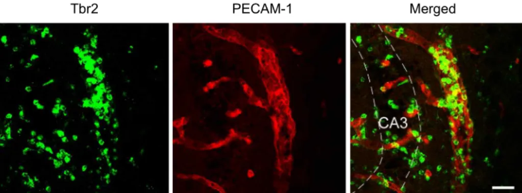
Considering the distribution and morphology of Tbr2-positive cells, it is conceivable that Tbr2-positive cells are infiltrating white blood cells. To confirm this, Tbr2 was double labeled with PECAM-1. PECAM-1 is constitutively
expressed on vascular cells; therefore, it is used as a marker
of blood vessels. As shown in Fig. 5, clustered Tbr2-positive
cells were observed inside the blood vessels in the CA3 area,
with scattered distribution pattern in the parenchyma at 2
days post SE. Taken together, these data strongly suggest
that Tbr2-positive cells in hippocampal CA3 region follow-
ing SE are infiltrating CD11b-positive white blood cells.
A
B
Tbr2 BrdU Merged
Tbr2 BrdU Merged
Fig. 3. Double labeling with Tbr2 and BrdU. Double immunofluorescence labeling with Tbr2 (green) and BrdU (red), a marker of actively proliferating cells, was performed at 2 days post SE. (A) In the CA3 region, there were no double-labeled cells with Tbr2 and BrdU. (B) In the piriform cortex, double-labeled cells with Tbr2 and BrdU were not observed. This data suggests that Tbr2-expressing cells in the CA3 and piriform cortex are not actively proliferating cells. For this study, BrdU was administered to mice at 2 days post SE and the mice were sacrificed 2 hr later. Scale bar=50 μm.
Discussion
In this study, the initial and unexpected finding was that Tbr2 is transiently expressed in the hippocampal CA3 region at 2 days after pilocarpine-induced SE. Double immunolabel- ing with several kinds of markers revealed that Tbr2-ex- pressing cells are CD11b positive. In addition, these dou- ble-labeled cells have amoeboid morphology, indicating that they are reactive microglia or infiltrating white blood cells, such as monocytes, CD8+ T cells, or natural killer cells. On the contrary, in the CA1 area, CD11b-positive cells, which were not colabeled with Tbr2, have a ramified shape, indicat- ing resting microglia. Further, clustered Tbr2-positive cells were found in the blood vessels near the CA3 area. This information reveals that Tbr2 is transiently expressed in in- filtrating white blood cells in vulnerable brain regions fol- lowing SE.
Tbr2 shows highly restricted spatiotemporal expression patterns during development. For example, Tbr2 is ex- pressed in multipotent trophoblast stem cells (TSCs) from early postimplantation stages, and Tbr2-positive TSCs con- stitute the cellular source for the embryonic part of the pla-
centa [23]. In developing neocortex, Tbr2 is upregulated dur- ing transition from radial glia to intermediate progenitor cells, which provide neurons of almost all cortical layers [8].
Recently, the function of Tbr2 during early brain develop- ment was well characterized by conditional inactivation [1].
Briefly, loss of Tbr2 leads to a reduced number of proliferat- ing cells in the subventricular zone concomitant with mark- edly reduced brain size, in cortex and olfactory bulbs, but without any influence on apoptosis.
In this study, the major and unexpected finding was that SE induces Tbr2 expression prominently in CA3 and piri- form cortex. Previously, it was reported that acute BrdU in- corporation (30 min before cervical dislocation) results in
~20% of double-labeled cells with BrdU and Tbr2 among
BrdU-labeled cells [8]. Therefore, although hippocampal
CA3 and piriform cortex are not known as neurogenic re-
gions in the adult brain, we employed BrdU to label actively
proliferating cells, thereby clarified the possibility of Tbr2
expression in intermediate progenitor cells. However, in our
study, although the subset of Tbr2-positive cells was co-la-
beled with BrdU in the subgranular zone (data not shown),
there were no double-labeled cells in CA3 and piriform cor-
A
B
C
Tbr2 CD11b Merged
Tbr2 CD11b Merged
Tbr2 CD11b Merged
CA1
CA3
PC
Fig. 4. Double labeling with Tbr2 and CD11b. Double immunofluorescence labeling with Tbr2 (green) and CD11b (red), a marker of microglia and a subset of white blood cells, such as monocytes, CD8+ T cells, or natural killer cells, was performed at 2 days post SE. (A) In the hippocampal CA1 area, CD11b-positive cells were not colabeled with Tbr2. (B) In the CA3 area CD11b-positive cells were co-labeled with Tbr2. (C) In the piriform cortex, CD11b-positive cells were colabeled with Tbr2. Moreover, double-labeled cells in the CA3 and piriform cortex have amoeboid or round morphology, suggesting reactive microglia or infiltrating white blood cells, such as monocytes, CD8+ T cells, or natural killer cells. Please note the existence of ramified CD11b-positive cells, which are not double-labeled with Tbr2, in CA3 and piriform cortex (arrows). Scale bar=20 μm.
tex, which suggests that Tbr2-positive cells in CA3 and piri- form cortex are not intermediate progenitor cells. Instead, almost all Tbr2-positive cells in CA3 and piriform cortex were CD11b positive. CD11b is a marker of microglia, natu- ral killer cells, and a subset of CD8+ T cells and monocytes.
Natural killer cells can be classified into four main devel- opmental stages according to their surface markers;
CD11b
+CD27
+and CD11b
+CD27
-natural killer cells are clas- sified as mature cells. In addition, CD8
+CD11b
+T cells com- prise less than 3% of naïve mouse splenocytes and its pop- ulation increases with viral infection [18]. Therefore, one po-
tential inference is that CD11b-positive cells in CA3 and piri- form cortex could be reactive microglia and/or infiltrating white blood cells such as CD8+ T cells, natural killer cells or monocytes.
Blood-brain barrier (BBB) is the most important vascular
structure of the central nervous system. The BBB regulates
the trafficking of immune cells and prevents free movement
of cytokines between the brain and the blood. Recently, in-
creasing evidences indicate that the intensity of BBB is dis-
rupted by various pathological events, including stroke and
SE [2, 19]. In addition, compelling evidence suggests that
Tbr2 PECAM-1 Merged
Fig. 5. Double labeling with Tbr2 and PECAM-1. Double immunofluorescence labeling with Tbr2 (green) and platelet endothelial cell adhesion molecule-1 (PECAM-1; red), a marker of blood vessels, was performed at 2 days post SE. In the hippocampal CA3 region, clustered Tbr2-positive cells were observed inside the blood vessels labeled PECAM-1. Scale bar=20 μm.
BBB leakage may be an etiological event contributing to the development of seizure [14]. Interestingly, several in- dependent research findings indicate that although some leukocytes are present in the vicinity of blood vessels after an acute seizure, leukocytes are not observed in the paren- chyma [14]. However, whether white blood cells, such as monocytes, CD8+ T cells, or natural killer cells, infiltrate into the brain following SE is still unclear; therefore, further study is required to clarify the identity of Tbr2-expressing cells in CA3 and piriform cortex following SE.
Next, the role of Tbr2 in the brain during inflammation is unknown. In the peripheral tissues, however, Tbr2 is re- portedly involved in the function of CD8+ T cells and natu- ral killer cells. Under normal conditions, low, but detectable, levels of Tbr2 mRNA were reported in naïve CD8+ T cells.
However, mice infected with lymphocytic choriomeningitis virus showed high levels of Tbr2 mRNA in CD8+ T cells, but low levels of Tbr2 in activated CD4+ T cells [22]. The major function of CD8+ T cells is to lyse target cells by se- creting granules containing perforin and granzymes.
Interestingly, it was reported that the production of IFN-γ, perforin, and granzyme B in CD8+ T cells and natural killer cells was impaired by the loss of the Tbr2 function, suggest- ing that Tbr2 plays a critical role in directing the lytic effector differentiation of CD8+ T cells [22]. Similarly, another study indicated that mice with compound mutations in Tbr2 and T-bet were nearly devoid of memory CD8+ T cells and ma- ture natural killer cells [13]. Given the proposed roles played by Tbr2 in peripheral tissues, elucidating the role of Tbr2 in neuronal damage, glial reaction, epileptogenesis, and even leukocyte infiltration following SE will provide a new in- sight into understanding epileptic events and development of new therapeutic tools.
In summary, this study is the first to reveal the involve- ment of Tbr2 in pilocarpine-induced SE and further studies will be required to clarify the identity of Tbr2-expressing cells and its role of in neuropathology by SE.
Acknowledgement
This research was supported by research grants (#20113027) from the Catholic University of Daegu in 2011.
References
1. Arnold, S. J., Huang, G. J., Cheung, A. F., Era, T., Nishikawa, S., Bikoff, E. K., Molnár, Z., Robertson, E. J. and Groszer, M. 2008. The T-box transcription factor Eomes/Tbr2 regu- lates neurogenesis in the cortical subventricular zone.
Genes Dev
22, 2479-2484.2. Asconape, J. J. and Penry, J. K. 1991. Poststroke seizures in the elderly.
Clin Geriatr Med
7, 483-492.3. Banerjee, A., Gordon, S. M., Intlekofer, A. M., Paley, M. A., Mooney, E. C., Lindsten, T., Wherry, E. J. and Reiner, S.
L. 2010. Cutting edge: The transcription factor eomeso- dermin enables CD8+ T cells to compete for the memory cell niche.
J Immunol
185, 4988-4992.4. Choi, Y. S., Cho, H. Y., Hoyt, K. R., Naegele, J. R. and Obrietan, K. 2008. IGF-1 receptor-mediated ERK/MAPK signaling couples status epilepticus to progenitor cell pro- liferation in the subgranular layer of the dentate gyrus.
Glia
56, 791-800.5. Choi, Y. S., Karelina, K., Alzate-Correa, D., Hoyt, K. R., Impey, S., Arthur, J. S. and Obrietan, K. 2012. Mitogen- and stress-activated kinases regulate progenitor cell proliferation and neuron development in the adult dentate gyrus.
J Neurochem
123, 676-688.6. Choi, Y. S., Lin, S. L., Lee, B., Kurup, P., Cho, H. Y., Naegele, J. R., Lombroso, P. J. and Obrietan, K. 2007. Status epi- lepticus-induced somatostatinergic hilar interneuron degen-
eration is regulated by striatal enriched protein tyrosine phosphatase.
J Neurosci
27, 2999-3009.7. Dudek, F. E., Hellier, J. L., Williams, P. A., Ferraro, D. J.
and Staley, K. J. 2002. The course of cellular alterations asso- ciated with the development of spontaneous seizures after status epilepticus.
Prog Brain Res
135, 53-65.8. Englund, C., Fink, A., Lau, C., Pham, D., Daza, R. A., Bulfone, A., Kowalczyk, T. and Hevner, R. F. 2005. Pax6, Tbr2, and Tbr1 are expressed sequentially by radial glia, in- termediate progenitor cells, and postmitotic neurons in de- veloping neocortex.
J Neurosci
25, 247-251.9. Glimcher, L. H., Townsend, M. J., Sullivan, B. M. and Lord, G. M. 2004. Recent developments in the transcriptional regu- lation of cytolytic effector cells.
Nat Rev Immunol
4, 900-911.10. Gordon, S. M., Chaix, J., Rupp, L. J., Wu, J., Madera, S., Sun, J. C., Lindsten, T. and Reiner, S. L. 2012. The tran- scription factors T-bet and Eomes control key checkpoints of natural killer cell maturation.
Immunity
36, 55-67.11. Hevner, R. F., Hodge, R. D., Daza, R. A. and Englund, C.
2006. Transcription factors in glutamatergic neurogenesis:
conserved programs in neocortex, cerebellum, and adult hippocampus.
Neurosci Res
55, 223-233.12. Hodge, R. D., Kowalczyk, T. D., Wolf, S. A., Encinas, J. M., Rippey, C., Enikolopov, G., Kempermann, G. and Hevner, R. F. 2008. Intermediate progenitors in adult hippocampal neurogenesis: Tbr2 expression and coordinate regulation of neuronal output.
J Neurosci
28, 3707-3717.13. Intlekofer, A. M., Takemoto, N., Wherry, E. J., Longworth, S. A., Northrup, J. T., Palanivel, V. R., Mullen, A. C., Gasink, C. R., Kaech, S. M., Miller, J. D., Gapin, L., Ryan, K., Russ, A. P., Lindsten, T., Orange, J. S., Goldrath, A. W., Ahmed, R. and Reiner, S. L. 2005. Effector and memory CD8+ T cell fate coupled by T-bet and eomesodermin.
Nat Immunol
6, 1236-1244.14. Janigro, D. 2012. Are you in or out? Leukocyte, ion, and neurotransmitter permeability across the epileptic blood- brain barrier.
Epilepsia
53, 26-34.15. Giulian, D. 1997. Immune responses and dementia.
Ann N Y Acad Sci
835, 91-110.16. Kallies, A. 2008. Distinct regulation of effector and memory T-cell differentiation.
Immunol Cell Biol
86, 325-332.17. Mao, C. A., Kiyama, T., Pan P., Furuta, Y., Hadjantonakis, A. K. and Klein, W. H. 2008. Eomesodermin, a target gene of Pou4f2, is required for retinal ganglion cell and optic nerve development in the mouse.
Development
135, 271-280.18. McFarland, H. I., Nahill, S. R., Maciaszek, J. W. and Welsh, R. M. 1992. CD11b (Mac-1): a marker for CD8+ cytotoxic T cell activation and memory in virus infection.
J Immunol
149, 326-333.19. Michalak, Z., Sano, T., Engel, T., Miller-Delaney, S. F., Lerner-Natoli, M. and Henshall, D. C. 2013. Spatio-tempo- rally restricted blood-brain barrier disruption after in- tra-amygdala kainic acid-induced status epilepticus in mice.
Epilepsy Res
103, 167-179.20. Mizuguchi, R., Naritsuka, H., Mori, K., Mao, C. A., Klein, W. H. and Yoshihara, Y. 2012. Tbr2 deficiency in mitral and tufted cells disrupts excitatory-inhibitory balance of neural circuitry in the mouse olfactory bulb.
J Neurosci
32, 8831-8844.21. Naiche, L. A., Harrelson, Z., Kelly, R. G. and Papaioannou, V. E. 2005. T-box genes in vertebrate development.
Annu Rev Genet
39, 219-239.22. Pearce, E. L., Mullen, A. C., Martins, G. A., Krawczyk, C.
M., Hutchins, A. S., Zediak, V. P., Banica, M., DiCioccio, C. B., Gross, D. A., Mao, C. A., Shen, H., Cereb, N., Yang, S. Y., Lindsten, T., Rossant, J., Hunter, C. A. and Reiner, S. L. 2003. Control of effector CD8+ T cell function by the transcription factor Eomesodermin.
Science
302, 1041-1043.23. Pimeisl, I. M., Tanriver, Y., Daza, R. A., Vauti, F., Hevner, R. F., Arnold, H. H. and Arnold, S. J. 2013. Generation and characterization of a tamoxifen-inducible Eomes(CreER) mouse line.
Genesis
51, 725-733.24. Pipkin, M. E., Sacks, J. A., Cruz-Guilloty, F., Lichtenheld, M. G., Bevan, M. J. and Rao, A. 2010. Interleukin-2 and in- flammation induce distinct transcriptional programs that promote the differentiation of effector cytolytic T cells.
Immunity
32, 79-90.25. Ross, G. D. and Vĕtvicka, V. 1993. CR3 (CD11b, CD18): a phagocyte and NK cell membrane receptor with multiple ligand specificities and functions.
Clin Exp Immunol
92, 181-184.26. Ryan, K., Garrett, N., Mitchell, A. and Gurdon, J. B. 1996.
Eomesodermin, a key early gene in Xenopus mesoderm differentiation.
Cell
87, 989-1000.27. Sessa, A., Mao, C. A., Hadjantonakis, A. K., Klein, W. H.
and Broccoli, V. 2008. Tbr2 directs conversion of radial glia into basal precursors and guides neuronal amplification by indirect neurogenesis in the developing neocortex.
Neuron
60, 56-69.28. Wolf, K., Mazo, I., Leung, H., Engelke, K., von Andrian, U. H., Deryugina, E. I., Strongin, A. Y., Bröcker, E. B. and Friedl, P. 2003. Compensation mechanism in tumor cell mi- gration: mesenchymal-amoeboid transition after blocking of pericellular proteolysis.
J Cell Biol
160, 267-277.29. Zhu, Y., Ju, S., Chen, E., Dai, S., Li, C., Morel, P., Liu, L., Zhang, X. and Lu, B. 2010. T-bet and eomesodermin are re- quired for T cell-mediated antitumor immune responses.