Effect of Salicornia herbacea L. Supplementation on Tissue Triglyceride Concentrations and PGC-1α & PPAR-γ Expression of Skeletal Muscle of Rats Fed a High-fat Diet
Hahyoung Cho1, Daekeun Kwon2, JinWoo Kim3 and Youngju Song2*
1Department of Sports Science, Graduate school, Sunmoon University, Asan 380-701, Korea
2Institute of Sports Health Science, Sunmoon University, Asan 380-701, Korea
3Department of Food Science, Sunmoon University, Asan 380-701, Korea Received March 2, 2018 /Revised June 11, 2018 /Accepted June 21, 2018
This study examined whether the supplementation of Salicornia herbacea L. (SH), a member of the Chenopodiaceae subfamily, affects tissue specific triglyceride (TG) accumulation and the peroxisome proliferator-activated receptor-γ coactivator-1α (PGC-1α) and peroxisome proliferator-activated re- ceptor-γ (PPAR-γ) protein expressions of skeletal muscle in rats with a high-fat diet. Sprague-Dawley male rats were randomly divided into three groups: control normal diet group (CD), high-fat diet group (HD), and 5.0% SH supplemented high-fat diet group (SD). The weights of fat tissue of the SD group were reduced by approximately 25%(p<0.01), while the skeletal muscle weight of the SD group increased approximately 5% compared to those in the HD group (p<0.01). The serum and hep- atic TG of the SD group decreased approximately 20% compared to those of the HD group (p<0.05).
In the protein expression levels in the skeletal muscle, the PGC-1α and PPAR-γ expressions of the SD group were 1.5-folds higher than those of the HD group (p<0.01). From these results, SH supple- mentation contributes to the improvement of the serum and hepatic TG concentrations, and the PGC-1 α and PPAR-γ protein expression levels in the skeletal muscle of fed a high-fat diet. Thus, SH supple- mentation was effective in reducing fat mass and increasing muscle mass.
Key words : Anti-obesity, PGC-1α, PPAR-γ, Salicornia herbacea L., triglyceride
*Corresponding author
*Tel : +82-41-530-2810, Fax : +82-41-530-2239
*E-mail : [email protected].
This is an Open-Access article distributed under the terms of the Creative Commons Attribution Non-Commercial License (http://creativecommons.org/licenses/by-nc/3.0) which permits unrestricted non-commercial use, distribution, and reproduction in any medium, provided the original work is properly cited.
Journal of Life Science 2018 Vol. 28. No. 7. 857~863 DOI : https://doi.org/10.5352/JLS.2018.28.7.857
Introduction
Obesity is becoming a major public health problem in af- fluent societies [8]. In recent years, some natural sources have been reported to reduce body weight effectively with- out significant side effects and may be an excellent alter- native strategy for developing effective and safe anti-obesity drugs [13, 24, 28]. The inhibitory effect of flavonoid com- pounds on adipocyte differentiation has been reported, and plant extracts and compounds have been intensely studied in order to fully understand their mechanisms [10, 27, 30].
Salicornia herbacea L. (SH), a holophyte, is a member of the Chenopodiaceae subfamily which grows mostly on mudflats with high salinity on the western and southern coasts of South Korea. SH consists of ~50% essential fatty acids, ~40%
essential amino acids, and natural minerals such as K+, Ca+, and Mg+ [1, 4, 29]. Reported studies of SH have focused on the improvement of physiologic functions such as osteo- blastogenesis [15], antioxidative activity [2, 17], and metabo- lism disorders [11, 20].
Most tissues that are involved in lipid metabolism, such as adipose tissue, skeletal muscle, and liver tissue, are quan- titatively more important than other tissues [23]. Each of these types of tissues has a store of triacylglycerol that can be mobilized in a regulated way to release fatty acids. In adipose tissue, fatty acids may be released into the circu- lation system for delivery to other tissues, while in skeletal muscle, fatty acids are a substrate for oxidation, and in the liver, fatty acids are a substrate for re-esterification within the endoplasmic reticulum to make triglycerol [3]. In skeletal muscle, peroxisome proliferator-activated receptor-γ co- activator-1α (PGC-1α) is abundant and plays an important role in the regulation of mitochondrial biogenesis, an insulin sensitivity [14]. PGC-1α acts as a transcription factor that has been shown to participate in pathways that control the pro- motion of fatty acid oxidation via increasing mitochondrial function and activity [5]. In the physiological role of PGC-1α
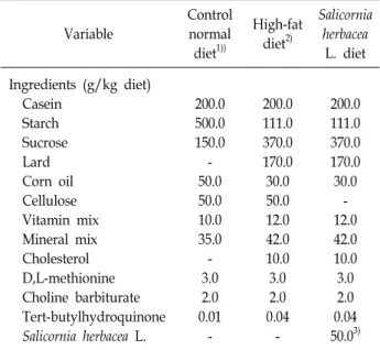
Table 1. Composition of experimental diets
Variable
Control normal diet1))
High-fat diet2)
Salicornia herbacea
L. diet Ingredients (g/kg diet)
Casein Starch Sucrose Lard Corn oil Cellulose Vitamin mix Mineral mix Cholesterol D,L-methionine Choline barbiturate Tert-butylhydroquinone Salicornia herbacea L.
200.0 500.0 150.0 - 50.0 50.0 10.0 35.0 - 3.0 2.0 0.01
-
200.0 111.0 370.0 170.0 30.0 50.0 12.0 42.0 10.0 3.0 2.0 0.04
-
200.0 111.0 370.0 170.0 30.0
- 12.0 42.0 10.0 3.0 2.0 0.04 50.03)
1)The control normal diet group (CD) was based on AIN-76G
2)The high-fat diet (HD and SD) were consisted of 20% protein, 48% carbohydrate, and 20% fat of total diet weight as modified diet from a previous study
3)The Salicornia berbacea L. supplemented group (SD) was re- ceived by substituting 5.0% of the fiber component of the high-fat diet
gene, the peroxisome proliferator-activated receptor-γ (PPAR- γ) regulates the development of fat cells and their capacity to store lipids [31, 32].
Considering the above, the present investigation was car- ried out to evaluate the effect of SH supplementation on changes in weight, lipid accumulation according to specific tissue, and changes in protein expression regulating lipid metabolism such as PGC-1α and PPAR-γ in rats fed a high- fat diet.
Materials and Methods
Experimental animals and diets
All of the experimental protocols were approved by the Animal Study Committee of Sunmoon University (2014-04-01). Six-week- old male Sprague-Dawley rats were obtained (Samtako Co., Osan, Korea) and individually housed in a controlled environment at 23±1℃ at 50±5% rela- tive humidity, under a 12 hr light-dark cycle. All animals were given free access to tap water and food. After an accli- matization period of 1 week, all rats were randomly divided into 3 groups: control normal diet group (CD, n=8), high-fat diet group (HD, n=8) [33], and Salicornia herbacea supple- mented high-fat diet group (SD, n=8). Each group was then monitored for 8 weeks. During this period, dietary intake was measured daily, and the change in the body weight of each animal was noted weekly.
Preparation of experimental diet
As shown in Table 1, the HD consisted of 20% protein, 48% carbohydrate, and 20% fat, as modified from a previous study [6] and based on AIN-76G. Dried SH powder was pur- chased from Buan Hamcho (Buan, Cheonranam-do, S.
Korea). The SD was prepared by using 5% SH and substitut- ing a portion of the fiber component with the HD.
Collection of serum and tissue samples
After 12 hr (20:00-08:00) of overnight fasting, the rats were sacrificed by exsanguination, and blood was drawn from the left ventricle under light diethyl ether anesthesia. Serum was obtained by centrifuging the blood at 700× g for 20 min at 4℃. The tissue samples such as liver and perirenal fat pad were dissected and immediately snap-frozen in liquid nitrogen. The triglyceride (TG) level of the tissue samples was analyzed, and 350~400 mg of the minced tissues were homogenized in 2 ml of the diluted standard diluents con-
taining protease inhibitors. The tissues were then centri- fuged at 10,000× g for 10 min at 4℃. The serum and tissue samples were stored at -80℃ until needed for analysis.
Analysis of triglycerides in serum, liver, and skeletal muscle
The serum TG level was analyzed using enzyme kits (Asan Pharmaceutical Co., Yongin, S. Korea). Hepatic TG and muscle TG were extracted using the following method.
Briefly, liver and gastrocnemius muscle were homogenized in 5% Triton X-100 solution and heated in an 80~100℃ water bath for 2~5 min to solubilize the TG. The samples were then centrifuged at 10,000 g for 10 min, and the resulting supernatant was used to determine the TG level following the manufacturer’s protocol (Cayman chemical, MI, USA).
Western blot analysis
In order to analyze the PGC-1α and PPAR-γ protein ex- pression levels, the gastrocnemius muscle was homogenized on ice with a polytron homogenizer in a 20 mmol/l Tris- HCl buffer (pH 7.5) containing 5 mmol/l ethylenediaminetetr- acetic acid, 2 mmol/l phenylmethylsulfonyl fluoride, and 1:200 protease inhibitor cocktail (Sigma, St Louis, MO, USA).
Fig. 1. Changes of body weight during total experimental period.
p<0.05. CD: control normal diet group, HD: high-fat diet group, SD: Salicornia herbacea supplemented high-fat diet group.
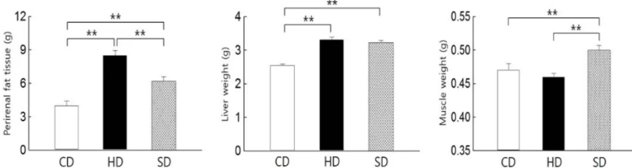
Fig. 2. Weight difference of tissues among groups. **p<0.01. CD: control normal diet group, HD: high-fat diet group, SD: Salicornia herbacea supplemented high-fat diet group.
The protein concentrations were determined using the Bradford method (Bio-Rad, Hercules, CA, USA) with bovine serum albumin (BSA) as the standard. An aliquot of tissue extract containing 20 ug (PGC-1α and PPAR-γ) of protein was separated on a 10% SDS-PAGE gel. After electro- phoresis, the proteins were transferred to a PVDF membrane (Millipore, Bedford, MA, USA) in a semi-dry blotting appa- ratus (Bio- Rad, Hercules, CA, USA). After treating with a blocking buffer (phosphate-buffered saline (PBS) containing 10% skim milk) for 90 min, the membrane was incubated with primary polyclonal antibodies for 2 hr, followed by five 10-minute washes with PBS (5% tween 20). The membranes were washed then incubated with horseradish peroxidase (HRP)-conjugated anti-goat IgG or anti-rabbit IgG (Santa Cruz, CA, USA) for 1 hr, followed by five 10-min washes with PBS (5% Tween 20). The target proteins were detected using an enhanced chemiluminescence (ECL) kit (Amersham Pharmacia Biotech, Piscataway, NJ, USA). The films were photographed, and the protein bands of interest were quan- tified with band analyzer software (Bio-Rad, Hercules, CA, USA).
Statistical analysis
The data were expressed as the means ± the standard er- ror of the mean (SE) using the SPSS/PC program (version 18.0 for Windows; SPSS Inc., USA). All statistical analyses were conducted using one-way ANOVA followed by the Least Significant Difference (LSD) post hoc test. Statistical significance was set at p<0.05.
Results and Discussion
It is well known that the lifestyle habits of humans in modern society, such as a high-fat diet and physical in- activity, promote weight gain, dyslipidemia, and profound systemic perturbations in lipid metabolism. In response to the small number of investigations carried out thus far to evaluate the effect of SH on lipid accumulation, in this study, we examined whether SH supplementation affects tis- sue-specific lipid metabolism characteristics such as weight and TG accumulation in rats with a high-fat diet.
The changes of body weight during the 8 week period, as the total experiment period, are presented in Fig. 1.
During the obesity-inducing period, regardless of SH sup- plementation, the body weights of the high-fat diet groups such as SD and HD groups significantly increased compared to those in the CD group (p<0.01). After the obesity-inducing period, the body weight of the SD group was significantly lower than that of the HD group, whereas the body weight of the HD group was significantly higher than that of the CD group (p<0.05). The differences of weight in the fat pad, liver, and skeletal muscle among the groups are illustrated in Fig. 2. The weights of the fat pad and liver in the HD group were significantly higher than those in the CD group (p<0.01), while the fat and liver weights in the SD group significantly reduced compared to those in the HD group (p<0.01, p<0.05). For skeletal muscle weight, the gastro-
Fig. 3. Levels of triglyceride in serum, liver, and muscle. *p<0.05, **p<0.01. CD: control normal diet group, HD: high-fat diet group, SD: Salicornia herbacea supplemented high-fat diet group.
cnemius weight in the SD group significantly increased com- pared to that in the CD and HD groups (p<0.01), whereas no significant difference was observed between the CD and HD groups.
Our results show the ability of SH supplementation to reduce the weight increase of the whole body and fat tissue while enhancing skeletal muscle mass. In other words, al- though the body and fat tissue weight increased, gastro- cnemius muscle mass reduced on the high-fat diet compared to the normal diet. These results indicate that, in the case of SH supplementation, even a high-fat diet with physical inactivity, body and fat tissue weights were reduced while muscle mass was enhanced. Although the inhibitory effect of flavonoid compounds on adipocyte differentiation has been reported, many studies of SH have focused on the im- provement of the metabolism of metabolites [1, 2, 11, 15, 17, 20, 29], but have not considered the effects of SH on TG levels and related lipid metabolism in specific tissues such as serum, fat, and skeletal muscle. Thus, for the first time, we attempted to investigate the possibility of SH sup- plementation to enhance muscle hypertrophy and improve lipid metabolism in skeletal muscle. We assume that SH might be useful in the utilization of fat as a substrate during supplementation and that SH feeding stimulated protein synthesis, which enhanced skeletal muscle mass. Therefore, even on a high-fat diet, SH supplementation could be recom- mended for the improvement of fat tissue with muscle hypertrophy.
The levels of TG in serum (p<0.05), liver (p<0.01), and skeletal muscle (p<0.05) in the HD group were significantly higher than those in the CD group. The serum (p<0.05) and hepatic TG (p<0.01) levels were found to have significantly reduced in the SD group compared to those in the HD group. However, a significant difference was not observed in the TG level of gastrocnemius muscle between the HD
and SD groups (Fig. 3). Our results showed strong inhibition on lipid accumulation in serum and liver by the SH supplementation. Regarding lipid metabolism, the adipose tissue, liver, and skeletal muscle are the main components that store TG in a regulated way and use hydrolysis to re- lease fatty acids, either for export of for internal con- sumption [7, 12, 22]. Dyslipidemia and intramuscular accu- mulation of triglyceride are increasingly recognized as an important feature of obesity and type 2 diabetes [16, 21].
Similar to the trends of tissue weights, the serum and hepatic TG levels also improved by the SH supplementation. SH supplementation has a positive role for the improvement of TG level in blood and liver. Interestingly, while muscle hy- pertrophy was shown in the SD group, muscle TG did not show significant improvement in the SD group compared to the HD group. From these results, it could be assumed that the TG utilization within muscles was limited by only SH supplementation. Thus, further studies, considering the muscle TG metabolism and catabolism, are needed to better elucidate the relationship between muscle weight and TG concentration.
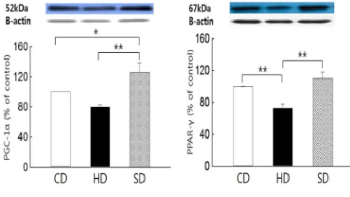
As shown in Fig. 4, the PGC-1α protein expression level of the SD group was significantly higher than that in the HS group (p<0.01) and CD group (p<0.05), respectively. In addition, the PPAR-γ protein expression level in the SD group was significantly higher than that in the HD group (p<0.01), while the PPAR-γ protein expression level in the HD group was significantly lower than that in the CD group (p<0.01).
PPAR-γ protein is reported to play an important role in the regulation of PGC-1α gene expression in skeletal muscle tissues [14]. Our study also showed that the PPAR-γ and PGC-1α protein expressions in the muscle of the SD group were significantly higher than those in the HD group.
Previous studies reported that adipogenesis induces a series
Fig. 4. Difference of PGC-1α and PPAR-γ protein expression in skeletal muscle. *p<0.05, **p<0.01 CD: control normal diet group, HD: high-fat diet group, SD: Salicornia herba- cea supplemented high-fat diet group.
of programmed changes in adipogenic gene expression that is activated by the transcription factors such as PGC-1α and PPAR-γ [18, 19]. Our finding is in accordance with previous studies which show that the PGC-1α and PPAR-γ protein expression in the SD group was significantly higher than that of the HD group. Skeletal muscle comprises a relatively large mass in the body and is an important target tissue for glucose metabolism by insulin, as reported in the pre- vious studies [9, 25, 26]. Additionally, as mentioned pre- viously, skeletal muscles have a store of triacylglycerol that can be mobilized in a regulated way to release fatty acids, while muscle fatty acids are a substrate for oxidation.
Therefore, although we could not precisely elucidate the mechanism of the PGC-1α and PPAR-γ protein expressions associated with SH supplementation in the present study, we can assume that the significant changes in the SH supple- mentation might have significant physiological effects be- cause the skeletal muscle is one of the major lipid metabo- lism target tissues.
Taken together, even on a high-fat diet, Salicornia herbacea L. supplementation plays a role in the suppression of lipid accumulation in serum and liver with muscle hypertrophy.
When Salicornia herbacea L. supplementation was accom- panied, the PGC-1α and PPAR-γ protein expressions in the skeletal muscle were enhanced compared to the high-fat diet group without S Salicornia herbacea L. supplementation.
In conclusion, SH supplementation may play a role in en- hancing muscle hypertrophy and improving the weights of fat tissue and liver in rats fed a high-fat diet. In addition, Salicornia herbacea L. supplementation was shown to sig- nificantly suppress lipid accumulation, which is mediated via PGC-1α and PPAR-γ activation. These results suggest that Salicornia herbacea L. supplementation may be useful as
an effective source for preventing lipid accumulation related disease.
References
1. Cho, H. D., Lee, J. H., Jeong, J. H., Kim, J. Y., Yee, S. T., Park, S. K., Lee, M. K. and Seo, K. I. 2016. Production of novel vinegar having antioxidant and anti-fatigue activities from Salicornia herbacea L. J. Sci. Food Agric. 96, 1085-1092.
2. Cho, J. Y., Kim, J. Y., Lee, Y. G., Lee, H. J., Shim, H. J., Lee, J. H., Kim, S. J., Ham, K. S. and Moon, J. H. 2016. Four new dicaffeoylquinic acid derivatives from glasswort (Salicornia herbacea L.) and their antioxidative activity.
Molecules 21, E1097.
3. Eu, C. H., Lim, W. Y., Ton, S. H. and bin Abdul Kadir, K. 2010. Glycyrrhizic acid improved lipoprotein lipase ex- pression, insulin sensitivity, serum lipid and lipid deposi- tion in high-fat diet induced obese rats. Lipids Health Dis.
9, 81.
4. Gurib-Fakim, A. 2006. Medicinal plants: traditions of yester- day and drugs of tomorrow. Mol. Aspects Med. 27, 1-93.
5. Hammarstedt, A., Anderson, C. X., Rotter, S. V. and Smith, U. 2005. The effects of PPAR gamma ligand on the adipose tissue in insulin resistance. Prostaglandins Leukot. Essent.
Fatty Acids. 73, 65-75.
6. Hariri, N. and Thibault, L. 2010. High-fat diet induced obe- sity in animal models. Nutr. Res. Rev. 23, 270-299.
7. Henriksson, J. 1995. Effect of training and nutrition on the development of skeletal muscle. J. Sports Sci. 13, S25-30.
8. Hofbauer, K. G., Nicholson, J. R. and Boss, O. 2007. The obesity epidemic: Current and future pharmacological treatments. Annu. Rev. Pharmacol. Toxicol. 47, 565-592.
9. Holloszy, J. O. 2011. Regulation of mitochondrial biogenesis and GLUT4 expression by exercise. Compr. Physiol. 1, 921- 940.
10. Huang, C. C., Tung, Y. T., Huang, W. C., Chen, Y. M., Hsu, Y. J. and Hsu, M. C. 2016. Beneficial effects of cocoa, coffee, green tea and garcinia complex supplement on diet induced obesity in rats. BMC Complement. Altern. Med. 16, 100.
11. Hwang, J. Y., Lee, S. K., Jo, J. R., Kim, M. E., So, H. A., Cho, C. W. and Kim, J. I. 2007. Hypolipidemic effect of Salicornia herbacea in animal model of type 2 diabetes mellitus. Nutr. Res. Pract. 1, 371-375.
12. Janesick, A. and Blumberg, B. 2012. Obesogens, stem cells and the developmental programming of obesity. Int. J.
Androl. 35, 437-448.
13. Jang, W. S. and Choung, S. Y. 2013. Antiobesity effects of the ethanol extract of Laminaria japonica Areshoung in high-fat-diet-induced obese rat. Evid. Based Complement.
Alternat. Med. 2013, 492807.
14. Kang, J. Y., Lee, J. H., Kwon, D. K. and Song, Y. J. 2013.
Effect of Opuntia humifusa supplementation and acute ex- ercise on insulin sensitivity and associations with PPAR-γ and PGC-1α protein expression in skeletal muscle of rats.
Int. J. Mol. Sci. 14, 7140-7154.
15. Karadeniz, F., Kim, J. A., Ahn, B. N., Kwon, M. S. and Kong, C. S. 2014. Effect of Salicornia herbacea on osteoblastogenesis and adipogenesis in vitro. Mar. Drugs 12, 5132-5147.
16. Kolka, C. M., Richey, J. M., Castro, A. V., Broussard, J. L., Ionut, V. and Bergman, R. N. 2015. Lipid-induced insulin resistance does not impair insulin access to skeletal muscle.
Am. J. Physiol. Endocrinol. Metab. 308, E1001-E1009.
17. Kong, C.S., Kim, Y. A., Kim, M. M., Park, J. S., Kim, J. A., Kim, S. K., Lee, B. J., Nam, T. J. and Seo, Y. 2009. Protective effect of 3-O-β-D-Glucoside from Salicornia herbacea against oxidation-induced cell damage. Food Chem. Toxicol. 47, 1914- 1920.
18. Kumar, P. M., Venkataranganna, M. V., Manjunath, K., Viswanatha, G. L. and Ashok, G. 2014. Methanolic extract of Momordica cymbalaria enhances glucose uptake in L6 myotubes in vitro by up-regulating PPAR-γ and GLUT4.
Chin. J. Nat. Med. 12, 895-900.
19. Kumar, R., Balaii, S., Uma, T. S. and Sehgal, P. K. 2009.
Fruit extracts of Momordica charantia potentiate glucose up- take and up-regulate Glut-4, PPAR gamma and PI3K. J.
Ethnopharmacol. 126, 533-537.
20. Lee, S. S., Seo, H. B., Ryu, S. P. and Kwon, T. D. 2015. The effect of swimming exercise and powdered- Salicornia her- bacea L. ingestion on glucose metabolism in STZ-induced diabetic rats. J. Exerc. Nutrition Biochem. 19, 235-245.
21. Li, L., Yang, G., Li, Q., Tang, Y. and Li, K. 2006. High-fat- and lipid-induced insulin resistance in rats: the comparison of glucose metabolism, plasma resistin and adiponectin levels. Ann. Nutr. Metab. 50, 499-505.
22. Lyssimachou, A., Santos, J. G., André, A., Soares, J., Lima, D., Guimarães, L., Almeida, C. M., Teixeira, C., Castro, L.
F. and Santos, M. M. 2015. The mammalian “Obesogen”
tributyltin targets hepatic triglyceride accumulation and the transcriptional regulation of lipid metabolism in the liver brain of Zebrafish. PLoS One 10, e0143911.
23. Maltin, C. A. 2008. Muscle development and obesity: Is there a relationship? Organogenesis 4, 158-169.
24. Moro, C. O. and Basile, G. 2000. Obesity and medicinal plants. Fitoterapia 71, S73-S82.
25. Morris, E. M., Meers, G. M., Booth, F. W., Fritsche, K. L., Hardin, C. D., Thyfault, J. P. and Ibdah, J. A. 2012. PGC-1α overexpression results in increased hepatic fatty acid oxida- tion with reduced triacylglycerol accumulation and secretion.
Am. J. Physiol. Gastrointest. Liver Physiol. 303, G979-G992.
26. Olson, A. L. 2012. Regulation of GLUT4 and insulin-depend- ent glucose flux. ISRN Mol. Biol. 2012, 856987.
27. Opala, T., Rzymski, P., Pischel, I., Wilczak, M. and Wozniak, J. 2006. Efficacy of 12 weeks supplementation of a botanical extract-based weight loss formula on body weight, body composition and blood chemistry in healthy, overweight subjects--a randomized double-blind placebo-controlled clinical trial. Eur. J. Med. Res. 11, 343-350.
28. Park, M. Y., Lee, K. S. and Sung, M. K. 2005. Effects of diet- ary mulberry, Korean red ginseng, and banaba on glucose homeostasis in relation to PPAR-alpha, PPAR-gamma, and LPL mRNA expressions. Life Sci. 77, 3344-3354.
29. Park, S. H., Ko, S. K., Choi, J. G. and Chung, S. H. 2006.
Salicornia herbacea prevents high fat diet-induced hyper- glycemia and hyperlipidemia in ICR mice. Arch. Pharm. Res.
29, 256-264.
30. Qin, G. W. and Xu, R. S. 1998. Recent advances on bioactive natural products from Chinese medicinal plants. Med. Res.
Rev. 18, 375-382.
31. Singh, S. and Bennett, R. G. 2010. Relaxin signaling activates peroxisome proliferator-activated receptor gamma. Mol. Cell Endocrinol. 315, 239-245.
32. Singh, S., Simpson, R. L. and Bennett, R. G. 2015. Relaxin activates peroxisome proliferator-activated receptor γ (PPAR γ) through a pathway involving PPARγ coactivator 1α (PGC1α). J. Biol. Chem. 290, 950-959.
33. Zhao, Y., Ling, F., Griffin, T. M., He, T., Towner, R., Ruan, H. and Sun, X. H. 2014. Up-regulation of the Sirtuin 1 (Sirt1) and peroxisome proliferator-activated receptor γ coactiva- tor-1α (PGC-1α) genes in white adipose tissue of Id1 pro- tein-deficient mice: implications in the protection against di- et and age-induced glucose intolerance. J. Biol. Chem. 289, 29112-29122.
초록:함초의 보충식이가 고지방식이 흰쥐의 혈청 및 조직의 중성지방 농도와 골격근 내 PGC-1α 및 PPAR-γ 단백질 발현에 미치는 영향
조하형1․권대근2․김진우3․송영주2*
(1선문대학교 일반대학원 체육학과, 2선문대학교 스포츠건강과학연구소, 3선문대학교 식품과학과)
본 연구는 함초의 보충식이가 고지방식이 흰쥐의 혈청 및 조직의 중성지방 농도와 골격근 내 PGC-1α와 PPAR- γ 단백질 발현에 미치는 영향에 대하여 연구하였다. SD계 수컷 흰쥐를 대조군(CD, n=8), 고지방식이군(HD, n=8), 고지방식이+ 5% 함초 보충식이군(SD, n=8)으로 분류하여 8주간 사육하였다. 그 결과 SD군의 지방조직 중량은 HD군에 비해 약 25%정도 유의하게 감소한 반면 골격근의 중량은 SD군이 HD군에 비해 약 5%정도 유의하게 증 가하였다(p<0.01). SD군의 혈청과 간장 내 중성지방은 HD군에 비해 약 20% 유의하게 감소하였다(p<0.05). SD군의 골격근 내 PGC-1α 와 PPAR-γ 단백질 발현은 HD군에 비해 1.5배 유의하게 높게 나타났다(p<0.01). 이상의 결과로 부터 함초 보충은 골격근 내 PGC-1α와 PPAR-γ 단백질 발현의 증가를 통하여 지방감소 및 근육량 증가에 효과적 이었음이 시사되었다.