Article
Paenibacillus kimchicus sp . nov., an antimicrobial bacterium isolated from Kimchi
A-rum Park, Ji-Sung Oh, and Dong-Hyun Roh*
Department of Microbiology, Chungbuk National University, Cheongju 28644, Republic of Korea
김치로부터 분리된 항균 활성 세균 Paenibacillus kimchicus sp . nov.
박아름 ・ 오지성 ・ 노동현*
충북대학교 미생물학과
(Received June 24, 2016; Revised July 12, 2016; Accepted July 13, 2016)
ABSTRACT: An antimicrobial bacterium to pathogenic microorganisms, strain W5-1
Twas isolated from Korean fermented-food Kimchi.
The isolate was Gram-staining-variable, strictly aerobic, rod-shaped, endospore-forming, and motile with peritrichous flagella. It grew at 15-40°C, at pH 6.0-10.0, and in the presence of 0–4% NaCl. Strain W5-1
Tcould hydrolyze esculin and xylan, and assimilate
D
-mannose, but not
D-mannitol. Strain W5-1
Tshowed antimicrobial activity against Listeria monocytogens, Pseudomonas aeruginosa, Staphylococcus aureus, and Salmonella typhi. The G+C content of the DNA of strains W5-1
Twas 52.6 mol%. The predominant respiratory quinone was menaquinone-7 (MK-7) and the major cellular fatty acids were C
16:0, antieiso-C
15:0, C
18:0, and C
12:0. The strain contained meso-diaminopimelic acid in cell-wall peptidoglycan. On the basis of 16S rRNA gene sequence and phylogenetic analysis, the strain W5-1 was shown to belong to the family Paenibacillaceae and was most closely related to Paenibacillus pinihumi S23
T(98.4% similarity) and Paenibacillus tarimensis SA-7-6
T(96.4%). The DNA-DNA relatedness between the isolate and Paenibacillus pinihumi S23
Twas 8.5%, indicating that strain W5-1
Trepresented a species in the genus Paenibacillus. On the basis of the evidence from this polyphasic study, it is proposed that strain W5-1
Tis considered to represent a novel species of the genus Paenibacillus, for which the name Paenibacillus kimchicus sp. nov. is proposed. The type strain is W5-1
T(=KACC 15046
T= LMG 25970
T).
Key words: Paenibacillus kimchicus, antimicrobial activity, new species, taxonomy
*For correspondence.
E-mail: [email protected];Tel.: +82-43-261-3368; Fax: +82-43-264-9600
The genus Paenibacillus (the family Paenibacillaceae;
De Vos et al., 2009) was proposed by Ash et al. (1993) to accommodate a phylogenetically coherent group of 11 species of the genus Bacillus and subsequently emended by Shida et al.
(1997). Members of the genus Paenibacillus are aerobic or facultative anaerobic, rod-shaped, and endospore-forming in swollen sporangia. Members of the genus possess anteiso-C
15:0as the major cellular fatty acid and MK-7 as the predominant menaquinone, and the DNA G+C content ranges from 39 to 54 mol% (Shida et al., 1997). At the time of writing, the genus Paenibacillus comprised over 182 species and 4 subspecies (Euzeby, 1997). Species belonging to the genus Paenibacillus
excrete many kinds of enzymes that degrade natural biopolymers
such as xylan (Choi et al., 2008) and produce antibacterial
compounds such as polymyxin, oxtoptin, and baciphelacin
(Slepecky and Hemphill, 1992) and an antifungal compound
(Chung et al., 2000). Especially, type species of the genus
Paenibacillus polymyxa was known to promote plant growth
and produce various peptide antibiotics, for example, polymyxin
group (Vogler and Studer, 1966), gatavalin (Nakajima et al.,
1972), LI-F complex (Kurusu et al., 1987), fusaricidin A
(Kajimura and Kaneda, 1996), gacaserin, and saltavalin (Pichard
et al., 1995). In this study, we report the polyphasic characteristics
of a Paenibacillus-like bacterium, having antimicrobial activity
against some pathogens, isolated from Kimchi, a traditional
food of Korea.
Materials and Methods
Bacterial isolation and culture conditions
In order to isolate antimicrobial bacteria against Staphylococcus aureus (KCCM 40881), Listeria monocytogenes (ATCC 15313), Salmonella typhi (KNIH 100), Pseudomonas aeruginosa (ATCC 27853), and Candida albicans (ATCC 10231), Kimchi samples were suspended and serially diluted in sterile distilled water and the suspensions were plated onto a hundred times diluted tryptic soy agar (TSA; Difco). After the plates were incubated at 25°C for 4 days, antimicrobial activity of every single colonies were assessed by agar spot test as described in Fleming et al. (1975). Bacterial strains having antimicrobial activity were selected and purified by repeated streaking on TSA.
Among those bacterial strains strain W5-1
Thad broad antibacterial activity to S. aureus, L. monocytogenes, S. typhi, and P. aeruginosa. Strain W5-1
Tstored at -80°C in TSB supplemented with 25% (v/v) glycerol. The related type strains of the genus Paenibacillus, P. pinihumi KACC 14199
T, P.
tarimensis KACC 14087
T, P. castaneae KACC 14162
T, P.
daejeonensis KACC 11453
T, and P. mendelii KACC 11472
Twere obtained from Korean Agricultural Culture Collection (KACC, Korea) and used for reference strains in polyphasic taxonomic tests. Strain W5-1
Tand reference strains were routinely grown on TSA or tryptic soy broth (TSB; Difco) at 30°C for 3 days.
16S rRNA gene sequencing and phylogenetic analysis The 16S rRNA gene was amplified from a single colony by PCR with 2X DyeMIX DNA Polymerase (Enzynomics
TM) and universal bacterial primer pair, 27F (5'-AGAGTTTGATCMT GGCTCAG-3') and 1492R (5'-TACGGYTACCTTGTTACG ACTT-3'). PCR was run for 30 cycles with a DNA thermal cycler (MJ Mini; Bio-Rad). The following thermal profiles were used for PCR: predenaturation at 95°C for 3 min and a repeated cycle of denaturation at 95°C for 20 sec, primer annealing at 55°C for 40 sec and extension at 72°C for 45 sec.
The final cycle included an extension at 72°C for 10 min. The PCR product was purified using HiYield Gel/PCR DNA Fragments Extraction Kit (RBC) and purified PCR products were sequenced by Solgent Co. Ltd. The full sequences of 16S rRNA gene were assembled using SeqMan software
(DNASTAR). The 16S rRNA gene sequences of related taxa were obtained from the GenBank database. The multiple alignments were performed by Clustal X2 program (Larkin et al., 2007). Gaps were edited in the BioEdit program (Hall, 1999). The evolutionary distances were calculated using the Kimura two-parameter model (Kimura, 1980). The phylogenic tree was constructed by using a neighbor-joining method (Saitou and Nei, 1987), maximum parsimony (Kluge and Farris, 1969) and maximum likelihood (Felsenstein, 1981) in MEGA7 Program (Kumar et al., 2016) with bootstrap values based on 1,000 replications.
Phenotypic and biochemical characterization Strain W5-1
Tand reference strains were routinely grown on TSA or tryptic soy broth (TSB; Difco) at 30°C for 3 days. Cell morphology, size and motility were observed by phase contrast microscopy (80i, Nikon) on cells under these conditions and flagella staining were carried out according to Heimbrook (1989). The production of endospores was examined on cells grown at 30°C for 7 days on NA and endospores were detected by using malachite green staining described as Schaeffer and Fulton (1933). Gram staining was performed with BD Gram stain kits according to the instructions of the manufacturer and with the non-staining method as described by Buck (1982).
Catalase activity was determined by bubble production in 3%
(v/v) H
2O
2and oxidase activity was assessed using oxidase
reagent (bioMérieux) according to the manufacturer’s instructions
after cultivation for 3 days. Acid production from glucose,
single-carbon-source assimilations and additional physiological
characteristics were determined using the API 20NE and API
ID32GN galleries (bioMérieux). All tests were performed
according to the instructions of the manufacturer. Hydrolylsis
of casein (1%, w/v), starch (1%, w/v), xylan (1%, w/v) and
Tween 80 (1%, v/v) were tested as described by Smibert and
Krieg (1994), using TSA as basal medium. DNase test was
performed using DNase test agar (Difco). The reference type
strains were included in these comparisons and all tests were
performed under the same conditions. Salt tolerance was
evaluated after incubation at 30°C for 7 days in nutrient broth
(NB; Difco) containing 0 –5% (w/v; interval of 1%) NaCl. The
temperature range for growth was assessed on NA incubated
for 7 days at 0, 4, 10, 15, 20, 25, 30, 37, 40, 45, and 50°C. The
pH range for growth was tested in NB adjusted with HCl and NaOH to pH values between 5.0 and 12.0 (interval of 1.0).
Antibiotic sensitivity test was performed on NA medium by disc-diffusion method with laboratory-prepared discs containing of ampicillin (50 µg), spectinomycin (20 µg) streptomycin (10 µg), tetracycline (10 µg), kanamycin (50 µg), penicillin G (50 µg), gentamycin (10 µg), and chloramphenicol (25 µg).
Chemotaxonomic and genomic analyses
For the analysis of cellular fatty acids, strain W5-1
Tand other type strains of the genus Paenibacillus were cultivated for 3 days on TSA. The cellular fatty acids were saponified, methylated, and extracted according to the protocol of the Sherlock Microbial Identification System (MIDI) by using the TSBA 40 method. Extracts were then analyzed by gas chromatograph (Hewlett Packard 6890N) using the Microbial Identification software package (Sasser, 1990). Isoprenoid quinones were extracted with chloroform/methanol (2:1, v/v), evaporated under vacuum conditions, and reextracted in n- hexane-water (1:1, v/v). Then, the crude quinone in n-hexane was purified using Sep-Pak
®Vac Cartridges Silica (Waters) and subsequently analyzed by HPLC, as previously described Hiraishi et al. (1996). Peptidoglycan analysis was determined according to Schleifer and Kandler (1972).
For the measurement of G+C content of the chromosomal DNA, genomic DNA of the strain was extracted and purified using a genomic DNA extraction kit (Solgent), and was enzymatically degraded into nucleosides, and then the G+C content of DNA was determined as described by Mesbah and Whitman (1989) using a reverse-phase HPLC.
To verify the new taxon of strain W5-1
T, DNA–DNA hybridization experiments were performed between a strain W5-1
Tand related five type strains of members of the genus Paenibacillus using photobiotin-labelled DNA probes and microdilution wells as described by Ezaki et al. (1989). The hybridizations were performed with five replications of each sample, where the highest and lowest values obtained were excluded, and the means of the remaining three values were quoted as the DNA-relatedness value.
Results and Discussion
Morphological and phenotypic characteristics Strain W5-1
Twas Gram-staining-variable depending on the used experimental methods. Cell was not strained by Gram straining, however, KOH method showed Gram-positive reaction.
Cells were strictly aerobic, rod-shaped approximately 0.5–1.1 μm in diameter and 2.8–4.3 μm in length, and motile by peritrichous flagella. Subterminal ellipsoidal spores were formed in swellen sporangia. Dimorphic colonies were formed on TSA and translucent, circular with slightly irregular margin measuring and ivory with faintly brown in center. Growth occurred at 15 –40°C (optimum, 37°C), pH 6.0–10.0 (optimum, pH 8) and in the presence of 0 –4% (w/v) NaCl (optimum, 0%).
The other phenotypic characteristics of W5-1
Tare given in the species description and compared to those of related other type strains of the genus Paenibacillus in Table 1.
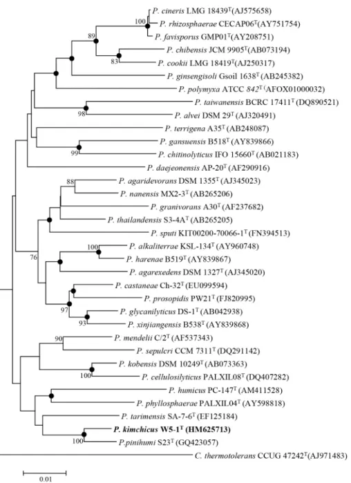
Phylogenetic analysis
The 16S rRNA gene sequence of strain W5-1
Tdetermined in this study was a continuous stretch of 1,524 bp. The NCBI/
EMBL/DDBJ accession number for the 16S rRNA sequence of strain W5-1
Tis HM625713. Strain W5-1
Tbelonged to the genus Paenibacillus in family Paenibacillaceae on the basis of 16S rRNA gene sequence analysis. The 16S rRNA gene sequence similarity values between strain W5-1
Tand the other related type strains of the genus Paenibacillus ranged from 91.5 – 98.4%. Strain W5-1
Tshowed the highest similarity to P. pinihumi KACC 14199
T(98.4%) and the second highest similarities was 96.4% to Paenibacillus tarimensis KACC 14087
T. Phylogenetic analyses based on the neighbour-joining, maximum parsimony and maximum-likelihood method revealed that strain W5-1
Tfell within the evolutionary radiation enclosed by the genus Paenibacillus and formed a cluster with P. pinihumi (Fig. 1).
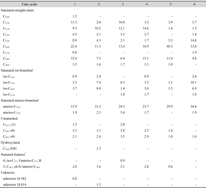
Chemotaxonomic and genomic analyses
The cellular fatty acid contents of strain W5-1
Tand other type strains of the genus Paenibacillus are shown in Table 2.
The major cellular fatty acids (>10%) of strain W5-1
Twere C
16:0(22.4%), anteiso-C
15:0(15.9%), C
18:0(12.4%), and C
12:0(11.3%). This fatty acid profile is characteristic of the genus
Table 1. Differential characteristics of strain W5-1T with closely related and representative type strains of the genus Paenibacillus species. Strain: 1, Paenibacillus kimchicus W5-1T; 2, Paenibacillus pinihumi KACC 14199T; 3, Paenibacillus tarimensis KACC 14087T; 4, Paenibacillus castaneae KACC 14162T; 5, Paenibacillus daejeonensis KACC 11453T; 6, Paenibacillus mendelii KACC 11472T. All data are from this study. All stains were able to assimilate maltose, but could not capric acid, adipic acid, malic acid, citric acid, phenylacetic acid, D-sorbitol, propionate, valerate, L-histidine, 2-ketogluconate, 3-hydroxy-butilate, 4-hydroxy-benzoate, L-proline, inositol, itaconate, suberate, molonate, acetate, DL-lactate, L-alanine, 5-ketogluconate, 3-hydroxy-benzoate,
L-serine. All strains were not detected reduction of nitrate, indole production, acidification of glucose, arginine dihydrolase, urease, and hydrolysis of starch and casein. +, positive reaction; w, weakly positive; -, negative reaction
Characteristics 1 2 3 4 5 6
Growth temperature (°C):
Range 15-40 20-37 10-30 25-45 15-37 15-30
Optimum 37 25 30 25 30 25
Hydrolysis of:
Esculin + - + + - -
Xylan + - - + + -
β-Galactosidase + - + + - -
Assimilation of:
D-Glucose + + + + - w
L-Arabinose + + + + - -
D-Mannose + - - + - -
D-Mannitol - + - + + -
N-Acetyl-glucosamine (PNPG) - - - - + +
Potassium gluconate - - + - - -
Salicin + - + + - +
D-Melibiose + w + + - +
L-Fucose - - + - + -
Rhamnose + - w - w +
D-Ribose + + - + - +
D-Sucrose + - w + - +
Glycogen - + + - + -
DNA G+C content (mol%) 52.6 49.5 53.7 46.0 53.0 50.8
Paenibacillus. Interestingly, anteiso-C
15:0was known as the major cellular fatty acid in the genus Paenibacillus (Shida et al., 1997), but C
16:0occupied first position in the proportion of cellular fatty acid in strain W5-1
Tand Paenibacillus daejeonensis AP-20
T. In addition, the proportions of C
18:0and C
12:0were somewhat higher in strain W5-1
Tthan those of other related type strains. Although the antieiso-C
15:0in strain W5-1
Ttook the second position in the proportion, it showed significant lower proportion than those of other type strains (Table 2).
Strain W5-1
Tcontained menaquinone-7 (MK-7) as the major respiratory quinone and meso-diamiopimelic acid in the cell- wall peptidoglycan as in most other members of the genus Paenibacillus (Shida et al., 1997). The DNA G+C base comp- osition of W5-1
Twas 52.6 mol%, placing it within the range other members of the genus Paenibacillus (Shida et al., 1997).
DNA-DNA relatedness values with type strains of the genus Paenibacillus have shown that strain W5-1
Thas 8.5% with P.
pinihumi KACC 14199
T, 6.5% with P. mendelii KACC 11472
T, 5.3% with P. castaneae KACC 14162
T, 4.2% with P. tarimensis KACC 14087
T, 2.0% with P. daejeonensis KACC 11453
T, respectively. These low DNA-relatedness values are enough for strain W5-1
Tto be classified as a novel species in the genus (Wayne, 1987).
Therefore, based on data from this polyphasic study, strain W5-1
Tshould be classified in the genus Paenibacillus as a representative of a novel species, for which the name Paenibacillus W5-1
Tsp. nov. is proposed.
Description of Paenibacillus kimchicus sp. nov.
Paenibacillus kimchicus (kimchicus: kimchi'cus. N.L. n.
Fig. 1. Neighbour-joining tree based on 16S rRNA gene sequences showing the phylogenetic relationship of W5-1T within the Paenibacillus. Bootstrap values (expressed as percentage of 1,000 replications) greater than 70% are given at the nodes. Filled circles indicate nodes also recovered reproducibly with the maximum parsimony and maximum-likelihood. The sequence of Cohnia thermotolerans CCUG 47242T was used as an outgroup and GenBank accession numbers are given in parentheses. Bar, 0.01 substitutions per nucleotide position.
kimchium, kimchi; L. masc. suff. -icus, suffix used with the sense of pertaining to; N.L. masc. adj. kimchicus, pertaining to kimchi, isolated from kimchi, a traditional Korean fermented- vegetable food made by Chinese cabbage)
Cells are Gram-staining-variable, strictly aerobic, rod-shaped approximately 0.5–1.1 μm in diameter and 2.8–4.3 μm in length, and motile by peritrichous flagella. Subterminal ellip-
soidal spores are formed in swellen sporangia. Dimorphic colonies are formed on TSA: translucent, circular with slightly irregular margin measuring and ivory with faintly brown in center. Growth occurs at 15 –40°C (optimum, 37°C), pH 6.0–
10.0 (optimum, pH 8) and in the presence of 0 –4% (w/v) NaCl
(optimum, 0%). Starch, esculin, casein, and xylan are hydro-
lysed, but Tween 80, gelatin, and DNA are not. Nitrate is not
Table 2. Fatty acids composition of strain W5-1T with closely related and representative type strains of the Paenibacillus species. Strain: 1, Paenibacillus kimchicus W5-1T; 2, Paenibacillus pinihumi KACC 14199T; 3, Paenibacillus tarimensis KACC 14087T; 4, Paenibacillus castaneae KACC 14162T; 5, Paenibacillus daejeonensis KACC 11453T; 6, Paenibacillus mendelii KACC 11472T. All data are from this study. -, Not detected; ECL, equivalent chain length; Fatty acids comprising less than 0.5% of the total were excluded. All strains were cultured on TSA at 30°C.
Fatty acids 1 2 3 4 5 6
Saturated straight-chain
C10:0 1.2 - - - - -
C12:0 11.3 2.8 10.0 3.5 3.9 3.7
C13:0 9.5 10.2 12.1 14.6 1.8 1.5
C14:0 4.5 2.1 3.5 2.7 - 1.8
C15:0 0.9 4.3 2.1 1.7 5.3 14.8
C16:0 22.4 11.3 13.4 16.9 40.3 13.6
C17:0 0.6 - - - - 1.0
C18:0 12.4 7.5 6.4 15.1 11.0 4.6
C20:0 3.5 1.6 1.7 3.3 1.0 -
Saturated iso-branched
iso-C14:0 0.9 2.4 - 0.9 - 2.6
iso-C15:0 1.5 7.4 6.5 3.2 1.1 10.1
iso-C16:0 3.7 8.0 1.4 3.0 1.5 6.9
iso-C17:0 - - 1.8 1.7 - 1.8
Saturated anteiso-branched
anteiso-C15:0 15.9 31.2 24.3 23.7 29.9 34.8
anteiso-C17:0 1.9 2.3 5.6 1.7 - 1.9
Unsaturated
C16:1 ω11c 1.5 - 2.0 - - -
C18:1 ω9c 3.5 1.1 2.8 2.5 1.6 -
C20:1 ω9c 2.1 2.4 3.5 2.9 1.0 1.0
Hydroxylated
C16:0 3OH - 1.5 - - - -
Summed features†
4; iso-C17:1 I/anteiso-C17:1 B - - 0.9 - - -
5; C18:2ω6,9c/anteiso-C18:0 2.0 1.6 2.1 2.8 0.6 -
Unknown
unknown 16.582 0.8 - - - - -
unknown 18.814 - 1.5 - - - -
† Summed features represent groups of two or three fatty acids that could not be separated by GLC with MIDI system.
reduced, indole is not produced and glucose is not fermented.
Catalase, oxidase and β-galactosidase (para-nitrophenyl β-
D
-galactopyranoside) activities are present, but arginine dihydrolase, urease activities are absent. Assimilates
D-glucose,
L-arabinose,
D
-mannose,
D-maltose, salicin,
D-melibiose, rhamnose,
D-ribose, and
D-sucrose, but does not assimilate
D-mannitol, N-acetyl- glucosamine, potassium gluconate, capric acid, adipate, malate, capric acid, trisodium citrate, phenylacetic acid,
L-fucose,
D
-sorbitol, propionate, valerate,
L-histidine, 2-ketogluconate,
3-hydroxy-butyrate, 4-hydroxy-benzoate,
L-proline, inositol,
itaconate, suberate, malonate, acetate,
DL-lactate,
L-alanine,
5-ketogluconate, 3-hydroxy-benzoate, and
L-serine (API 20 NE
and API 32 GN galleries). Susceptible to ampicillin, spectinomycin,
streptomycin, tetracycline, penicillin G, gentamycin, and chlo-
ramphenicol, but resistant to kanamycin. The major cellular
fatty acids are C
16:0, antieiso-C
15:0, C
18:0, and C
12:0. The major
respiratory quinone is menaquinone-7. DNA G+C content is
52.6 mol%.
The type strain, W5-1
T(=KCTC 15046
T=LMG 25970
T), was isolated from Kimchi, a traditional food of Korea.
적 요
병원성 미생물들에 대해 항균활성을 보이는 W5-1
T균주가
한국의 발효식품인 김치에서 분리되었다. 이 분리주는 그람염 색변이성, 절대호기성, 간균, 내생포자형성과 주모성의 편모 를 가지고 운동성을 나타내었다. 균주는 15–40°C, pH 6.0–
10.0, 0–4% NaCl 조건에서 생육하였다. 균주는 esculin과 xylan 을 가수분해하였고,
D-mannose 을 동화하였으나
D-mannitol 은 동화하지 못하였다. W5-1
T균주는 Listeria monocytogens, Pseudomonas aeruginosa, Staphylococcus aureus, Salmonella typhi에 항균활성을 보였다. W5-1
T균주의 DNA의 G+C 함량 은 52.6 mol%였다. 주요 호흡성 퀴논은 menaquinone-7 (MK-7) 였고, 주요 세포성 지방산은 C
16:0, antieiso-C
15:0, C
18:0, and C
12:0였다. 균주는 세포벽 펩티도클리칸으로 meso-diaminopimelic acid을 함유하였다. 16S rRNA 유전자서열 분석에 근거하여 W5-1
T균주는 Paenibacillaceae 과로 분류되었으며 Paenibacillus pinihumi S23
T(98.4% similarity), P. tarimensis SA-7-6
T(96.4%) 균주와 높은 연관성을 보였다. 분리주와 P. pinihumi S23
T는 8.5% 의 DNA-DNA 관련성을 보임으로 W5-1
T균주가 Paenibacillus 속의 한 종임을 보여주었다. 이러한 다각적 연구의 증거로 볼 때 W5-1
T균주는 Paenibacillus 속의 신종으로 사료되어 Paenibacillus kimchicus로 명명을 제안하며, 표준균주는 W5-1
T(=KACC 15046
T=LMG 25970
T)이다.
References
Ash, C., Priest, F.G., and Collins, M.D. 1993. Molecular identification of rRNA group 3 bacilli (Ash, Farrow, Wallbanks and Collins) using a PCR probe test. Proposal for the creation of a new genus Paenibacillus. Antonie van Leeuwenhoek 64, 253–260.
Buck, J.D. 1982. Nonstaining (KOH) method for determination of Gram reactions of marine bacteria. Appl. Environ. Microbiol.
44, 992–993.
Choi, J.H., Im, W.T., Yoo, J.S., Lee, S.M., Moon, D.S., Kim, H.J., Rhee, S.K., and Roh, D.H. 2008. Paenibacillus donghaensis sp.
nov., a xylan-degrading and nitrogen-fixing bacterium isolated from East Sea sediment. J. Microbiol. Biotechnol. 18, 189–193.
Chung, Y.R., Kim, C.H., Hwang, I., and Chun, J. 2000. Paenibacillus koreensis sp. nov., a new species that produces an iturin-like antifungal compound. Int. J. Syst. Evol. Microbiol. 50, 1495–
1500.
De Vos, P., Ludwig, W., Schleifer, K.H., and Whitman, W.B. 2009.
Family IV. Paenibacillaceae fam. nov. In Bergey'sManual of Systematic Bacteriology, 2nd edn, vol. 3 (The Firmicutes), pp.
269. In De Vos, P., Garrity, G.M., Jones, D., Krieg, N.R., Ludwig, W., Rainey, F.A., Schleifer, K.H., and Whitman, W.B.
(eds.) Springer, New York, USA.
Euzeby, J.P. 1997. List of bacterial names with standing in nomenclature: a folder available on the Internet. Int. J. Syst.
Bacteriol. 47, 590–592. (List of prokaryotic names with standing in nomenclature. http://www.bacterio.net).
Ezaki, T., Hashimoto, Y., and Yabuuchi, E. 1989. Fluorometric deoxyribonucleic acid-deoxyribonucleic acid hybridization in microdilution wells as an alternative to membrane filter hybridization in which radioisotopes are used to determine genetic relatedness among bacterial strains. Int. J. Syst. Bacteriol.
39, 224–229.
Felsenstein, J. 1981. Evolutionary trees from DNA sequences: a maximum likelihood approach. J. Mol. Evol. 17, 368–376.
Fleming, H.P., Etchells, J.L., and Costilow, R.N. 1975. Microbial inhibition by an isolate of Pediococcus from cucumber brines.
Appl. Microbiol. 30, 1040–1042.
Hall, T.A. 1999. BioEdit: a user-friendly biological sequence alignment editor and analysis program for Windows 95/98/NT. Nucleic Acids Symp. Ser. 41, 95–98.
Heimbrook, M.E., Wang, W.L., and Campbell, G. 1989. Staining bacterial flagella easily. J. Clin. Microbiol. 27, 2612–2615.
Hiraishi, A., Ueda, Y., Ishihara, J., and Mori, T. 1996. Comparative lipoquinone analysis of influent sewage and activated sludge by high performance liquid chromatography and photodiode array detection. J. Gen. Appl. Microbiol. 42, 457–470.
Kajimura, Y. and Kaneda, M. 1996. Fusaricidin A, a new depsipeptide antibiotic produced by Bacillus polymyxa KT-8. Taxonomy, fermentation, isolation, structure elucidation and biological activity. J. Antibiot. (Tokyo) 49, 129–135.
Kimura, M. 1980. A simple method for estimating evolutionary rates of base substitutions through comparative studies of nucleotide sequences. J. Mol. Evol. 16, 111–120.
Kluge, A.G. and Farris, J.S. 1969. Quantitative phyletics and the evolution of anurans. Syst. Zool. 18, 1–32.
Kumar, S., Stecher, G., and Tamura, K. 2016. MEGA7: Molecular Evolutionary Genetics Analysis version 7.0 for bigger datasets.
Mol. Biol. Evol. 33, 1870–1874.
Kurusu, K., Ohba, K., Arai, T., and Fukushima, K. 1987. New peptide antibiotics LI-F03, F04, F05, F07, and F08, produced by Bacillus polymyxa. I. Isolation and characterization. J. Antibiot.
(Tokyo) 40, 1506–1514.
Larkin, M.A., Balckshields, G., Brown, N.P., Chenna, R., McGettigan, P.A., McWilliam, H., Valentin, F., Wallace, I.M., Wilm, A., Lopez, R., et al. 2007. Clustal W and Clustal X version 2.0.
Bioinformatics 23, 2947–2948.
Mesbah, M. and Whitman, W.B. 1989. Measurement of deoxyguanosine/
thymidine ratios in complex mixtures by high-performance liquid chromatography for determination of the mole percentage guanine + cytosine of DNA. J. Chromatogr. 479, 297–306.
Nakajima, N., Chihara, S., and Koyama, Y. 1972. A new antibiotic, gatavalin. I. Isolation and characterization. J. Antibiot. (Tokyo) 25, 243–247.
Pichard, B., Larue, J.P., and Thouvenot, D. 1995. Gavaserin and saltavalin, new peptide antibiotics produced by Bacillus polymyxa.
FEMS Microbiol. Lett. 133, 215–218.
Saitou, N. and Nei, M. 1987. The neighbor-joining method: a new method for reconstructing phylogenetic trees. Mol. Biol. Evol. 4, 406–425.
Sasser, M. 1990. Identification of bacteria by gas chromatography of cellular fatty acids. MIDI Technical Note 101. MIDI Inc., Newark, DE, USA.
Schaeffer, A.B. and Fulton, M.D. 1933. A simplified method of staing endospores. Science 77, 194.
Schleifer, K.H. and Kandler, O. 1972. Peptidoglycan types of bacterial cell walls and their taxonomic implications. Bacteriol. Rev. 36, 407–477.
Shida, O., Takagi, H., Kadowaki, K., Nakamura, L.K., and Komagata,
K. 1997. Transfer of Bacillus alginolyticus, Bacillus chondroitinus, Bacillus curdlanolyticus, Bacillus glucanolyticus, Bacillus kobensis, and Bacillus thiaminolyticus to the genus Paenibacillus and emended description of the genus Paenibacillus. Int. J. Syst.
Bacteriol. 47, 289–298.
Slepecky, R. and Hemphill, E. 1992. The genus Bacillus – nonmedical.
The Prokaryotes, 2nd edn, pp. 1663–1696. In Balows, A., Trüper, H.G., Dworkin, M., Harder, W., and Schleifer, K.H.
(eds.). Springer, New York, USA.
Smibert, R.M. and Krieg, N.R. 1994. Phenotypic characterization.
Methods for General and Molecular Bacteriology, pp. 607–655.
In Gerhardt, P., Murray, R.G.E., Wood, W.A., and Krieg, N.R.
(eds.). American Society for Microbiology, Washington, DC, USA.
Vogler, K. and Studer, R.O. 1966. The chemistry of the polymyxin antibiotics. Experientia 22, 345–354.
Wayne, L.G., Brenner, D.J., Colwell, R.R., Grimont, P.A.D., Kandler, O., Krichevsky, M.I., Moore, L.H., Moore, W.E.C., Murray, R.G.E., Stackebrandt, E., et al. 1987. Report of the ad hoc committee on reconciliation of approaches to bacterial systematics.
Int. J. Syst. Bacteriol. 37, 463–464.