약학희 지 제 54 권 제 1 호 55~61 (2010)
Yakhak Hoeji Vol. 5^ No. 1
사염화탄소 유도 급성 간득성 모델에서 처자의 간 보호 효과
신 전 규 • 김 효 연 * • 이 선 미 *'*
경 희 대 학 교 생 명 과 학 부 , *성 균 관 대 학 교 약 학 대 학
(Received December 28, 2009; Revised January 5, 2010; Accepted January 5, 2010)
Protective Effect of Gardenia jasminoides Against Carbon Tetrachloride-Induced Acute Hepatotoxicity
Jun-Kyu Shin, Hyo-Yeon K im * and Sun-Mee Lee*'*
College of Life Science, Kyung Hee University, Yongin 446-701, Korea
"^School of Pharmacy, Sungkyunkwan University, Suwon 440-746, Korea
Abstract — — Gardenia jasminoides is one of the most widely used herbal preparations for the treatment of liver disorders.
This study evaluated the potential beneficial effect of G. jasminoides in a mouse model of carbon tetrachloride (CCI
4)- induced liver injury. The mice were treated intraperitoneally with CCI
4(10 m 모 /kg). They received G. jasminoides (30, 100, 300 mg/kg) 48 h, 24 h and 2 h before and
6h after administering CCI
4. The serum activities of aminotransferase and the hepatic level of malondialdehyde were significantly higher 24 h after the CCI
4treatment, while the concentration of reduced glutathione was lower. These changes were attenuated by G. jasminoides. CCI
4increased the level of circulating tumor necrosis factor-a (TNF-a) markedly, which was reduced by G. jasminoides. The levels of hepatic inducible nitric oxide syn
thase (iNOS) and cyclooxygenase-2 (COX-2) protein expression were markedly higher after the CCI
4treatment. G. jas
minoides diminished these alterations. CCI
4increased the level of TNF-a, iNOS and COX-2 mRNA expressions, and these increases were attenuated by G. jasminoides. These results suggest that G. jasminoides alleviates CCl
4-induced liver injury, and this protection is likely due to the reduced oxidative stress and the downregulation of proinflammatory mediators.
Keywords □ carbon tetrachloride, Gardenia jasminoides, hepatotoxicity, inflammation
간질환의 원인은 다양한 것으로 알려져 었으며 병인학적^
볼 때 바이러스에 기인된 간질환, 약물(득성물질)중득에 기인된 간질환 및 담도기능부전에 의한 간질환 등으로 분류할 수 있다.
이러한 간질환들은 간세포 파괴에 의한 해득능력저하와 담즙분 비억제에 의한 독성물질 배설장애, 비타민 B i,B
2및 C의 흡수 억제 및 측적저하에 의한 전신 권태감, 소양감, 식욕부진 및 피 로 등의 임상증상을 나타낸다.^^ 현재 국내외적으로 시용되고 있 는 간질환 치료제로는 합성 약물인 인터페론과 malotilate가 있 으나 이들은 간득성 물질에 대한 보호작용만으로 그의 유효성을 인정 받고 있는 수준이며, 부작용 또한 빈번히 나타난다. 또한 마 리아 엉검퀴의 활성성분인 silymarin과 천연물 유래 합성 간질환 치료제인 biphenyl dimethyl dicarboxylate의 개발로 천연물에 대 한 관심이 고조되고 있으나,2> 대다수의 천연물의 경우 유효 용
본 논문에 관한 문의는 저자에게로 ( 견화 ) 031-290-7712 ( 팩스 ) 031-292-8800 (E-mail) [email protected]
량뿐 아니라 정확한 약리효과에 대한 기초연구 결과가 미약하다.
사염화탄소는 지방성 뢰행 (fatty degeneration),^* 섬유증 (fib ro sis),간세포 사멸 및 간암 등을 유발시키며,^^ 독성물질이 간세포에 미처는 영향과 그 대사과정을 규명하는 데 대표물질로 서 주로 사용되어 오고 있다. 사염화탄소는
1차적으로 일산소 첨 가 효소계(mixed function oxidase system)의 활성에 의해 CCI
3라디칼로 활성화 되어 세포막의 지질과산화를 일으켜 간득성을 유발하며,® 2차적으로는 간장 대식세포인 Kupffer cell을 활성화 시켜 염증 매개인자를 생성한다7^
치자는 꼭두서니과에 속한 상록관목인 처자나무(Garakm'a
jasminoides)^ 성숙한 과실을 건조한 것이다. 치자의 효능은 열
득을 없애고 황달을 낫게 하며 소같을 딪게 하며 눈이 붓고 아
픈 것, 문둥병, 창 잉 을 낫게 하고 지층(tM i)의 득을 없엔
다고 알려져 있다. 처자의 추출물은 hexobarbital 의 수면효과를
현저히 연장시키고,생쥐의 운동성을 감소시키며 체온강하와 부
교감 신경계 활성화를 통하식 혈압강하작용도 나타낸다. 처자의
활성성분인 geniposide는 a-naphthylisothiocyanatesL 유도한 간
56 신견규 • 김효연 • 이선미
득성 모델에서 혈중 bilirubin, ALT 및 AST 등의 생화학적 수치 뿐 만 아니라 병리학적 조직 병번을 개선시켰으며,®^ genipin과 crocetin도 담즙분비를 증가시켰다/®* 또한 genipin은 위산의 분 비률 족진하고 항산화작용 및 nitric oxide(NO) 생성 저해를 통 해 소염작용을 나타낸다."«
따라서 , 저자 등은 본 연구에서 사염화탄소 급성 간득성 모델 을 사용하식 처자 추출물의 간장 보호 작용을 확인하고, 더 나아 가 산화적 손상과 염증메개인자틀 중심으로 작용기전도 알아 보 고자 하였다.
실험방법
실험동물
실험동물은 대한바이오링크(주)로부터 생후
8주 수컷 ICR 생 쥐를 공급 받아 온도 2 3 ± rC , 상대습도 55±15%, 300-500 Lux 및 12시간 간격으로 명임■이 조절되는 성균관대학교 약학대 학 동물 사육설에서 7일 이상 순화시킨 후 육안적 증상을 관찰 하여 정상적인 동물만을 실험에 시용하였으며 , 실험동물용 고형 사료(Dae-Han Biolink, Korea) 및 물은 자유롭게 섬취시켰다.
시료의 추출 및 제조
실험에 사용된 치자는 약초약업사(주)에서 구입하식 시료로 사 용하였다. 조절(1*W)한 처자 500 요을 70% EtOH 2.5/에 가한 후 실온에서 24시간 냉침하였다. 냉침 후 70% EtOH 2.5/로 3 회 반 복 환 류 추출하였고, 추출하식 얻은 총 추출액을 여파 후 감압 농측 후 동결건조하식 처자시료 125 g(수율 25%) 얻어 기밀용기에 보존 후 실험에 사용하였다.
약물 및 사염화탄소 투여
약물투여는 치자(30, 100 및 300mg/kg)를 생리식염수에 용해 시킨 후 , 사염화탄소 투여 48시간, 24시간, 2시간 전 및 투여 후
6
시간 후에 경구투여 하였다. 사염화탄소는 올리브유에 희석 (1:999, v/v)하여 복강투여 하였으며 , 최종 용량은 10 나/kg이었 다. 모든 실험동물은 사염화탄소 투여 후 24시간에 채혈한 후 간 을 적출하여 실험에 시용하였다.
혈청 아미노산 전어효소 촬성
혈청 중 alanine 전이효소(ALT)와 aspartate 전이효소(AST)의 활성도는 각각 ALT 및 AST assay kit(IVDLab Co., Ltd., Korea)률 이용하여 UV spectrophotometer(Shimadzu, Japan)으 로 흡광도룰 측정하였다.
조직학적 분석
간의 좌엽 부분을
1 0% 중성 완층 포르말린^ 고정시킨 후
파라핀에 넣고 5 나m의 관상 절편으로 제작하였다. Xylene으로 파라핀을 제거시키고, 알코을로 친수화시킨 후 hematoxylin과 eosin으로 염색하여 광학현미경을 통해 조직병리학으로 간조직 을 관찰하였다.
지질 과산화울 및 glutathione 함량
간장 내 지질 과산화 함량은 thiobarbituric acid reactive substances의 형광법으로 측정하였으며, 표준물질로서는 malondialdehyde(l,l,3,3-tetraethoxypropane, Sigma, U SA )#
시용하였다.조으오 총 glutathione은 간조직을 1% picric acid 5배 부 피 에 glutathione reductase, yeast glutathione reductase, 5,5'- dithio-bis(2-nitrobenzoic acid), 및 NADPH를 가하의 흡광도 번 화량을 이용하여 측정하였고/ 3* 단백질 농도는 BCA™ Protein assay kit(Pierce, USA)률 사용하여 죽정하였다.비
혈중 TNF-a 농도
Tumor necrosis factor-a(TNF-a) 농도는 TNF-a ELISA assay kit(BD Biosciences, USA)를 시용하여 정량하였다.
Western blot 분석
정량한 단백질을 SDS-PAGE로 분리한 후 Semi-Dry Trans- Blot Cell(Bio-Rad Laboratories, USA)를 이용하여 PVDF (Polyvinyllidene fluoride) membrane(Millipore, USA)에 전기영 동하고 membrane을 TBS/T(Tris-Buffered Saline/Tween-20)로 세척한 후 5%(w/v) skim milk률 넣은 TBS/T로 상온에서 1시간 동안 blocking하였다. 1차항체와 4°C에서 12시간 반응시킨 후, horseradish peroxidase-conjugated 2차항체에 반응시 켜 ECL detection system(iNtRON Biotechnology Co., Ltd., Korea) 를 사용하여 발색시 켰다. 각각의 밴드는 ImageQuant™ TL (Amersham Biosciences/GE Healthcare, USA) 밀도죽정법으로 평 가하였다. 사용한
1차항체는 다옴과 같다: inducible nitric oxide synthase(iNOS, 1:500 호)석, Transduction Laboratories, USA), cyclooxygenase-2(COX-2, 1:1000 희 석, Cayman, USA) 및 P-actin(l: 5000 희석 , Sigma, USA).
역전사축합효소 연쇄 반응(RT-PCR)
Total RNA 2 |ig을
0 1igo(dT)i
2_i
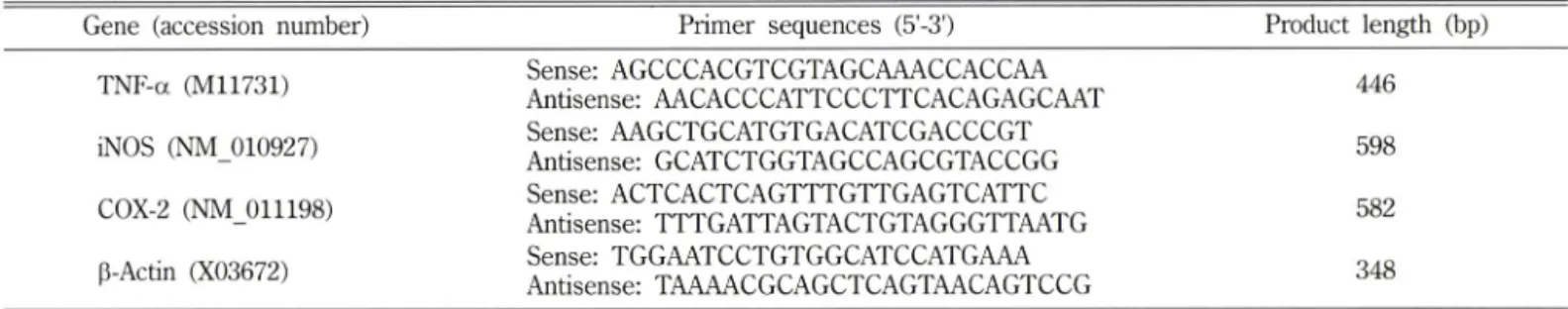
8prime와 Superscript™ ii RNase H' Reverse Transcriptase(Invitrogen Tech-Line™, USA)를 이용하여 역?i사하였다. Primer의 종류 및 서열은 Table I에 표시한 바와 같다. PCR 반응 조건은 GeneAmp 2700 thermocycler(Applied Biosystems, USA)에서 94°C, 5분간 변성,
72% 7분간 연장하였으며, 각각의 primer에 대한 증폭 주기의
조건은 다옴과 같다.: TNF-a, 28주기 94°C 30초, 65 X 30초,
72°C 60초; iNOS, 35주기 94T 30초, 65°C 30초, 72X 30초;
치자의 간보호 효과 57
Table I - Characteristics of specific primers used for RT-PCR analysis
Gene (accession number) Primer sequences (5'-3') Product length (bp) TNF-a (M11731) Sense: AGCCCACGTCGTAGCAAACCACCAA
Antisense: AACACCCATTCCCTTCACAGAGCAAT 446 iNOS (NM_010927) Sense: AAGCTGCATGTGACATCGACCCGT
Antisense: GCATCTGGTAGCCAGCGTACCGG 598
COX-2 (NM_011198) Sense: ACTCACTCAGTTTGTTGAGTCATTC
Antisense; TTTGATTAGTACTGTAGGGTTAATG 582
p-Actin (X03672) Sense; TGGAATCCTGTGGCATCCATGAAA
Antisense; TAAAACGCAGCTCAGTAACAGTCCG 348
COX-2, 35주기 9 4 T 30초, 60°C 30초, 72°C 30초; P-actin, 25 주기 94°C 30초, 56°C 30초, 72°C 30초. 이후 반응 생성물을 ethidium bromideS. 염색된 1.5% agarose gel을■ 이용하여 100 V 에서 견기 영동 하였고, 각 PCR 산물은 SLB MylmagerOJVP Inc., USA)와 ImageQuant™ TL(Amersham Biosciences/GE Healthcare, USA)률 사용하여 반정량적^ 분석하였다_
통계 처리
모든 실험 걸과는 one-way ANOVA를 사용하였으며 P <0.05 일 때 유의성 있는 차이가 었는 것^ 관정하였다.
실험 결과 및 고찰
혈중 ALT 및 AST 찰성도
아미노산 견이효소는 세포질에 존재하며, ALT 및 AST 활성 증가는 알코을, 유기용매 및 기타 득소에 의해 간장해가 발생할 때 혈중으로 유리되어 총 혈중농도가 증가하므로 득성 지표로 널 리 이용되고 있다.^® ALT 및 AST는 세포 내 위치에 따라 다른 동종효소로 존재하는데, 특히 ALT는 간에 많이 존재하며 AST 는 심장, 간, 골격근에 많아 그 특이성이 인정된다. 혈중 ALT 및 AST 활성도를 측정한 결과는 Table II와 같다. 사염화탄소 단득 투여군의 혈중 ALT 수치는 2115.6 ± 235.0 IU /; 으로 대조군에 비 해 현저히 증가하였으나 치자 30, 100 및 300 mg/kg 투여군의 경우 각각 903.0±80.0 IU//, 1012.7±76.5 IU// 및 1112 .5 ± 5 3 .9 IU//로 사염화탄소 단득 투여군에 비해 현저히 감소하였다. 혈중 AST 수처는 ALT 수치와 유사하게 사염화탄소 단득 투여시 대
조군에 비해 현저히 증가하였으나, 치자 30, 100 및 300 mg/kg 투여군 모두에서 사염화탄소 단득 투여군에 비해 현저히 감소하 였다. 따라서 간득성 지표인 혈중 ALT 및 A ST의 수처가 처자 30, 100, 300 mg/kg 모두에서 대조군에 비해 현저하게 감소한 것 으로 보아 처자가 간 보호에 탁월한 효과가 있음을 알 수 있었 다. 이률 바탕^ 이후 조직학적 분석 및 작용기전 연구에서는 치자 30 mg/kg 용량을 선택하쉬 실험을 진행하였다.
조직학적 분석
Fig. 1 는 사염화탄소 유도 급성 간득성 모델에서 치자가 간의 조직학적 번화에 머처는 영향을 살펴본 것이다. 그림에서 보는 바와 같어 간조직의 염색 결과, 사염화탄소 단득 투여군(Fig. IB) 에서는 문맥 주위 염증, 간세포 괴사 및 Kupffer cell 비대가 관 찰 되었으나 치자 30 mg/kg 투여군(Fig. 1C)에서 이러한 번화가 현저히 완화되었다. 이는 처자의 혈중 ALT 및 A ST 결과률 일 관성 있게 뒷받침하였으며, 이로서 처자가 사염화탄소로 유도된 급성 긴득성에 있어 보호효과가 있옴을 확인할 수 있었다.
지질 고 I 산화물 및 glutathione 정량
지질 과산화는 활성산소가 세포막의 불포화지방산을 공격함으 로써 일어나며, 이로 인하여 세포막뿐 아니라 세포막으로 둘러 싸인 소기관, 즉 미토콘드리아 및 소포체막 등에도 기능 손상을 유발한다. 최근 연구에 따르면 사염화탄소 그 자체는 득성이 강 하지 않으나 간장 내 일산소 첨가 효소계에 의해 활성화 되어 산 화적 손상과 세포막 인지질 내 불포화 지방산을 공격하여 간세 포의 기능과 구조를 파괴시킨다고 한다. Table III에서와 같이
Table II - Effects of G. jasminoides on the serum aminotransferase activities in the CC^-induced mice
Group Dose (mg/kg) ALT (IU//) AST (IU/ ii)
Control 41.6±3.4 30.2 ±5.5
CCI
4Vehicle 2115.6±235.0** 1629.6±127.4*="
G. jasminoides 30 903.0 ±80.0** 662.6±63.1 **'사
10 0
1012.7 : t76.5 **'■나 861.1 ±85.4 *=나 300 1112.5±53.9**''" 979.5±115.0**'^^
Silymarin
2001365.0+176.5*=^ 832.2 ±94.7 **■ ■ "우
The results are presented as the mean±S.E.M. of
8mice per group. ** Denotes significant differences (P<0.01) versus control group;
우 ' 우 사 " denote significant differences (P<0.05, P<0.01) versus
CCI4group.
C o n tro l C C I 4 G . Jasminoides
+
C C I4
Fig. 2 - Effect of G. jasminoides (30 mg^g) on the serum TNF-ct level of the CCl^-treatcd mice. The values are reported as the means±S.E.M. of
8mice per group. ** Denotes significant differences (P<
0.
0 1) versus control group;
나denotes significant differences (PcO.Ol) versus CCL group.
사염화란소 단득 투여군의 MDA 함량온 대조군의 약 5.1 배로 현 저히 증가되었으나, 처자 30 mg/kg 투여군에서 사*염화탄소 단득 투여군의 MDA 함량의 약 65.0% 정도로 현저히 억제되었다.
Glutathione은 세포의 산화적 손상에 대해 1차적 방어역할을 하 는 중요한 비단백성물질로서 , 세포가 산화적 손상을 받으면 GSH 가 GSSG로 산화됨으로써 활성산소룰 무 득 화 시 킨 다 본 실험 에서 사염화탄소 단득 투여 후 간 조직에 ■있는 GSH의 앙은 대조군의 82.9%로 감소하였으나, 처자 30 mg/kg 투여는 이러한 감소틀 억제하였다. 이는 처자의 간보호 작용의 일부가 사염화 탄소로 인한 세포막의 파괴룰 유발하는 지 질 파산화물의 생성 억 제와 간장 내 glutathione pool을 증가시킴으로써 세포의 산화적 손상에 대한 항산화 방어 기작을 통해 나타냄을 알 수 있었다.
혈중 TNF-a 농도 및 유전자 발현
간은 인체 주요 염증기관(inflammatory organ)으로 다양한 간 득소 노출 후 염증 반응은 일련의 병리학적 번화에 관여한다. 간
2 0 0
Fig. 1 - Liver sections stained with hematoxylin and eosin. Mice were treated orally with vehicle or G. jasminoides (30 kg) 48 h, 24 h and 2 h before
6h after CCI
4injection. (A):
Control group, showing normal hepatic architecture; (B):
CCI
4group, showing hepatocellular degeneration and necrosis with inflammatory infiltration; (C): G. jasminoides (30 mg/kg) + CCI
4group, showing mild hepatocellular necrosis and inflammatory infiltration. Original magnification x400.
Table III - Effects of G. jasminoides on the lipid peroxidation and hepatic glutathione content in the CC^-induced mice
Group Dose (mg/kg) MDA (nmol/mg protein) GSH (fimol/g liver)
Control 0.34±0.01 7.76+0.31
CCI4
Vehicle 1.76±0.14 =*^=*='■"구 6.43 ±0.03*=^
G. jasminoides 30 1.16±0.06 ==*'■ 느 우 7.17±0.16 *'■"우 The results are presented as the mean±S.E.M. of
8mice per group. 늙 * Denote significant differences (P<0.05, P<0.01) versus control group; 우두 denotes significant differences (PcO.Ol) versus
CCI4group.
58 신전규 • 김효연 • 이선미
++ * *
X
0
0
0 5
0
5 1
1
{IE /T o co
? d z l E E 0 W
I I C o n tro l
'(
혜CCI
4BS§
88S
88i G.
ja sm ino id es+ CCI
4J L
iNOS COX-2
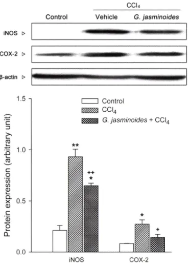
Fig. 3 - Effect of G. jasminoids (30 mg/kg) on the protein expression of iNOS and COX-2. Whole Hepatic proteins were extracted from mice 24 h after CCI
4injection. iNOS and COX-2 were detected by western blotting as described in Materials and Methods and p-actin was used as a loading control. The results are presented as the mean±S.E.M. of
8
mice per group. " 뉴 Denote significant differences (P<
0.05, P<0.01) versus control group : '
우' 우우denote significant differences (P<0.05, PcO.Ol) versus CCI
4group.
장내 대식세포인 Kupffer cell은 간세포 괴사 혹은 다성한 간 득 소에 반응하식 염증매개 인자를 유리하며 이는 사염화탄소로 유 도된 간 손상의 악화에도 관여된다.^®* TNF-a는 조직 손상시 대 식세포에서 빠르게 생성되는 대표적인 염증성 사미토카인이다.
사염 화탄소 노출 후 활성화된 Kupffer cell에서 TNF-a는 TNF- a 수용체와 함께 작용하여 간조직 괴사률 일으킨다.^® 본 연구 에서 사염화탄소 투여 후 혈중 TNF-a 농도는 149.3±0.5F®^m/
로 대조군에 비해 현저히 증가하였으나 처자 투여군에서 127.1±
1.3 pg/m/로 감소하였고, 이와 유사하게 유전자 발현량도 사염화 탄소 단득 투여군에서 대조군에 비해 약 3.8배로 증가하였으나 치자 투여는 이러한 증가를 현저히 억제하였다(Figs. 2 및 3).
iNOS 및 C0X-2 단백질과 유전자 발현
NO는 반응성이 메우 높은 산화제로 iNOS의 작용에 의해 L- Control
________ ecu__________
Vehicle G.
ja sm in oide s iNOS >COX-2 >
^-actin >
^ ' ■■■■■서
2 .0
1.5
o
ci 5
^IjunAJroJi!CIJro}Uojwwalclx0<NdLU
0.0
C ontrol
_________ ecu___________
V e h icle G. jasminoides TNF-a >
iNOS
COX-2
p-actIn >
C o n tro l
CCI
4G.
ja sm ino id es+ CCI
4T N F -a iNOS COX-2
Fig. 4 - Effect of G. jasminoides (30 mg/kg) on the mRNA expression of TNF-a, iNOS and COX-2. The levels of TNF-a, iNOS and COX-2 mRNA expression were measured by using RT- PCR as described in Materials and Methods. Also, p-actin was used as a loading control. The result is presented as the mean±S.E.M. of
8mice per group. Denote sig
nificant differences (P<0.05, F<0.01) versus control group : denote significant differences (P<0.05, P<0.01) versus CCI
4group.
arginine으로부터 간장의 설질세포와 비실질세포에서 생성된다.
정상시 NO는 세포 내에 낮은 농도로 존재하나 염증 만응 시 TNF- a에 의해서 iNOS가 유도되면 장시간 동안 다량의 NO률 생성하 며, 생성된 NQ는 혈관확장, 세포득성 및 조직손상 등과 같온 생 체유해작용을 나타낸다.^^^ 또한 iNOS에 의해 생성된 N O 는 superoxide anion과 반응하여 좀 더 강력한 산화물질인 peroxy- nitrite를 생성하며, 세포 내 oxidant-sensitive견사인자인 NF-
kB 을 활성화 시켜 다른 염증매개체의 생성을 촉진하쉬 염증 반응 을 심화시킨다.^^^ 다수의 염증 억제 약물돌의 작용기전은 prostaglandin 함성 억제이며, 이는 C0X-2의 생성 및 활성저해 에 의한 것이다. C0X-2는 동물이나 인간의 염증 반응 부위에서 발현되는 효소로, C0X-2에 의한 prostaglandin의 합성은 염증 반 응을 메개한다. 연구보고에 따르면 염증 반응에 있어 C0X-2 외
처자의 ?}보호 효과 59
olcrlAJalilclJro}col^waldx
이
60 신전규 • 김효연 ■ 이선머
생성은 사염화탄소로 유도된 간득성의 이차적 효과에도 관여한 다고 한다.2^^ Figs. 3 및 4에서 보는 바와 같이 iNOS 및 COX-
2
의 단백질 발현량은 대조군에 비해 사염화탄소 단득 투여군에 서 각각 약 4.4배, 및 3.3배로 현저히 증가하였으나 치자 투여 군 에서 이러한 증가가 현저히 감소되었다. 이와 유사하게 유견자 발현량에 있어서도 iNOS 및 C0X-2의 유전자 발현량이 사염화 탄소 단득 투여군에서 각각 약 3.8배 및 5.8배로 현저히 증가하 였으나 치자 투여 군에서 이러학 증가가 현저히 감소되었다. 이 는 아마도 치자가 견사 단계(transcriptional level)에서 사염화탄 소로 유도된 염증 매개인자인 iNOS 및 C0X-2의 생성을 조절하 여 간보호 작용을 나타내는 것으로 여겨진다.
결 론
처자의 간보호작용과 산화성 스트레스 및 염증매개인자 발현 억제에 대한 치자의 역할을 규명하기 위하식 사염화탄소룰 처치 한 생쥐의 간조직에서 ALT, AST, 지질과산화 생성과 염증매개 인자 TNF-a, iNOS 및 C0X-2의 혈중 농도, 유전자 및 단백질 발현량을 관찰하였다.
혈중 ALT 및 AST의 수치는 사•염화탄소 단득 투여군에 비해 치자 투여 군에서 더 낮은 활성도를 나타내었고, 지질과산화량 은 처자 투여군에서 감소하였고 이와는 반대로 GSH는 처자 투 여군에서 증가하였다. 처자 투여군에서 염증매개인자인 TNF-a 혈중 농도 및 유전자 발현량이 사염화탄소 단득 투여군에 비해 낮게 나타났으며, iNOS 및 C0X-2의 단백질 및 유전자 발현량 역시 사염화탄소 단득 투여군에 비해 현저히 감소하였다.
이상의 결과듬을 중합하쉬 볼 때 치자는 사염화탄소로 유도된 간득성 모델에서 산화성 스트레스와 염증매개인자 발현 억제률 통하여 간보호 작용을 나타내는 것으로 여겨진다.
감사의 말씀
본 파제(결과물)는 교육과학기술부 • 지식경제부의 출연금으로 수행한 산학협력 중심대학육성사업의 연구결 파입니다.
참고문헌
1) Di Pascoli, L., Lion, A., Milazzo, D. and Caregaro, L. : Acute liver damage in anorexia nervosa. Int. J. Eat. Disord. 36, 144 (2004).
2) Wang, G. X., Ben, C. E. and Ye, B. K. : Reparative effects of biphenyl dimethyl dicarboxylate on experimental liver injury in rats with histochemical and electronmicroscopy study. Zhong Xi Yi Jie He Za Zhi
8, 158 (1988).
3) Bergamini, A., Bendandi, A., Maggi, G. and Chierego, G. :
Effect of lipotropic substances on the enzyme picture of liver tissue in fatty degeneration induced by carbon tetrachloride.
Boll Soc. Ital Biol Sper. 31, 800 (1955).
4) Aterman, K .: Studies in fibrosis of the liver induced by carbon tetrachloride. III. Pantothenic acid and liver fibrosis. AMA Arch. Pathol. 57, 26 (1954).
5) Simler, M., Maurer, M. and Mandard, J. C .: Cancer of Liver on Cirrhosis Due to Carbon Tetrachloride. Strasb. Med. 15, 910 (1964).
6
) Clawson, G. A. : Mechanisms of carbon tetrachloride hepatotoxicity. Pathol Immunopathol Res.
8, 104 (1989).
7) Ding, H., Huang, J. A., Tong, J., Yu, X. and Yu, J. P : Influence of Kupffer cells on hepatic signal transduction as demonstrated by second messengers and nuclear transcription factors. World J. Gastroenterol 9, 2519 (2003).
8