• 검색 결과가 없습니다.

Electrophysiological Study of Medial Plantar Nerve in Idiopathic Tarsal Tunnel Syndrome

N/A
N/A
Protected

Academic year: 2021

Share "Electrophysiological Study of Medial Plantar Nerve in Idiopathic Tarsal Tunnel Syndrome"

Copied!
6
0
0

로드 중.... (전체 텍스트 보기)

전체 글

(1)

Introduction

The tarsal tunnel syndrome (TTS) is the most common form of entrapment neuropathy of the

tibial nerve, and compression neuropathy of the posterior tibial nerve and its branches (medial or lateral plantar nerve) under the flexor retinacu- lum behind and below the medial malleolus of the ankle. No population-based study exists to deter- mine the incidence or prevalence of TTS. The most common cause of TTS is idiopathic. The common symptoms of TTS are burning pain, numbness and paresthesia in the sole of the foot.

The pain may worsen, or occur only after pro- longed standing, walking, jogging, or running.

특발성 발목터널 증후군에서 내측 발바닥 신경의 전기 생리학적 검사

가톨릭대학교 의과대학 신경과학교실, 성균관대학교 의과대학 삼성서울병원 신경과�
안재영∙김병준�

Electrophysiological Study of Medial Plantar Nerve in Idiopathic Tarsal Tunnel Syndrome

Jae Young An, M.D., Byoung Joon Kim, M.D.*

Department of Neurology, College of Medicine, The Catholic University of Korea

Department of Neurology, Sungkyunkwan University School of Medicine, Samsung Medical Center*

Background: Tarsal tunnel syndrome (TTS) is an entrapment neuropathy of the tibial nerve within fibrous tunnel on the medial side of the ankle. The most common cause of TTS is idiopathic. This is a retrospective study to define the electrophysiological characteristics of idiopathic TTS. Methods: We reviewed the medical and electrophysiological records of consecutive patients with foot sensory symptoms referred to electromyography laboratory. Inclusion of patients was based on clinical findings suggestive of TTS. Among them, patients with any other possible causes of sen- sory symptoms on the foot were excluded. Control data were obtained from 19 age-matched people with no sensory symptoms or signs. Routine motor and sensory nerve conduction study (NCS) including medial plantar nerve (MPN) using surface electrodes were performed. Result: Twenty one patients (13 women, 8 men, 9 unilateral, 12 bilateral) were enrolled to have idiopathic TTS (total 31 feet). Tinel’s sign was positive in 16 feet (51.6%) of TTS and four feet (10.5%) in control group. The statistically significant electrophysiological parameter was difference of sensory conduc- tion velocity (SCV) between sural nerve and MPN. Amplitude of sensory nerve action potential and SCV of MPN were not different significantly between idiopathic TTS feet and controls. Conclusion: Bilateral development in idiopathic TTS was more common. Tinel’s sign and difference of SCV between sural nerve and MPN may be helpful for the diag- nosis of idiopathic TTS.

Key Words: Tarsal tunnel syndrome, Medial plantar nerve, Tinel’s sign, Sural nerve, Sensory conduction velocity

Address for correspondence Byoung Joon Kim, M.D.

Department of Neurology, Samsung Medical Center,

Sungkyunkwan University School of Medicine,

Ilwon-Dong 50, Gangnam-Gu, Seoul, Korea, 135-710

Tel: +82-2-3410-3594 FAX: +82-2-3410-0052

E-mail : [email protected]

(2)

The clinical features of TTS have been described in detail.

1-4

The electrophysiological methods used to measure distal motor latency and sensory con- duction velocities in the plantar nerves and the results of surgical release in TTS vary consider- ably throughout the literature. In this study, we retrospectively reviewed clinical and electrophys- iological features in clinically suspected idiopathic TTS patients.

Methods

Patients

We retrospectively reviewed the medical and electrophysiological records of patients who had referred to electromyography laboratory due to foot sensory symptoms between 1995 and 2004.

We selected 21 patients with clinically suggestive of idiopathic TTS. Case inclusion criteria were based on clinical history and symptoms suggest- ing TTS. They included any kind of paresthesia or pain in all or part of the foot supplied by the plantar nerves. These symptoms may be triggered or increased by prolonged walking and standing or at night, and rarely radiated to the calf.

Symptoms may be relieved by rest and removal of shoes. Possible physical signs included Tinel’ s sign (elicited by percussion just proximal to upper edge of tarsal tunnel behind medial malleolus), sensory loss over plantar nerve territory and weakness, hypotrophy or contractures of the plantar foot muscles. Patients with any possible causes of sensory symptoms on the foot, such as diabetes, rheumatoid arthritis, gout, hypothy- roidism, back pain and lower lumbosacral herni- ated intervertebral disc, history of ankle or foot trauma, were excluded based on medical history, physical examination, routine blood test and electromyography (EMG). Also, patients were excluded if the nerve conduction study showed peripheral neuropathy inconsistent with TTS.

Body mass index (BMI), which is a good general indicator of body fat content, was calculated as weight/height

2

(kg/m

2

). Control data were obtained from 19 age-matched people with no sensory symptoms or signs.

Electrophysiological method

Motor conduction velocity (MCV) of tibial nerve was performed, stimulating the nerve supramaxi- mally at the popliteal fossa and above the flexor retinaculum. Recording surface electrodes (Ag/AgCl) were placed on the motor point of the abductor hallucis muscle for the medial plantar nerve (MPN). Distal motor latency (DML) was determined at a distance of 10 cm between stimu- lating (above the flexor retinaculum) and record- ing points. Amplitude of compound muscle action potential (CMAPs) was calculated peak to peak.

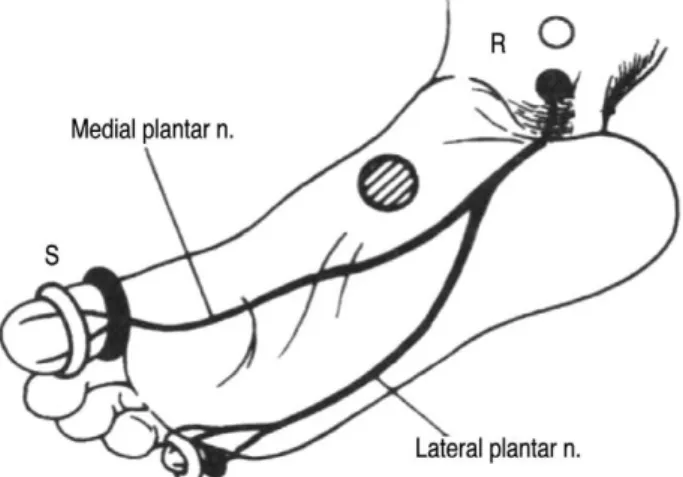
Maximum MCV was measured from the popliteal fossa to the medial malleolus. Orthodromic sen- sory conduction velocity (SCV) was obtained stimulating the big toe with ring electrodes for MPN (Fig. 1). Recording surface electrodes were placed above the flexor retinaculum. Latency was calculated from the start of the electrical artifact to the first positive peak of the nerve potential.

Amplitude of sensory nerve action potential (SNAP) was measured peak to peak. SNAP was considered absent when no constant potential was averaged and distinguished from noise after 80~100 stimuli in two successive trials. The SCV of sural nerve was measured antidromically from calf to lateral malleolus. The foot skin tempera- ture was measured at the start of the assessment, and if lower than 30� C was warmed using warm water.

The electrophysiological values of each subject

Figure 1. Position of the stimulating and recording electrode

for sensory nerve conduction of the plantar nerve.

(3)

were considered abnormal if they were 2 SD below or above the mean of age-matched controls. And it was considered abnormal if it was 8 m/sec slower than the asymptomatic MPN and/or the distal SCV of the sural nerve of the same side.

5

Statistical analysis for differences in clinical and electrophysiological data between TTS patients and the control group was performed with Chi-square test or Mann-Whitney U test.

Needle EMG of the intrinsic foot muscles inner- vated by the plantar nerves was not performed.

Results

Twenty one patients, 13 women and 8 men, were enrolled to have idiopathic TTS. Nine uni- lateral and 12 bilateral idiopathic TTS (total 31 feet) were included. Two feet were excluded due to incomplete nerve conduction study. Mean age of patients with bilateral idiopathic TTS (52±11.1 years) was significantly older than unilateral (44

±8.8 years)(p=0.012). Among idiopathic TTS feet, Tinel’ s sign was positive in 16 feet (51.6%). Four feet in control group were positive for Tinel’ s sign (10.5%). BMI and low-density lipoprotein (LDL) were not significantly different between idiopathic TTS and control group. Control group was consisted of 19 patients (9 men, 10 women;

mean age, 50±9 years, total 38 feet). Demo graphic and clinical features were showed in Table 1.

Electrophysiological findings

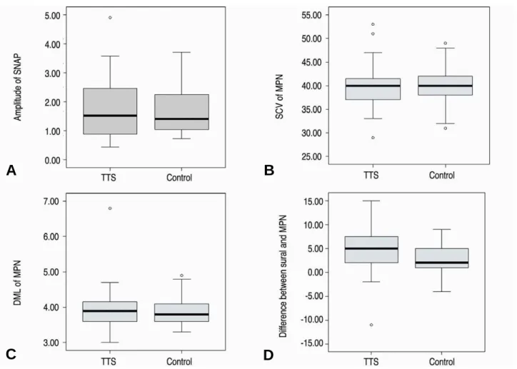
SNAP of MPN was absent in 4 feet (12.9%). All control group provoked sensory action potential of MPN. The NCS findings of MPN and difference of SCV between sural nerve and MPN are shown in Fig. 2. SCV of MPN was reduced in 5 feet (16.1%) and amplitude of SNAP of MPN was reduced in 4 feet (12.9%). Amplitude of CMAP of MPN was reduced in 8 feet (25.8%), while DML was delayed in 1 foot (3.2%) and MCV was reduced in 2 feet (6.5%). Among clinically idiopathic TTS, the number of foot showing difference of SCV of more than 8 m/s between MPN and sural nerve was 11 (35.5%) and between symptomatic MPN and asymptomatic MPN was 1 (12.5%). In compar- ison between idiopathic TTS and control group, findings showing statistical significance were dif- ference of SCV between sural nerve and MPN (p=0.04)(Table 2).

Discussion

The incidence of TTS in the EMG laboratories is 0.4~0.5% of all EMG examinations.

5,6

Idiopathic cases are the most frequent and women are more often affected than man, although the gender difference is not as marked as in CTS. Generally, unilateral idiopathic TTS is more common than bilateral. TTS may be determined by the direct or indirect consequences of ankle trauma, intra- neural ganglion, synovial sarcoma, benign and malignant tibial nerve neoplasm, retinaculum

Table 1. Demographic and clinical findings of patients with idiopathic tarsal tunnel syndrome and control group

Idiopathic TTS feet (n=31 feet) Control group (n=38 feet)

Age (year) 50 11.0 50 9.0

BMI (kg/m

2

)* 22.6 2.06 (n=18) 22.5 2.67 (n=22)

LDL (mg/dL) 124 48.9 (n=10) 117 15.5 (n=8)

Tinel’s sign (p<0.001) 16 (51.6%) 4 (10.5%)

Idiopathic TTS patients (n=21)

Site of symptoms Unilateral (n=9) Bilateral (n=12)

Age (year)(p=0.021) 44 8.8 52 11.1

Duration of symptoms (months) 016 27.3 25 30.2

* BMI, body mass index = weight/height

2

(kg/m

2

) Chi square test

Mann-Whitney U test

(4)

fibrosis, muscle anomalies, tendon sheath or syn- ovial cyst, external compression by a cast, ski- boots or high-heeled shoes and by foot abnor- malities (pes planus, cavus or rectus). TTS has also been reported in rheumatoid arthritis, dia-

betes, hypothyroidism, acromegaly, progressive systemic sclerosis, hyperlipidemia, chronic thrombophlebitis, chronic venous stasis and some sports.

1,7-9

In our study, idiopathic TTS is more common in women, but, bilateral idiopathic TTS

Figure 2. Amplitude of sensory nerve action potential (A), sensory conduction velocity (B) and distal motor latency (C) of medial plantar nerve and difference of conduction velocity between sural nerve and medial plantar nerve (D) in TTS feet and control group.

A B

C D

Table 2. Electrophysiological findings in feet with idiopathic TTS and in a control group

TTS (n=31 feet) Control (n=38 feet) p-value Medial plantar nerve

SCV (m/sec) 39.8 5.2 39.8 3.8 NS

Amplitude of SNAP ( ) 01.7 1.1 01.7 0.8 NS

DML (ms) 03.9 0.6 03.9 0.4 NS

MCV (m/sec) 48.2 3.7 49.4 3.3 NS

Sural nerve

SCV (m/sec) 44.2 3.0 42.7 1.8 NS

Amplitude of SNAP ( ) 20.9 9.0 21.4 7.0 NS

Difference of SCV between sural and MPN (m/sec) 04.6 4.9 02.9 3.3 0.04

Difference of SCV between symptomatic and asymptomatic MPN (m/sec) 3.16 4.87 (n=8) 0.94 0.911 (n=19) NS SCV, sensory conduction velocity: SNAP, sensory nerve action potential; DML, distal motor latency; MCV, motor conduction veloc- ity; MPN, medial plantar nerve; NS, non significant

Mann-Whitney U test

(5)

is more common than unilateral. Bilateral idio- pathic TTS patient is significantly older than uni- lateral idiopathic TTS patients. We thought that idiopathic TTS may be associated with aging process. And so, unilateral idiopathic TTS may be developed to bilateral TTS over time. Hyperc holesterolemia and BMI were associated to other entrapment neuropathy, such as idiopathic carpal tunnel syndrome.

10,11

But in idiopathic TTS, BMI and LDL were not risk factor. The report in the literature about the sensitivity of Tinel’ s sign was variable. However, Tinel’ s sign is not pathog- nomic of nerve entrapment syndromes, and can also be elicited in the normal population and in patients with polyneuropathy.

12,13

Our study showed that Tinel’ s sign was more predominant in TTS patients than in control group. This signs may be helpful to diagnosis of TTS.

No international guidelines for clinical and electrophysiological diagnosis criteria have yet been published.

14

The pathophysiology of TTS may be similar to that of CTS and indeed a relation- ship has been demonstrated between idiopathic CTS and idiopathic TTS.

15

Like CTS, in which a recent review of the literature showed that com- parative tests of the median/ulnar nerves follow- ing stimulation of the ring finger had the highest sensitivity and the second highest specificity,

16

comparative tests (digit-malleolus tibial and dis- tal sural SCVs) emerge as the most sensitive elec- trophysiological tests in TTS. Mondeli et al.

5

found to be able to increase the diagnostic sensi- tivity to 91% by comparing the NCV from the unaffected side MPN or distal sural nerve: more than 8 m/sec difference was considered abnormal.

EMG of the intrinsic muscles of the foot sole may be useless in TTS. In fact, electromyographic signs of denervation activity at rest have been observed in normal feet.

17

Great diagnostic cau- tion is needed if tibial SAP is found to be absent in subjects over 65 years and SAP of the con- tralateral unaffected nerve has not been checked, because SAP of the plantar nerves may be unde- tectable in healthy, clinically normal, elderly sub- jects.

18,19

A possible explanation of widely debated incidence of TTS is that electrophysiological eval- uation of suspected TTS is not simple due to vari-

ous technical difficulties. In this research, differ- ences of SCV and amplitude between symptomatic and asymptomatic MPN is not helpful for the diagnosis of idiopathic TTS. But differences of SCV between sural and MPN was statistically sig- nificant, and so may helpful for the electrophysi- ological diagnosis of idiopathic TTS. The reason for non-significant difference of SCV of MPN between idiopathic TTS and control group may be due that asymptomatic foot may be subclinical TTS or aging effect to MPN. Response of MPN is reduced in older age than in younger age.

20-22

Due to retrospective character of our study, there were several limitations. First, only medial plantar nerve was included because lateral plan- tar nerve conduction study was not performed.

Second, imaging studies of foot to identify cause of TTS were not performed, and so other possible cause for TTS was not completely excluded.

Third, previous report of plantar nerve conduc- tion study showed variable range of normal for each age decade.

22

But our study included control group of a relatively wide age range. Fourth, while pervious study used SCV of distal sural nerve in comparing NCV, our study used SCV, was measured from calf to lateral malleolus.

In conclusion, some observations drawn from our study using surface electrode are as follows.

First, idiopathic TTS may be associated to aging process. Second, Tinel’ s sign is helpful for diag- nosis of idiopathic TTS. Third, in the electrophys- iological evaluation of idiopathic TTS, difference of SCV between sural nerve and MPN must be included.

REFERENCES

01. Mahn KT, Rock JJ, Hillstrom HJ. Tarsal tunnel syndrome.

A retrospective study. J Am Podiatr Med Assoc 1996;

86:81-91.

02. Goodgold J, Kopell HP, Spielholz NI. The tarsal tunnel syndrome: objective diagnostic criteria. N Eng J Med 1965;273:742-745.

03. Edwards WG, Lincoln CR, Bassett FH, Goldner JL. The tarsal tunnel syndrome: diagnosis and treatment. J Am Med Assoc 1969;207:716-720.

04. Mann RA. Tarsal tunnel syndrome. Othop Clin North Am

1974;5:109-115.

(6)

05. Mondelli M, Giannini F, Reale F. Clinical and electro- physiological findings and follow-up in tarsal tunnel syn- drome. Electroencephalogr Clin Neurophysiol 1998;

109:418-425.

06. Oh SJ, Sarala PK, Kuba T, Elmore RS. Tarsal tunnel syn- drome: electrophysiological study. Ann Neurol 1979;

5:327-330.

07. Ferraresi S, Leidi P, Leidi M, Ubiali E, Bortolotti G, Cassinari V. Tarsal tunnel syndrome. Report of a case and review of clinical and surgical aspects. Ital J Neurol Sci 1992;13:47-51.

08. Jackson DL, Haglund B. Tarsal tunnel in athletes. Case reports and literature review. Am J Sports Med 1991;

19:61-65.

09. Lorei MP, Hershman EB. Peripheral nerve injuries in ath- letes. Sports Med 1993;16:130-147.

10. Nakamichi K, Tachibana S. Hypercholesterolemia as a risk factor for idiopathic carpal tunnel syndrome. Muscle Nerve 2005;32:364-367.

11. Moghtaderi A, Izadi S, Sharafadinzadeh N. An evaluation of gender, body mass index, wrist circumference and wrist ratio as independent risk factors for carpal tunnel syn- drome. Acta Neurol Scand 2005;112:375-379.

12. de Kroma MCTF, Knipschild PG, Kester ADM, Spaans F.

Efficacy of provocative tests for diagnosis of carpal tunnel syndrome. Lancet 1990;335:393-395.

13. Jabre J, Dillard JW, Salzsieder BT, Guidos AR, Guidos PT. The use of multiple Tinel’s sign in the identification of patients with peripheral neuropathy. Electromyogr Clin Neurophysiol 1995;35:131-136.

14. Bilbao A, Wilcox MS. Tarsal tunnel revisited. Muscle

Nerve 1995;18:791.

15. Mondelli M, Cioni R. Electrophysiological evidence of a relationship between idiopathic carpal and tarsal tunnel syndromes. Neurophysiol Clin 1998;28:391-397.

16. American Association of Electrodiagnostic Medicine, American Academy of Neurology, and American Academy of Physical Medicine and Rehabilitation.

Practice parameters for electrodiagnostic studies in carpal tunnel syndrome: summary statement. Muscle Nerve 2002;25:918-922.

17. Fack B, Alaranta H. Short report: fibrillation potentials, positive sharp waves and fasciculation in the intrinsic muscles of the foot in healthy subjects. J Neurol Neursug Psychiatry 1983;45:681-683.

18. Guiloff RJ, Sherratt RM. Sensory conduction in medial plantar nerve. J Neurol Neurosurg Psychiatry 1977;

40:1168-1181.

19. Ponsford SN. Sensory nerve conduction in the medial and lateral plantar nerves. J Neurol Neurosurg Psychiatry 1988;51:188-191.

20. Antunes AC, Nobrega JA, Manzano GM. Nerve conduc- tion study of the medial and lateral plantar nerves.

Electromyogr Clin Neurophysio 2000;40:135-138.

21. Guiloff RJ, Sherratt RM. Sensory conduction in medical plantar nerve: normal values, clinical applications, and a comparison with the sural and upper limb sensory nerve action potentials in peripheral neuropathy. J Neurol Neurosurg Psychiatry 1977;40:1168-1181.

22. Ponsford SN. Sensory conduction in medial and lateral plantar nerves. J Neurol Neurosurg Psychiatry 1988;

51:188-191.

수치

Figure 1. Position of the stimulating and recording electrode for sensory nerve conduction of the plantar nerve.
Table 1. Demographic and clinical findings of patients with idiopathic tarsal tunnel syndrome and control group
Figure 2. Amplitude of sensory nerve action potential (A), sensory conduction velocity (B) and distal motor latency (C) of medial plantar nerve and difference of conduction velocity between sural nerve and medial plantar nerve (D) in TTS feet and control g

참조

관련 문서

Detection of Organic Vapors and Nerve Agent Simulant Vapors Based on Photoluminescent Bragg-Reflective Porous Silicon Interferometer.. Preparation of

(4)the spiral ganglion (5)the auditory nerve fibres.. The Basilar Membrane is a Mechanical Analyzer of Sound Frequency The Basilar Membrane is a Mechanical Analyzer of

Underground Sewage Treatment Plant, Trondheim, Norway 19.. Underground Sewage Treatment Plant,

The purpose of this study was to analyze the impaction pattern of the impacted mandibular third molar and the relationship with the inferior alveolar nerve

Kinematics : study of the geometry of motion. Relates displacement, velocity, acceleration, and time without reference to the cause of motion. Kinetics : study of

- Prove or give a counter-example of the following statement: In three space dimensions and time, given an initial velocity field, there exists a vector velocity and

1 John Owen, Justification by Faith Alone, in The Works of John Owen, ed. John Bolt, trans. Scott Clark, &#34;Do This and Live: Christ's Active Obedience as the

This study confirmed that the characteristics of cyberspace, hypermedia and multi-sensory experience of new media in the fourth industrial revolution era can