• 검색 결과가 없습니다.

Interleukin-13 Increases Podocyte Apoptosis in Cultured Human Podocytes

N/A
N/A
Protected

Academic year: 2021

Share "Interleukin-13 Increases Podocyte Apoptosis in Cultured Human Podocytes"

Copied!
6
0
0

로드 중.... (전체 텍스트 보기)

전체 글

(1)

DOI: https://doi.org/10.3339/jkspn.2018.22.1.22 ISSN 2384-0250 (online)

Interleukin-13 Increases Podocyte Apoptosis in Cultured Human Podocytes

Purpose: Podocytes are important architectures that maintain the crucial roles of glomerular filtration barrier functions. Despite this structural importance, how- ever, the mechanisms of the changes in podocytes that can be an important pa- thogenesis of minimal change nephrotic syndrome (MCNS) are not clear yet. The aim of this study was to investigate whether apoptosis is induced by interleukin (IL)-13 in cultured human podocytes.

Methods: Human podocytes were treated with different IL-13 doses and apoptotic cells were analyzed using terminal deoxynucleotidyl transferase dUTP nick-end labeling (TUNEL assay) and fluorescence-activated cell sorting (FACS).

Results: The IL-13 increased the number of TUNEL-positive cells in a dose-depen- dent manner at 6 and 18 hours (P<0.05 and P<0.05, respectively). The apop tosis rate was appeared to be increased slightly in the IL-13-stimulated podocytes (8.63

%, 13.02%, and 14.46%; 3, 10 and 30 ng/mL, respectively) than in the control cells (7.66%) at 12 hours by FACS assay.

Conclusion: Our study revealed that IL-13 expression may increase podocyte apoptosis. Blocking the IL-13 signal pathway can potentially play an important role in regulating the apoptosis of podocytes.

Key words: Interleukin (IL)-13, Podocytes, Apoptosis, Minimal change nephrotic syndrome (MCNS)

Keum Hwa Lee, M.D. 1,2 Ji Young Oh, M.D. 1 Su-Bin Seong, M.D. 3 Tae-Sun Ha, M.D., Ph.D. 3 * Jae Il Shin, M.D., Ph.D. 1,2,4 *

1

Department of Pediatrics, Yonsei University College of Medicine, Seoul, Korea,

2

Department of Pediatric

Nephrology, Severance Children’s Hospital, Seoul, Korea,

3

Department of Pediatrics, Chungbuk National University College of Medicine, Cheongju, Korea,

4

Institute of Kidney Disease Research, Yonsei University College of Medicine, Seoul, Korea

*These authors are co-correspondence to this work.

Corresponding author 1: Tae Sun Ha, M.D., Ph.D.

Department of Pediatrics, Chungbuk National University College of Medicine, 410 Sung Bong-Ro, Heungduk-gu, Cheongju 28644, Korea

Tel: +82-43-269-6374, Fax: +82-43-264-6620 E-mail: [email protected]

Corresponding author 2: Jae Il, Shin, M.D., Ph.D.

Department of Pediatrics, Yonsei University College of Medicine, Seoul50 Yonsei-Ro, Seodaemun-gu, Seoul 03722, Korea Tel: +82-2-2228-2050, Fax: +82-2-393-9118 E-mail: [email protected]

Received: 28 March 2018 Revised: 3 April 2018 Accepted: 3 April 2018

This is an open-access article distributed under the terms of the Creative Commons Attribu tion Non-Commercial License (http://

crea tivecom mons.org/licenses/by-nc/4.0/) which permits unrestricted non-commercial use, distribution, and reproduction in any medium, provided the original work is properly cited.

Copyright © 2018 The Korean Society of Pediatric Nephrology

Introduction

Minimal change nephrotic syndrome (MCNS) accounts for 84.5% of idio­

pathic nephrotic syndrome (INS) in children, and 10 to 25% of INS in adults

1,

2)

. About 80–90% of children with childhood INS are steroid­sensitive ne­

phrotic syndrome (SSRS) and around 60% of MCNS children show SSRS

3,4)

. Even though initial response rate to steroids is 90% to 95%, 20–60% of SSRS children relapse and about 60–90% of them will have five or more relapses with increased morbidity

4,5)

. Resistance to therapy, also called as steroid­re­

sistant nephrotic syndrome (SRNS), occurs in 50% of focal segmental glo­

merulosclerosis (FSGS) and 10% of MCNS often progress to renal failure re­

quiring dialysis and transplantation despite various immunosuppressive treatments

6­9)

.

MCNS is composed of two important pathologic features: 1) absence of glomerular immune complex deposition and 2) foot process effacement (podo­

cytes seem to be fused together a flattened morphology)

8)

. Foot process

(2)

effacement is closely related the changes in the selective barrier in the slit diaphragm, which is composed of a number of proteins: nephrin, P­cadherin, CD2­asso ciated protein, zona occludens (ZO)­1, Fat cad herin, podo cin, and Neph1

10,11)

.

Interleukin (IL)­13 is a kind of T cell­derived cytokine

12)

. It is also reported to be an important cytokine in MCNS and we previously reported that IL­13 may increase podo­

cyte permeability through the modulation of ZO­1

13,14)

. How ever, there have been no reports about podocyte apop­

tosis related to IL­13. The aim of this study was to investi­

gate whether apoptosis is induced by IL­13 in cultured human podocytes.

Materials and methods

1. Cell culture of human podocytes

Human conditionally immortalized podocytes (AB8/23) was cloned from human glomerular cultures. Dr. Moin A.

Saleem (University of Bristol, Bristol, UK) cha racterized and generously provided them. Then human podocytes were maintained in RPMI 1640 (WelGENE Inc., Daegu, South Korea) supplemented with 10% heat­inacti vated fetal bovine serum (FBS), Insulin­Transferrin­Sele nium­

Pyruvate Supplement (ITSP; WelGENE Inc.), and anti­

biotics. Once every 2 days, fresh media was supplied.

Cells were cultivated at 33℃ (permissive conditions) in a culture medium supplemented with human recombinant ITSP to induce expression of temperature­sensitive large T antigens and to stimulate human podocyte proliferation.

To induce differentiation, podocytes were maintained at 37℃ (non­permissive conditions) for at least 2 weeks, and for subcultures, 0.05% trypsin was used to detach cells from the culture dishes.

2. IL-13 treatment conditions

To imitate MCNS­like conditions, cells were incubated with various concentrations of IL­13 (Peprotech Inc., Rocky Hill, NJ, USA) during the indicated time periods (6, 12 and 18 hours). IL­13 was administered with various concentra­

tions (3, 10, 30, and 100 ng/mL) into 0.5% RPMI at 37℃.

3. Measurement of apoptosis

1) Terminal deoxynucleotidyl transferase dUTP nickend labeling (TUNEL assay)

TUNEL assay was done using In situ Cell Death Detec­

tion Kit (Roche Molecular Biochemicals, Mannheim, Germany). Podocytes that were grown on type I collagen­

coated glass coverslips incubated for 24 hours were fixed in 4% paraformaldehyde for 1 hour, followed by permea­

bilization with 0.1% Triton X­100 for 10 minutes at room temperature. After coverslips were mounted in mountant, the samples were immediately evaluated using a fluores­

cence microscope. The TUNEL index (apoptotic podocytes) was determined by counting the positively and negatively stained cells in each of 10 fields of vision. Cell numbers were converted to percent apoptotic cells for statistical analysis.

2) Fluorescence­activated cell sorting (FACS)

We conducted a flow cytometric analysis to elucidate whether IL­13 has apoptotic effects on podocytes. The ap­
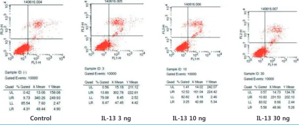
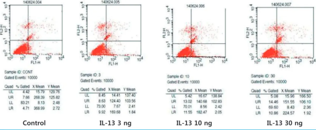
optotic rate was calculated by a single­color flow cytometric analysis depending upon the IL­13 concentrations (control, 3, 10, and 30 ng/mL) and apoptotic hours (6 and 12 hours).

Equal numbers of podocytes were cultivated on 6 cm tissue culture plates in medium. After IL­13 modulation, cell cul­

ture medium was collected and saved. Cells were washed once with 1.5 ml of phosphate­buffered saline (PBS). PBS used for washing was combined with the saved culture medium. Podocyte cells were analyzed with the Mofro Astrios flow cytometer (FACSCalibur­S System, Becton Dickinson Biosciences, San Jose, CA, USA). The number of apoptotic cells was calculated by multiplying the percen­

tages of apoptosis and necrosis as determined by FACS.

4. Statistical analysis

Results are described as mean ± standard deviation, as appropriate under different conditions. Statistical signifi­

cance was evaluated by the non­parametric Kruskal­Wallis

analysis or Student’s t­test. P­values <0.05 were considered

significant.

(3)

Results

1. Apoptotic rate on TUNEL assay

Apoptosis is a programmed cell death which results the series of events including alterations in the plasma mem­

brane, activation of enzymes

15)

. DNA fragmentation can be detected in terminal stages of apoptosis by labeling with fluorescent tagged nucleotides through a technique called as TUNEL

16)

. Apoptotic podocytes were identified as TUNEL positive cells while the normal cells lacked nuclear staining. IL­13 increased the number of TUNEL­positive cells in a dose­dependent manner at 6 and 18 hours ( P<0.05 and P<0.05, respectively) (Fig. 1, 2).

2. FACS assay for apoptosis

The apoptotic effects of IL­13 on podocytes were an­

alyzed by FACS. There was no significant difference in the rate of apoptosis in controls and podocytes stimulated by different concentrations (3, 10 and 30 ng/mL) at 6 hours (9.73% vs. 13.89%, 12.52%, 10.83%, respectively, Fig. 3); at 12 hours, however, the rate of apoptosis was slightly in­

creased in IL­13 stimulated podocytes (8.63%, 13.02%, 14.46%) than control cells (7.66%, Fig. 4).

Discussion

Podocytes are important structures which maintain the crucial roles of glomerular filtration barrier functions

9,10)

. In 2007, Lai et al. suggested that IL­13 overexpression could

lead to podocyte injury with downregulation of proteins such as nephrin, podocin, and dystroglycan and a concur­

rent upregulation of B7­1 in MCNS induced rat

17)

. After that, our previous studies reported that IL­13 was involved in the changes of ZO­1 proteins in podocytes which could

* *

0.000 0.020 0.040 0.060 0.080 0.100 0.120 0.140 0.160

(-) 3ng 10ng 30ng 100ng

Apo pt oti c r at e

IL-13 concentrations

Fig. 1. TUNEL assay for apoptosis (6 hours). IL-13 increased the number of TUNEL-positive cell in a dose-dependent manner at 6 hours (* P<0.05). IL-13, interleukin-13; TUNEL, terminal deoxynu- cleotidy1 transferase dUTP nickend labeling.

*

* *

*

0.000 0.020 0.040 0.060 0.080 0.100 0.120 0.140 0.160

(-) 3ng 10ng 30ng 100ng

Apo pt oti c r at e

IL-13 concentrations

Fig. 2. TUNEL assay for apoptosis (18 hours). IL-13 increased the number of TUNEL-positive cell in a dose-dependent manner at 18 hours (* P<0.05). IL-13, inter leukin-13; TUNEL, terminal deoxy- nucleotidy1 transferase dUTP nickend labeling.

Control IL-13 3 ng IL-13 10 ng IL-13 30 ng

Fig. 3. FACS assay for apoptosis (6 hours). FACS, fluorescence-activated cell sorting; IL-13, interleukin-13.

(4)

be a pathogenesis of MCNS

14,18)

. Other recent studies have shown that increase in IL­13 production was to cause fre­

quent relapse in SSNS

13,19)

. In this point of view, the aim of this study was to identify whether apoptosis in podocytes could be induced by IL­13.

There have been two suspected modes of podocyte apop­

tosis: 1) intrinsic apoptosis and 2) extrinsic apoptosis. In­

trinsic apoptosis is related to a mitochondrion­centered control mechanism

20)

. On the other hand, extrinsic apop­

tosis is a caspase­dependent cell death related to activation of the caspase­9­caspase­3 cascade

21,22)

. Generally, caspase­

3 is known as a key mediator as a leading enzyme in cell death

23)

. Pro­caspase­3 is usually processed by autoproteo­

lytic cleavage and activated caspase­3 has been identified that are related to mitochondrial cytochrome c release and caspase­9 function

24,25)

. Manna et al. reported that IL­13 is a potent inhibitor of TNF­mediated activation and caspase­

3 related apoptosis in 1998

26)

. In contrast, recent studies show IL­13 induces apoptotic pathways of cell death in head and neck cancer models

27,28)

.

IL­13 has been known as a causative factor of cell apop­

tosis in several previous studies. Borowski et al. reported that IL­13 can be a mediator on lung epithelial cells as an apoptotic effector, which leads to loss of functional airway tissue and finally results in asthma

29)

.

Several studies have already reported about the role of IL­13 as a pathogen in other cells and diseases other than podocytes. Heller et al. found that IL­13 can effect on in­

testinal epithelial barrier dysfunction and eventually cause ulcerative colitis

30)

. For this explanation, signal transduc­

tion qualities mediated by the respective receptors for IL‐

13 may result in distinct patterns of transcription regula­

tion, ultimately explaining the unique role of IL‐13 in asthma pathogenesis. JAK/STAT activation pathway can be an optional mechanism to explain these results

31)

. This pathway is induced by the IL‐13 receptor complex and then eventually causes cell apoptosis and destruction of cell junc­

tions

31)

. Exact mechanisms of these mediators, however, are unclear.

In this study, it is implicated that IL­13 can induce po­

docyte apoptosis. Our results and previous studies above indicate that IL­13 can be related to extrinsic apop tosis of podocyte, blocking this signal pathway.

Our study has several limitations: First, we were unable to reveal all exact mechanism of IL­13 in podocytes. Second, we did not include treatment attempt to prevent podocyte apoptosis into our experiments. Despite these limitations, our study is the first attempt to demonstrate the IL­13 as a key marker and potential treatment target to prevent changes related to cell death in human podocytes.

In conclusion, our preliminary results suggest that IL­13 may increase podocyte apoptosis. Such alterations can be relevant to the foot process changes in podocytes related to pathogenesis of proteinuria in the IL­13­induced MCNS model. Further studies about the methods of suppression of IL­13 are needed, and it will be noteworthy to explore whether IL­13 modulation can modify podocytes from MCNS.

Control IL-13 3 ng IL-13 10 ng IL-13 30 ng

Fig. 4. FACS assay for apoptosis (12 hours). FACS, fluorescence-activated cell sorting; IL-13, interleukin-13.

(5)

Conflict of interest

The authors of the manuscript declare no conflict of interest.

Role of funding source

This research was supported by the Basic Science Research Program through the National Research Foundation of Korea (NRF) and funded by the Ministry of Education, Science and Technology (2013R1A1A1012112 to J.I. Shin and 2016R1D1A1B03931888 to T.S. Ha).

References

1. Churg J, Habib R, White RH. Pathology of the nephrotic syndrome in children: a report for the International Study of Kidney Disease in Children. Lancet 1970;760:1299-30.

2. Haas M, Meehan SM, Karrison TG, Spargo BH. Changing etiolo- gies of unexplained adult nephrotic syndrome: a comparison of renal biopsy findings from 1976-1979 and 1995-1997. Am J Kidney Dis 1997;30:621.

3. Trompeter RS, Lloyd BW, Hicks J, White RH, Cameron JS. Long- term outcome for children with minimal-change nephrotic syndrome. Lancet 1985;1:368-70.

4. A report of the International Study of Kidney Disease in Children.

The primary nephrotic syndrome in children. Identification of patients with minimal change nephrotic syndrome from initial response to prednisone. J Pediatr 1981;98:561-4.

5. Teeninga N, Kist-van Holthe JE, van Rijswijk N, de Mos NI, Hop WC, Wetzels JF, et al. Extending prednisolone treatment does not reduce relapses in childhood nephrotic syndrome. J Am Soc Nephrol 2013; 24:149-59.

6. Tarshish P, Tobin JN, Bernstein J, Edelmann CM Jr. Prognostic sig- nificance of the early course of minimal change nephrotic synd- rome: Report of the International Study of Kidney Disease in Children. J Am Soc Nephrol 1997;8:769-76.

7. Gipson DS, Chin H, Presler TP, Jennette C, Ferris ME, Massengill S, et al. Differential risk of remission and ESRD in childhood FSGS.

Pediatr Nephrol 2006;21:344-9.

8. D’Agati VD. Pathologic classification of focal segmental glome- rulosclerosis. Semin Nephrol 2003;23:117-34.

9. Schnaper HW. Idiopathic focal segmental glomerulosclerosis.

Semin Nephrol 2003;23:183-93.

10. Pavenstadt H, Kriz W, Kretzler M. Cell biology of the glomerular podocyte. Physiol Rev 2002;83:253-307.

11. Asanuma K, Mundel P. The role of podocytes in glomerular pa- thobiology. Clin Exp Nephrol 2003;7:255-9.

12. McKenzie AN, Culpepper JA, de Waal Malefyt R, Briere F, Punnonen J, Aversa G, et al. Interleukin 13, a T-cell-derived cytokine that regulates human monocyte and B-cell function. Proc Natl Acad Sci USA 1993; 90:3735-40.

13. Cheung W, Wei CL, Seah CC, Jordan SC, Yap HK. Atopy, serum IgE, and interleukin-13 in steroid-responsive nephrotic syndrome.

Pediatr Nephrol 2004;19:627-32.

14. Park SJ, Saleem MA, Nam JA, Ha TS, Shin JI. Effects of interleukin- 13 and montelukast on the expression of zonula occludens-1 in human podocytes. Yonsei Med J 2015;56:426-32.

15. Majno G, Joris I. Apoptosis, oncosis, and necrosis. An overview of cell death. Am J Pathol 1995;146:3-15.

16. Gavrieli Y, Sherman Y, Ben-Sasson SA. Identification of pro-gram- med cell death in situ via specific labeling of nuclear DNA frag- mentation. J Cell Biol 1992;119:493-501.

17. Lai KW, Wei CL, Tan LK, Tan PH, Chiang GS, Lee CG, et al. Overex- pression of interleukin-13 induces minimal-change-like nephro- pathy in rats. J Am Soc Nephrol 2007;18:1476-85.

18. Ha TS, Nam JA, Seong SB, Saleem MA, Park SJ, Shin JI. Montelukast improves the changes of cytoskeletal and adaptor proteins of human podocytes by interleukin-13. Inflamm Res 2017;66:793- 802.

19. Yap HK, Cheung W, Murugasu B, Sim SK, Seah CC, Jordan SC. Th1 and Th2 cytokine mRNA profiles in childhood nephrotic synd- rome: evidence for increased IL-13 mRNA expression in relapse. J Am Soc Nephrol 1999;10:529-37.

20. Kroemer G, Galluzzi L, Brenner C. Mitochondrial membrane permeabilization in cell death. Physiol Rev 2007;87:99-163.

21. Ryu M, Mulay SR, Miosge N, Gross O, Anders HJ. Tumour necrosis factor-alpha drives Alport glomerulosclerosis in mice by promo- ting podocyte apoptosis. J Pathol 2011;226:120-31.

22. Galluzzi L, Vitale I, Abrams JM, Alnemri ES, Baehrecke EH, Blago- sklonny MV, et al. Molecular definitions of cell death subroutines:

recommendations of the Nomenclature Committee on Cell Death 2012. Cell Death Differ 2012;19:107-20.

23. Faleiro L, Kobayashi R, Fearnhead H, Lazebnik Y. Multiple species of CPP32 and Mch2 are the major active caspases present in- apoptotic cells. EMBO J 1997;16:2271-81.

24. Polverino AJ and Patterson SD. Selective activation of caspases during apoptotic induction in HL-60 cells. J Biol Chem 1997;272:

7013-21.

25. Zou H, Henzel WJ, Liu X, Lutschg A, Wang X. Apaf-1, a human protein homologous to C. elegans CED-4, participates in cyto- chrome c-dependent activation of caspase-3. Cell 1997;90:405- 13.

26. Manna SK, Aggarwal BB. IL-13 suppresses TNF-induced activation of nuclear factor-kappa B, activation protein-1, and apoptosis. J Immunol 1998;161:2863-72.

27. Kawakami M, Kawakami K, Puri RK. Apoptotic pathways of cell

(6)

death induced by an interleukin-13 receptor-targeted recom- binant cytotoxin in head and neck cancer cells. Cancer Immunol Immunother 2002;50:691-700.

28. Kawakami M, Kawakami K, Puri RK. Tumor regression mechanisms by IL-13 receptor-targeted cancer therapy involve apoptotic pa- thways. Int J Cancer 2003;103:45-52.

29. Borowski A, Kuepper M, Horn U, Knüpfer U, Zissel G, Höhne K, et al. Interleukin-13 acts as an apoptotic effector on lung epithelial cells and induces pro-fibrotic gene expression in lung fibroblasts.

Clin Exp Allergy 2008;38:619-28.

30. Heller F, Fromm A, Gitter AH, Mankertz J, Schulzke JD. Epithelial apoptosis is a prominent feature of the epithelial barrier distur- bance in intestinal inflammation: effect of pro-inflammatory in- terleukin-13 on epithelial cell function. Mucosal Immunol 2008;1 Suppl 1:S58-61.

31. Friedrich K, Brändlein S, Ehrhardt I, Krause S, Luttmann W. Inter-

leukin‐4 and Interleukin‐13 receptors trigger distinct JAK/STAT

activation patterns in mouse lymphocytes. Signal Transduction

2003;3:26-32.

수치

Fig. 3. FACS assay for apoptosis (6 hours). FACS, fluorescence-activated cell sorting; IL-13, interleukin-13.
Fig. 4. FACS assay for apoptosis (12 hours). FACS, fluorescence-activated cell sorting; IL-13, interleukin-13.

참조

관련 문서

Apoptotic index(AL, 세포사멸 지수)는 terminal deoxynu- cleotidyl transferase (TdT)-mediated dUTP-biotin nick end labeling (TUBEL)에 의하여 측정되었다.. The

Also, ferrerol induced apoptotic bodies of MCF-7 cells as evaluated by TUNEL assay and Annexin V/PI staining using FACS.. By mechanism of action, ferrerol regulated the activation

Terminal deoxynucleotidyl transferase dUTP nick end labeling (TUNEL) and phospho-histone 3 (PH3) staining confirmed the association of gpx3 knockdown with increased cell

To verify whether the cytotoxicity of Vitisin A may be associated with apoptosis, TdT-mediated-dUTP Nick-End Labeling (TUNEL) assay and cell cycle analysis were

change in ZO-1 protein production in a puromycin amino- nucleoside (PAN)-treated rat model, we found that ZO-1 proteins were redistributed and reduced in MCNS. In addi- tion,

In scratch based wound healing assay, IL-6 treated group and HA treated group significantly increased cell migration compared to control group at 24 and 48 hr,