Protective Effect of Hypoxic Preconditioning on Hypoxic-Ischemic Injured Newborn Rats
Brief episodes of cerebral hypoxia-ischemia cause transient ischemic tolerance to
subsequent ischemic events that are otherwise lethal. This study was conducted to evaluate the protective effect of hypoxic preconditioning on hypoxic-ischemic injury in the neonatal rat and the persistence of a protective window after hypoxic preconditioning. The rats were preconditioned with hypoxia (8% oxygen, 92% nitrogen) for three hours, subjected to ischemia using ligation of the right common carotid artery, and then exposed to another three hours of hypoxia. Using proton magnetic resonance spectroscopy, terminal deoxynucleotidyl transferase-mediated dUTP-biotin nick end-labeling (TUNEL) staining, and morphologic scores, this study shows that hypoxic preconditioning 6-hr to 1-day before hypoxic-ischemic injury increases survival rates and has neuroprotective effects against subsequent hypoxic-ischemic injury. The mechanism of the protective effects of hypoxic preconditioning in the newborn rat brain may involve downregulation of apoptotic cell death.
Key Words: Hypoxia-Ischemia, Brain; Magnetic Resonance Spectroscopy Hyun-Kyung Park1, In-Joon Seol1
and Ki-Soo Kim2,3
1Department of Pediatrics, Hanyang University College of Medicine, Seoul; 2Department of Pediatrics, Asan Medical Center, University of Ulsan College of Medicine, Seoul; 3NMR Laboratory, Asan Institute for Life Science, Seoul, Korea
Received: 29 April 2011 Accepted: 5 September 2011 Address for Correspondence:
Ki-Soo Kim, MD
Department of Pediatrics, University of Ulsan College of Medicine, Asan Medical Center, 88 Olympic-ro 43 gil, Songpa-gu, Seoul 138-736, Korea
Tel: +82.2-3010-3386, Fax: +82.2-3010-6978 E-mail: [email protected]
This work was supported by the research fund of Hanyang University (HY-2008-000-0000-8123).
http://dx.doi.org/10.3346/jkms.2011.26.11.1495 • J Korean Med Sci 2011; 26: 1495-1500
INTRODUCTION
Hypoxic-ischemic encephalopathy due to birth asphyxia is the major factor that induces neuronal cell injury before, during, and after birth. Although there has been marked development in the fields of obstetrics and neonatology, the incidence of and mortality caused by hypoxic-ischemic encephalopathy remain elevated (1, 2).
Low birth weight infants and infants with intrauterine growth retardation, who have already experienced intrauterine repeti- tive hypoxic distress, seem to have milder hypoxic-ischemic brain damage than full-term newborns who are exposed to a lethal ischemic insult in the perinatal period. Therefore, some have suggested the possibility that sublethal hypoxic episodes afford a protective effect against further hypoxic exposure (3, 4).
Hypoxic preconditioning has been described in the brain, heart, retina, and other tissues. Gidday et al. (5) first showed that exposure of neonatal rat pups to hypoxia alone (8% oxygen for 3 hr) protected these animals 1 day later from a stroke induced by combined hypoxia/ischemia.
Although the mechanisms of preconditioning are still unde- termined, it appears to require synthesis of new RNA and pro- teins during the reoxygenation period before permanent ligation (6, 7). In addition, Miller et al. (8) explained that the difference in the degree of hypoxia and the delay between precondition- ing and ischemia determines the efficacy of preconditioning.
This study was conducted to evaluate the protective effect of hypoxic preconditioning on hypoxic-ischemic injury in the neonatal rat and the persistence of a protective window after hypoxic preconditioning. Furthermore, this study investigated the relationship between the downregulation of apoptosis and its possible role in the protective effect of hypoxic precondition- ing. The survival rates, as well as ratios of lipid to N-acetyl aspar- tate (NAA) and lipid to creatine (Cr), were investigated in posi- tive cells, and morphologic changes were investigated with pro- ton (1H) magnetic resonance spectroscopy (MRS) and terminal deoxynucleotidyl transferase-mediated dUTP-biotin nick end- labeling (TUNEL) staining.
MATERIALS AND METHODS Subjects
A total of 231 seven-day-old Sprague-Dawley newborn rats weigh- ing 12-18 grams were divided into six groups: preconditioned 6 hr before hypoxic-ischemic injury (Pre-6 hr, n = 30), 12 hr be- fore (Pre-12 hr, n = 32), 1 day before (Pre-1 day, n = 34), 3 days before (Pre-3 day, n = 41), 6 days before (Pre-6 day, n = 43), and control without preconditioning (n = 51). All experimental ani- mal protocols were approved by the University of Ulsan College of Medicine IACUC Committee (2008-12-011).
The rats were preconditioned by hypoxic exposure (8% oxy- gen/92% nitrogen) for three hours at 37˚C. Exhaled carbon di-
oxide was removed through a one-way valve in the lower part of a hypoxic chamber. Rats were returned to their home cages at room temperature immediately after the hypoxic precondi- tioning. Control group rats were isolated during precondition- ing, and then they were also returned to their home cages.
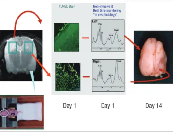
Using ligation of the right common carotid artery, 6 hr, 12 hr, 1, 3, and 6 days after receiving three hours of hypoxic precondi- tioning, all rats, including the control group, were subjected to ischemia. The right common carotid artery was dissected and ligated using silk (No. 5) under isoflurane anesthesia, and the rats were submitted to hypoxia (8% oxygen, 92% nitrogen) for 3 hr under the same conditions as in the preconditioning proce- dure. After the hypoxic-ischemic insult, the specimens were re- turned to their home cages at room temperature. The schematic diagram in Fig. 1 illustrates our hypoxic-ischemic animal model.
Measurements
1H MRS was performed 24 hr after hypoxic-ischemic insult, and the results were examined using Bruker Biospec 4.7 Tesla MRI/
MRS system (Bruker, Fallanden, Switzerland). The voxel for MRS was located in the parietotemporal area, and the volume of a single voxel was 3 × 3 × 4 µL. The spin echo signal was detected by TR = 3,000 msec, TE = 30 msec, and NS = 128, and the H2O signal was inhibited by a chemical shift selective (CHESS) se- quence. Spectra were analyzed semi-quantitively using an As- pect 3000 computer and Tomikon software (Bruker, Fallanden, Switzerland). In order to measure the ratios of lipid to NAA and lipid to Cr as early predictors of apoptosis, the 1H spectra of NAA, creatine, choline, and lipids were assessed at 2.02, 3.03, 3.24, and 1.3 ppm, respectively. To differentiate between lipids and lactate at 1.3 ppm, the presence of the peak at TE = 135 msec was analyzed.
One day after the hypoxic-ischemic injury, the rats were anes-
thetized with an intra-abdominal ketamine (2 mg/kg) injection following 1H MRS examination. The rats were then perfused through the left ventricle of the heart with heparinized saline (2 unit heparin/1 mL saline, 2.5 mL/g) followed by buffered para- formaldehyde (4% in phosphate buffer, pH 7.4). The brains were extracted, frozen, and enveloped in paraffin.
Two rats in each group were sacrificed one day after hypoxic- ischemic injury, and these brains were examined for histologic study using TUNEL staining. TUNEL staining for the detection of DNA fragmentation produced during cell apoptosis began with the digestion of deparaffinized sections using proteinase K, blocking solution, and permeabilization solution. These sec- tions were then stained with the TUNEL reaction mixture (50 µL) of an in situ cell death detection kit (Boehringer-Mannheim, Germany). Tissue sections were evaluated using immunofluo- rescence microscopy.
The remaining rats were morphologically evaluated on the fourteenth day after hypoxic-ischemic injury and scored accord- ing to the following scale of modified Palmer’s classification (9):
0, no difference between the two hemispheres in shape and size;
1, mild reduction of the right hemisphere volume only; 2, vol- ume reduction and atrophic changes with small cysts in the right hemisphere; 3, moderate atrophic changes of the right hemi- sphere; and 4, severe atrophic changes of the right hemisphere.
The results were expressed as mean ± standard deviation, and the statistics were calculated using an unpaired t-test, one way ANOVA, Tukey’s test, Dunn’s method, Mann-Whitney U test, and Pearson’s correlation with the threshold of significance at 0.05.
RESULTS
Here we confirmed the preconditioning groups, followed by 8%
oxygen hypoxia, reduced brain injury in newborn rats. The Pre-
Day 1 Day 1 Day 14
Fig. 1. Schematic diagram illustrating the hypoxic-ischemic animal model. One day after hypoxic-ischemic injury, the number of TUNEL-positive cells and the lipid peak on 1H MRS in the right brain markedly increase. Fourteen days after the insult, severe atrophy of the right hemisphere is observed.
TUNEL Stain Non-invasive &
Real time monitoring
“in vivo histology”
Survival rates (%)
Control Pre-6 days Pre-3 days Pre-1 day Pre-12 hr Pre-6 hr 100
80 60 40 20 0
* * *
Fig. 2. The effect of hypoxic preconditioning on survival rates after hypoxic-ischemic brain injury in newborn rats. The survival rates are higher in the Pre-1 day, Pre-12 hr, and Pre-6 hr groups than in the control group. The values are expressed as mean ± standard deviation.
6 hr, Pre-12 hr, and Pre-1 day hypoxic preconditioning groups had significantly higher survival rates when evaluating 152 rats among 231: control (25 rats, 47%); Pre-6 hr (26 rats, 86%); Pre- 12 hr (32 rats, 100%); Pre-1 day (32 rats, 94%); Pre-3 day (19 rats, 45%); and Pre-6 day (18 rats, 42%) (Fig. 2).
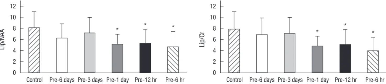
We performed 1H MRS study after hypoxic-ischemic insult;
and then lipid/NAA (N-acetyl aspartate) and lipid/Cr (creatine)
ratios were measured as early predictors of apoptosis. The Pre- 6 hr, Pre-12 hr, and Pre-1 day hypoxic preconditioning groups also had lower lipid/NAA and lipid/Cr ratios on 1H MRS in com- parison with the Pre-3 day and Pre-6 day (P < 0.05, Fig. 3). The lipid/NAA ratios in control, Pre-6 hr, Pre-12 hr, Pre-1 day, Pre-3 day and Pre-6 day ratios were 8.05 ± 2.95, 4.64 ± 2.78, 5.25 ± 2.58, 5.05 ± 1.86, 7.09 ± 2.87, and 6.19 ± 2.62, respectively.
Fig. 3. The ratios of lipid/N-acetyl aspartate (Lip/NAA) and lipid/creatine (Lip/Cr) on 1H MRS in the control group and hypoxic preconditioned groups one day after hypoxic-isch- emic brain injury in the newborn rat. The ratios of lipid/NAA and lipid/Cr at the hypoxic-ischemic injured right brain decrease significantly in the Pre-1 day, Pre-12 hr, and Pre- 6 hr groups. The values are expressed as mean ± standard deviation. *P < 0.05.
Lip/NAA
Control Pre-6 days Pre-3 days Pre-1 day Pre-12 hr Pre-6 hr 12
10 8 6 4 2 0
* * *
Lip/Cr
Control Pre-6 days Pre-3 days Pre-1 day Pre-12 hr Pre-6 hr 12
10 8 6 4 2 0
* *
*
Lt. Cerebral cortex Rt. Hippocampus
Left hemisphere
Control
Pre-6 days
Pre-3 days
Pre-1 day
Pre-12 hr
Pre-6 hr
Right hemisphere Lt. Hippocampus Rt. Cerebral cortex MRS
Fig. 4. Histologic findings of TUNEL staining in the newborn rat brain obtained on the first day after hypoxic-ischemic injury. Increased TUNEL-positive cells are noted in the right hemisphere in the control, Pre-6 day, and Pre-3 day groups. Increased 1H MRS lipid peaks were observed in the control, Pre-6 day, and Pre-3 day groups.
Results of the histologic evaluation demonstrated that the hypoxic preconditioning attenuated hypoxic-ischemic induced apoptosis in newborn brain. On day 1, the groups that received hypoxic preconditioning 6 hr, 12 hr, and 1 day prior to hypoxic- ischemic injury had fewer TUNEL-positive apoptotic cells pres- ent than in the control group, which received no precondition- ing. The apoptotic cells were found predominantly in cortex and hippocampus. In addition, the 1H MRS characteristics are shown and lower lipid peaks are observed in the preconditioning groups in Fig. 4.
We monitored the structural damage in rats of different groups on the fourteenth day after hypoxic-ischemic injury. The mor- phologic scores of the preconditioning groups were also signifi- cantly lower than those of the control group (P < 0.05, Fig. 5).
The morphologic scores in the control, Pre-6 hr, Pre-12 hr, Pre-1 day, Pre-3 day, and Pre-6 day groups were 3.28 ± 0.84, 1.12 ± 1.21, 1.59 ± 1.46, 1.69 ± 1.15, 2.95 ± 1.08, and 2.61 ± 1.54, respective- ly, at day 14.
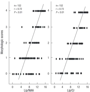
The high lipid/NAA and lipid/Cr ratios on 1H MRS were ob- tained on the first day after hypoxic-ischemic insult and were predictive of morphologic changes (Fig. 6).
However, the 1H MRS and morphologic scores of the Pre-3 day and Pre-6 day preconditioning groups did not show a protective effect on survival rates or on lipid/NAA and lipid/Cr ratios.
DISCUSSION
Both fetal and neonatal asphyxia can cause cerebral hypoxic- ischemic injury, resulting in severe neurologic sequelae and death. Survivors of perinatal asphyxia frequently have moder- ate to severe brain injury for which there is currently no prom- ising therapy.
Reports published in the last decade have made increasing use of the term hypoxic or ischemic preconditioning to describe
an adaptive response to subsequent lethal events (3). Focusing on protective strategies to prevent and reduce hypoxic-ischemic injury, sublethal stimuli, such as hypoxia (4), hyperthermia (10), hypothermia (11), glutamate and seizure (12), oxygen-glucose deprivation (13), receptor blockade/activation (14), and cyto- kine (15), have been reported.
The mechanism of the brain tolerance induced by hypoxic preconditioning has not been fully elucidated, but three major mechanisms have been proposed.
First, the formation of various types of prolonged neuronal adaptive responses is associated with the activation of the cell genome and protein synthesis. These include stress proteins (heat shock proteins [HSP]) (10); immediate early genes (c-fos, c-jun, krox-24 [NGFI-A]) (16); transcription factors (hypoxia-in- ducible factor-1α [HIF-1α]) (17); NFκB (nuclear factor κB) (18);
cAMP response element-binding protein (CREB) (19); neuro- modulatory peptides for survival, differentiation, and plasticity;
growth factors (IGF-1, brain-derived neurotrophic factor [BDNF], NGF) (20); and anti-apoptotic genes (bcl-2, bcl-xl, bax) (3). In particular, an important role is played by immediate early gene superfamilies whose protein products are transcription factors that regulate the expression of phenotype-specific late genes.
A rather long time (24 hr or more) between mild precondi- tioning episodes and severe hypoxia is needed for the maximal protective effect of preconditioning to appear, which suggests the involvement of neuronal gene expression and de novo pro- tein synthesis in the mechanisms of brain tolerance (7). For ex- ample, Kitagawa et al. (21) found that sublethal ischemia at 1 and Fig. 5. The effect of hypoxic preconditioning on morphologic scores of the newborn
rat brain fourteen days after hypoxic-ischemic brain injury. The morphologic scores are significantly lower in the Pre-1 day, Pre-12 hr, and Pre-6 hr groups than in the control group (*P < 0.05). The values are expressed as mean ± standard deviation.
Morphologic scores
Control Pre-6 days Pre-3 days Pre-1 day Pre-12 hr Pre-6 hr 4
3
2
1
0
* *
*
Fig. 6. Correlation between the morphologic scores and 1H MRS findings. The high lipid/NAA and lipid/Cr ratios on the first day after hypoxic-ischemic injury were pre- dictive of morphologic changes.
Morphologic scores
4
3
2
1
0
4
3
2
1
0
Lip/NAA Lip/Cr
0 4 8 12 16 0 4 8 12 16
n= 152 r = 0.72 P < 0.01
n= 152 r = 0.73 P < 0.01
2 days prior to an ischemic insult that is normally lethal to neu- rons was neuroprotective in vivo.
Our data, however, showed significant neuroprotective effects in the Pre-6 hr hypoxic preconditioning group. This suggests that the factors mentioned above could be induced to influence the tolerance of the brain in as little as 6 hr, and the persistence of the protective effect lasted 3 days after preconditioning.
The second possibility involves the roles of signaling proteins (NO, ERK1/2, Akt) (22). NO, an important proximal component of the transduction pathway of adaptive genomic changes, was shown to induce diverse paracrine actions in the central nervous system. NO production and activity were critical to the induc- tion of ischemic tolerance.
Third, the roles of molecules, including adenosine A1 recep- tors (23) and manganese superoxide dismutase (MnSOD) (24) have suggested a possible mechanism.
Finally, activation of N-methyl-D-aspartate (NMDA) recep- tors has been shown to be involved in the development of both adaptive and pathological brain reactions. Glutamate has long been known to kill neurons by NMDA receptor-mediated mech- anisms. Paradoxically, subtoxic concentrations of NMDA pro- tect neurons against glutamate-mediated excitotoxicity (1, 14).
The major mechanism of NMDA receptor-medicated tolerance after preconditioning involves HSP induction; BCL-2 gene ex- pression; activation of adenosine A1 receptors; release and syn- thesis of BDNF by activation of NFκB; TrkB receptor, Akt activa- tion through phosphoinositide 3-kinase, and calmodulin-de- pendent protein kinase; and downregulation of the mixed–lin- eage kinase 3 (MLK3)-c-Jun N-terminal kinase (JNK) signaling pathway via Akt activation (22).
According to data from the present study, the Pre-6 hr, Pre-12 hr, and Pre-1 day hypoxic preconditioning groups had signifi- cantly higher survival rates than those of the control group. These results suggest that preconditioning was effective in myocardial cells as well as cells in the brain. Mild, short ischemic events in myocytes contributed to the resistance to subsequent, lethal ischemic exposure, which reduced cardiac infarction and im- proved cardiac function during reperfusion after ischemic insult.
The survival rates showed the possibility of cardiac tolerance through preconditioning.
It is well known that apoptosis plays an important role in hy- poxic-ischemic brain injury and that the apoptotic cell counts are closely related to the degree of brain injury (25). Evidence of fragmented and condensed DNA is apparent using the TUNEL method of in situ DNA end labeling. Cantagrel et al. (3) found a decrease in the number of apoptotic cells 24 and 48 hr after the insult using a newborn rat model of hypoxic preconditioning (8% oxygen/92% nitrogen, 3 hr, 37˚C) on day 6 followed by ca- rotid ligation and hypoxic insult on day 7, suggesting that regu- lation of apoptotic cell death is one of the mechanisms involved in the tolerance to hypoxia-ischemia that is induced by hypoxic
preconditioning. Using the same experimental model, we inves- tigated the preconditioning effects in the present study.
MRS allows for the study of brain biochemistry and metabo- lism and for indirect characterization of the composition of brain tissue in vivo. MRS has become an important adjunct to diag- nostic structural imaging that permits more accurate and earlier determination of prognosis after hypoxia-ischemia in the new- born (26-28). Furthermore, non-invasive in vivo 1H MRS of a hypoxic-ischemic brain might reveal apoptosis (29). In 1H MRS of hypoxic-ischemic newborn rat brains, the prominent lipid peaks at 1.3 ppm appear early, and the apoptotic cell counts might be correlated with the intensity of the lipid peak and the ratios of lipid/NAA and lipid/Cr. Therefore, the ratios of lipid/
NAA and lipid/Cr could be used as an early indicator to diag- nose hypoxic-ischemic injury by apoptosis and also to assess the effect of preconditioning on decreasing apoptosis.
We have used 1H MRS in vivo for the purpose of early detec- tion in the hypoxic-ischemic brain injury model (30). Addition- ally, the results of the current study demonstrate that the increase of the lipid peak at 24 hr following hypoxic-ischemic injury is correlated with morphologic scores. We suggest a promising future for early in vivo detection of apoptosis using 1H MRS, which can be used to develop new therapeutic methods to attenuate apoptosis in the hypoxic-ischemic injured brain.
In conclusion, using 1H MRS, TUNEL staining, and morpho- logic scores, this study showes that hypoxic preconditioning 6 hr to 1 day before hypoxic-ischemic injury produces higher sur- vival rates and the presence of neuroprotective effects against subsequent hypoxic-ischemic injury. However, hypoxic precon- ditioning in the Pre-3 day & Pre-6 day groups does not show these protective effects. The downregulation of apoptotic cell death may be a mechanism underlying the protective effects of hypoxic preconditioning in the newborn rat brain.
REFERENCES
1. Lee JM, Grabb MC, Zipfel GJ, Choi DW. Brain tissue responses to isch- emia. J Clin Invest 2000; 106: 723-31.
2. Pin TW, Eldridge B, Galea MP. A review of developmental outcomes of term infants with post-asphyxia neonatal encephalopathy. Eur J Paedi- atr Neurol 2009; 13: 224-34.
3. Cantagrel S, Krier C, Ducrocq S, Bodard S, Payen V, Laugier J, Guilloteau D, Chalon S. Hypoxic preconditioning reduces apoptosis in a rat model of immature brain hypoxia-ischaemia. Neurosci Lett 2003; 347: 106-10.
4. Sanders RD, Manning HJ, Robertson NJ, Ma D, Edwards AD, Hagberg H, Maze M. Preconditioning and postinsult therapies for perinatal hypoxic- ischemic injury at term. Anesthesiology 2010; 113: 233-49.
5. Gidday JM, Fitzgibbons JC, Shah AR, Park TS. Neuroprotection from isch- emic brain injury by hypoxic preconditioning in the neonatal rat. Neu- rosci Lett 1994; 168: 221-4.
6. Cadenas S, Aragonés J, Landázuri MO. Mitochondrial reprogramming through cardiac oxygen sensors in ischaemic heart disease. Cardiovasc
Res 2010; 88: 219-28.
7. Wrang ML, Moller F, Alsbo CW, Diemer NH. Changes in gene expression following induction of ischemic tolerance in rat brain: detection and ver- ification. J Neurosci Res 2001; 65: 54-8.
8. Miller BA, Perez RS, Shah AR, Gonzales ER, Park TS, Gidday JM. Cere- bral protection by hypoxic preconditioning in a murine model of focal ischemia-reperfusion. Neuroreport 2001; 12: 1663-9.
9. Palmer C, Vannucci RC, Towfighi J. Reduction of perinatal hypoxic-isch- emic brain damage with allopurinol. Pediatr Res 1990; 27: 332-6.
10. Sivaswamy S, Neafsey EJ, Collins MA. Neuroprotective preconditioning of rat brain cultures with ethanol: potential transduction by PKC isoforms and focal adhesion kinase upstream of increases in effector heat shock proteins. Eur J Neurosci 2010; 32: 1800-12.
11. Nishio S, Chen ZF, Yunoki M, Toyoda T, Anzivino M, Lee KS. Hypother- mia-induced ischemic tolerance. Ann N Y Acad Sci 1999; 890: 26-41.
12. Schurr A, Payne RS, Tseng MT, Gozal E, Gozal D. Excitotoxic precondi- tioning elicited by both glutamate and hypoxia and abolished by lactate transport inhibition in rat hippocampal slices. Neurosci Lett 2001; 307:
151-4.
13. Khaspekov L, Shamloo M, Victorov I, Wieloch T. Sublethal in vitro glu- cose-oxygen deprivation protects cultured hippocampal neurons against a subsequent severe insult. Neuroreport 1998; 9: 1273-6.
14. Meloni BP, Majda BT, Knuckey NW. Evaluation of preconditioning treat- ments to protect near-pure cortical neuronal cultures from in vitro isch- emia induced acute and delayed neuronal death. Brain Res 2002; 928:
69-75.
15. Liu J, Ginis I, Spatz M, Hallenbeck JM. Hypoxic preconditioning protects cultured neurons against hypoxic stress via TNF-alpha and ceramide.
Am J Physiol Cell Physiol 2000; 278: C144-53.
16. Rybnikova E, Vataeva L, Tyulkova E, Gluschenko T, Otellin V, Pelto-Hui- kko M, Samoilov MO. Mild hypoxia preconditioning prevents impair- ment of passive avoidance learning and suppression of brain NGFI-A ex- pression induced by severe hypoxia. Behav Brain Res 2005; 160: 107-14.
17. Hieber S, Huhn R, Hollmann MW, Weber NC, Preckel B. Hypoxia-induc- ible factor 1 and related gene products in anaesthetic-induced precondi- tioning. Eur J Anaesthesiol 2009; 26: 201-6.
18. Blondeau N, Widmann C, Lazdunski M, Heurteaux C. Activation of the nuclear factor-kappaB is a key event in brain tolerance. J Neurosci 2001;
21: 4668-77.
19. Lee HT, Chang YC, Wang LY, Wang ST, Huang CC, Ho CJ. cAMP response
element-binding protein activation in ligation preconditioning in neo- natal brain. Ann Neurol 2004; 56: 611-23.
20. Wang X, Deng J, Boyle DW, Zhong J, Lee WH. Potential role of IGF-I in hypoxia tolerance using a rat hypoxic-ischemic model: activation of hy- poxia-inducible factor 1alpha. Pediatr Res 2004; 55: 385-94.
21. Kitagawa K, Matsumoto M, Tagaya M, Hata R, Ueda H, Niinobe M, Handa N, Fukunaga R, Kimura K, Mikoshiba K, Kamada T. ‘Ischemic tolerance’ phenomenon found in the brain. Brain Res 1990; 528: 21-4.
22. Yin XH, Zhang QG, Miao B, Zhang GY. Neuroprotective effects of pre- conditioning ischaemia on ischaemic brain injury through inhibition of mixed-lineage kinase 3 via NMDA receptor-mediated Akt1 activation. J Neurochem 2005; 93: 1021-9.
23. Pugliese AM, Latini S, Corradetti R, Pedata F. Brief, repeated, oxygen-glu- cose deprivation episodes protect neurotransmission from a longer isch- emic episode in the in vitro hippocampus: role of adenosine receptors. Br J Pharmacol 2003; 140: 305-14.
24. Arthur PG, Lim SC, Meloni BP, Munns SE, Chan A, Knuckey NW. The protective effect of hypoxic preconditioning on cortical neuronal cultures is associated with increases in the activity of several antioxidant enzymes.
Brain Res 2004; 1017: 146-54.
25. Nakajima W, Ishida A, Lange MS, Gabrielson KL, Wilson MA, Martin LJ, Blue ME, Johnston MV. Apoptosis has a prolonged role in the neuro- degeneration after hypoxic ischemia in the newborn rat. J Neurosci 2000;
20: 7994-8004.
26. Li YK, Liu GR, Zhou XG, Cai AQ. Experimental hypoxic-ischemic enceph- alopathy: comparison of apparent diffusion coefficients and proton mag- netic resonance spectroscopy. Magn Reson Imaging 2010; 28: 487-94.
27. Hüppi PS, Lazeyras F. Proton magnetic resonance spectroscopy ((1)H- MRS) in neonatal brain injury. Pediatr Res 2001; 49: 317-20.
28. da Silva LF, Höefel Filho JR, Anés M, Nunes ML. Prognostic value of 1H- MRS in neonatal encephalopathy. Pediatr Neurol 2006; 34: 360-6.
29. Blankenberg FG, Katsikis PD, Storrs RW, Beaulieu C, Spielman D, Chen JY, Naumovski L, Tait JF. Quantitative analysis of apoptotic cell death using proton nuclear magnetic resonance spectroscopy. Blood 1997; 89:
3778-86.
30. Park SJ, Yoon HS, Lim KH, Lee JH, Kim KS, Pi SY. Early prediction of hy- poxic-ischemic brain damage in newbon rats using proton magnetic resonance spectroscopy and neuroprotective effect of EGb 761. J Korean Soc Neonatol 2001; 8: 133-40.
AUTHOR SUMMARY
Protective Effect of Hypoxic Preconditioning on Hypoxic-Ischemic Injured Newborn Rats
Hyun-Kyung Park, In-Joon Seol and Ki-Soo Kim
Brief episodes of hypoxia before subsequent lethal ischemic events could be helpful. The protective effect of hypoxic preconditioning on hypoxic-ischemic brain injury was investigated in the neonatal rat. Our study showed that hypoxic preconditioning 6-hr to 1-day before hypoxic-ischemic injury increased survival rates and had neuroprotective effects against subsequent hypoxic-ischemic injury.