http://dx.doi.org/10.5090/kjtcs.2013.46.6.413 ISSN: 2233-601X (Print) ISSN: 2093-6516 (Online)
1
Department of Thoracic Surgery, Dongnam Institute of Radiological and Medical Science, Departments of
2Thoracic and Cardiovascular Surgery and
3Physiology, Chungnam National University School of Medicine
Received: July 15, 2013, Revised: August 5, 2013, Accepted: August 5, 2013
Corresponding author: Min-Woong Kang, Department of Thoracic and Cardiovascular Surgery, Chungnam National University Hospital, Chungnam National University School of Medicine, 282 Munhwa-ro, Jung-gu, Daejeon 301-721, Korea
(Tel) 82-42-280-7375 (Fax) 82-42-280-7373 (E-mail) [email protected]
Cuk-Seong Kim, Department of Physiology, Chungnam National University School of Medicine, 282 Munwha-ro, Jung-gu, Daejeon 301-721, Korea
(Tel) 82-42-580-8216 (Fax) 82-42-585-8440 (E-mail) [email protected]
C
The Korean Society for Thoracic and Cardiovascular Surgery. 2013. All right reserved.
CC
This is an open access article distributed under the terms of the Creative Commons Attribution Non-Commercial License (http://creative- commons.org/licenses/by-nc/3.0) which permits unrestricted non-commercial use, distribution, and reproduction in any medium, provided the original work is properly cited.
Nafamostat Mesilate: Can It Be Used as a Conduit Preserving Agent in Coronary Artery Bypass Surgery?
Yoo Sang Yoon, M.D., Ph.D.
1, Hyunkong Oh, M.D.
2, Yonghwan Kim
2,
Seung Pyung Lim, M.D., Ph.D.
2, Cuk-Seong Kim, Ph.D.
3, Min-Woong Kang, M.D., Ph.D.
2Background: Graft vessel preservation solution in coronary artery bypass surgery is used to maintain the graft conduit in optimal condition during the perioperative period. Nafamostat mesilate (NM) has anticoagulation and an- ti-inflammatory properties. Therefore, we investigated NM as a conduit preservative agent and compared it to papaverine. Methods: Sprague–Dawley (SD) rat thoracic aortas were examined for their contraction–relaxation ability using phenylephrine (PE) and acetylcholine (ACh) following preincubation with papaverine and NM in standard clas- sical organ baths. Human umbilical vein endothelial cells (HUVECs) were cultured to check for the endothelial cell viability. Histopathological examination and terminal deoxynucleotidyl transferase dUTP nick end labeling assay were performed on the thoracic aortas of SD rats. Results: The anti-contraction effects of papaverine were superior to those of NM at PE (p<0.05). The relaxation effect of NM on ACh-induced vasodilatation was not statistically dif- ferent from that of papaverine. Viability assays using HUVECs showed endothelial cell survival rates of >90% in various concentrations of both NM and papaverine. A histopathological study showed a protective effect against ne- crosis and apoptosis (p<0.05) in the NM group. Conclusion: NM exhibited good vascular relaxation and a reason- able anti-vasocontraction effect with a better cell protecting effect than papaverine; therefore, we concluded that NM is a good potential conduit preserving agent.
Key words: 1. Coronary artery bypass surgery 2. Coronary artery bypass conduits 3. Anticoagulants
INTRODUCTION
Coronary artery bypass surgery (CABG) is effective in cor- onary artery diseases that are not effective by intervention [1]. CABG is known to be particularly effective for treating high-risk group patients who may have left main coronary ar-
tery disorders, severe ventricular functional defects, or dia- betic conditions [2,3].
Preservative solution for a graft conduit in a CABG proce-
dure is used mainly in a mixture with antispastics and
anticoagulants. The antispastic drug prevents the contraction of
blood vessels, and the anticoagulant drug prevents the for-
mation of blood clots. A component that maintains high via- bility of vascular cells is also added to the preservative solution for a graft conduit before the solution is used in CABG [4,5].
In order to prevent the formation of blood clots within blood vessels, the interior of the free graft is cleansed with a saline solution or an organ-maintaining solution. To enhance the anticoagulant effect of these solutions, heparin is often added to these solutions [5].
In the interest of elevating the viability of cells of the graft conduit, various types of solutions—autologous whole blood, histidine-tryptophan-ketoglutarate (HTK), or University of Wisconsin solution (UWS)—are used, or the graft conduit cells are stored at a temperature of <4
oC [5,6].
The ideal antispastic drug for CABG should have no effect on the hemodynamics of the entire body, should suppress the contraction of blood vessels postoperatively, and should mini- mize apoptosis of the vascular cells of the graft conduit [7].
Therefore, a solution that is used only in the affected region is useful because it does not have any effect on the hemody- namics of the entire body and can easily be employed during an operation. Various drugs—such as nitroglycerin, nicardi- pine, verapamil, levosimendan, phenoxybenzamine, and papa- verine—have been used for minimizing the spasms of blood vessels—graft conduit radial coronary artery, gastroepiploic artery, and great saphenous vein—that are used in the CABG procedure [7-11]. Of the antispastic drugs, papaverine is often used because of its powerful vasodilation effect on the region of interest.
When performing CABG, a dilute papaverine solution is either directly or indirectly applied on the exterior of the graft. Papaverine is widely used as a preservative agent dur- ing the CABG procedure because of its short-lived effect and its ability to maximally diminish the resistibility of the coro- nary artery [8]. Furthermore, when papaverine is injected di- rectly into the coronary artery, it is reported that papaverine causes very minimal changes in the heart rate and the blood vessel resistance of the entire body [12]. However, papaverine is reported to reduce the function of endotheliocytes in the internal thoracic artery and to induce apoptosis of human en- dotheliocytes and smooth muscle cells of blood vessels [13].
Nafamostat mesilate (NM), the main drug of the experi- ment, is a proteinase inhibitor and is known to exhibit an an-
ticoagulant effect when it acts on the blood coagulators IX, X, XIIa, and VIIa [14]. NM has the physicochemical charac- teristic of being quickly metabolized in the liver and blood, thereby having a short half-life of 5 to 8 minutes. Further, because of its low molecular weight of 539.6 M, NM is easi- ly removed from the body via extracorporeal circulation [15].
Therefore, NM is used for improving the conditions of acute pancreatitis and for preventing blood clot formation in the ex- tracorporeal circulation in patients suffering from dissem- inative blood vessel coagulation, hemorrhagic lesion, or hem- orrhagic tendency [16,17]. NM has been used mainly for he- modialysis or continuous renal replacement therapy [17], but the recent trend revealed an increasing use of NM in heart surgery involving heart-and-lung machines or in extracor- poreal membrane oxygenation equipment [18,19].
Further, NM has an anti-inflammatory effect and sup- presses nitric oxide (NO) overexpression, apoptosis, and inter- leukin (IL)-6 and IL-8 [20,21]. An enzyme, kallikrein, acts on kininogen, a molecule that exists in the fractional layer of al- pha-2 globulin, to separate bradykinin and kallidin. The sepa- rated kinins have a tendency to expand peripheral blood ves- sels to control the blood pressure, and they also induce in- flammation. Further, kallikrein can convert plasminogen into plasmin; thus, it is capable of triggering the fibrinolysis effect [22]. NM is known to prevent these actions of kallikrein, which is a type of serine protease, and thereby, works as an anti-inflammatory or antifibrinolytic drug [14,23]. Ryu et al.
[24] reported that because of the suppression of the kallikrein –kinin system, when used as a pretreatment, NM can reduce the postperfusion syndrome, which is a condition of a sudden drop of blood pressure seen in the reperfused graft liver after a liver transplant.
Because of its anticoagulant, anti-inflammatory, and anti- fibrinolytic effects, NM was believed to act as a preservative solution for the graft conduit. However, the vasodilator ability of NM, one of its most important effects, needs to be studied. Therefore, we hypothesized that NM may be useful as a preserving agent for the graft conduit. Various tests—
contractile, relaxation, and viability of vascular endothelial
cells—were performed on NM and papaverine concomitantly
to carry out a comparative experiment. Because NM is
known to have an anti-inflammatory effect, apoptosis-reducing
function, and anticoagulation effect, we hypothesized that NM will be useful as a preserving agent for the graft conduit if it also has vasodilation action. For the investigation of NM as a conduit preservation solution, we performed in vitro and in vivo experiments as follows.
METHODS 1) Experimental animals
Upon approval from the Chungnam National University Animal Experiment Committee Board, the appropriate ex- perimental animals were carefully chosen according to the laboratory standards. The experiment was carried out with ap- proximately 6-week-old and about 150 to 170-g Sprague–
Dawley (SD) male rats. All the rats began fasting 12 hours before the experiment. No significant problem was observed in any of the animals.
2) Tension test of thoracic aorta of white mice (1) Tissue collections and organ bath: The SD rats (n=6) were anesthetized by injecting 80 mg/kg of ketamine and 12 mg/kg of xylazine intraperitoneally. Thoracotomy was per- formed, 300 U/mL of heparin was injected into the left ven- tricle to prevent blood clot formation, and then the thoracic aorta was carefully extracted. The extracted thoracic aorta was placed in a laboratory dish containing the modified Krebs–Henseleit bicarbonate buffer solution at 4
oC. The fat and connective tissue around the aorta was removed using stereomicroscopy. Taking care not to damage the vascular en- dothelial cells, the descending thoracic aorta was cleanly trimmed and cut into 5-mm-long aorta segments. Both ends of the excised aorta segments were hung on the stainless loops of an isometric force transducer (MultiMyograph 610 M; Danish Myo Technology, Copenhagen, Denmark), and they were installed in 7.5 mL of the modified Krebs–
Henseleit bicarbonate buffer solution tank. By mixing 100 mM of NaCl, 4.7 mM of KCl, 1.9 mM of CaCl
2, 1.2 mM of MgSO
4, 1.03 mM of HK
2PO
4, 23 mM of NaHCO
3, and 11.1 mM of glucose, we prepared the modified Krebs–Henseleit bicarbonate buffer solution. The buffer solution was placed in the organ bath and was maintained at 37
oC, supplied with 95% O
2and 5% CO
2, and kept at a pH of 7.4. Tension was
applied gradually to the excised aortic vessel installed on the isometric force transducer. When the stabilized tension level was sustained at 2 g, the buffer solution was exchanged at 15-minute intervals for 60 minutes, and then, the equilibrium state was maintained. Using the method above, which is used for investigating the soundness of vascular endothelial cells of aorta segments, we examined the contraction reaction of the thoracic aorta using a high concentration of 60 mM of K
+. Then, the results were used to revise the numerical value of the contraction and relaxation measurements.
(2) Contraction and relaxation of the thoracic aorta: In the interest of checking the contractile ability of the thoracic aorta, the buffer solution was exchanged three times in 1 hour and was stabilized. Phenylephrine (PE) was administered incrementally into the buffer solution, thereby raising the con- centration level of PE from 10
-8M to 3×10
-7M and leading to an incremental increase of PE-induced contractions of the thoracic aorta. The degree of contraction was measured at the maximum level, and the contraction level achieved an equili- brium state. After the submaximally contracted aorta seg- ments using PE reached the climax, acetylcholine (ACh) was administered incrementally and its concentration was in- creased from 10
-8M to 10
-5M. This was done to record the relaxation reaction of the aorta. The degree of contractile and relaxation reactions displayed on the polygraph in the unit of tension was measured. The relaxation reaction was expressed as a percentage according to the numerical submaximal con- traction value of PE. This PE value was revised into the nu- merical submaximal contraction value induced via KCl.
(3) Contraction and relaxation of papaverine and nafa-
mostat mesilate: After checking the contractile and relaxation
abilities of the thoracic aorta, the buffer solution was ex-
changed three times to regain a stable status. Then, 3 μg/mL
of papaverine and 10 μg/mL of NM were used as a pretreat-
ment for 20 minutes. After continuously exchanging the buf-
fer solution three times to remove the drugs, PE was ad-
ministered incrementally by increasing its concentration from
10
-8M to 3×10
-7M in order to induce contraction in the
aorta. The maximum contraction reaction point occurrence
was observed, and the equilibrium point was attained. When
the submaximally contracted aortic ring using PE reached the
climax point, a relaxation reaction was recorded after increas-
ing the concentration of ACh incrementally from 10
-8M to 10
-5M. The degree of the contractile and relaxation reactions displayed on the polygraph in the unit of tension was measured. The relaxation reaction was expressed as a percent- age according to the numerical submaximal contraction value of PE. This PE value was revised into the numerical sub- maximal contraction value induced via KCl.
3) Examination of the viability of vascular endothelial cells
(1) Cytotoxicity test of vascular endothelial cells: After 1×10
5human umbilical vein endothelial cells (HUVECs) were sprayed onto the 12 well plates, they were stabilized for 24 hours. NM and papaverine with increasing concentrations from—100 ng/mL to 100 μg/mL, increasing in multiples of 10—were administered to the HUVECs in the order of the different concentration levels, and the cells were cultivated for an additional 24 hours. An automatic cell counter (ADAM-MC; Digital Bio, Seoul, Korea) was used for con- ducting the cytotoxicity test, and both live and dead cells were quantified after adding propidium iodide (PI). PI is known to couple with double-stranded nucleic acids. In our experiment, PI did not couple with the double-stranded nu- cleic acids of the live cells; however, it coupled with those of the dead or dying cells. Each well plate was sorted according to its concentration level, and the ratio between the number of cells that coupled with PI and the total number of cells was expressed as a percentage.
4) Histopathology and immunopathology tests on the thoracic aorta of white mice
(1) Collection of tissue culture: Approximately 6-week- old and about 150 to 170-g SD male rats (n=6) were anes- thetized by injecting 4 mL/kg of 25% urethane into the ab- dominal cavity. They were put to death by dislocating their cervical vertebrae, and the thoracic aorta was extracted imme- diately after death. Then, the thoracic aorta was cut into 2-cm-long segments and immersed in a saline solution, NM solution (10 μg/mL), or papaverine solution (10 μg/mL) for 1 hour. Then, each segment was fixed in a 10% formalin sol- ution for three days, and histopathology and immunopathol- ogy tests were carried out simultaneously.
(2) Hematoxylin and eosin stain: The thoracic aorta seg- ments, which were fixed in 10% formalin after the extraction, were cut along the cross-section line of the thoracic aorta into 4-mm-thick segments. Microscope slides were prepared for each segment. A paraffin-embedded block was made and sliced with 5 μm. Thereafter, the hematoxylin and eosin (H&E) stain was performed.
(3) Pathological assessment: One pathologist, who was not aware of the content of the experimental groups, carried out the pathological assessment for all cases. The pathologist observed the nuclei and the cytoplasm of the cells of the in- tima and media membranes, searched for any structural dam- age, and studied the overall morphology of the cells. The im- ages of the blood vessel fragment dyed with H&E staining were scanned and transferred to a computer, and a quantita- tive morphometric analysis was performed using the Scion Image Analysis software (ver. 1.0). We evaluated the thick- ness of the intima and media membranes. The thickness of the media membrane was measured at eight places that were 45
oapart and were located around the central site of the blood vessel as a reference point. The results were averaged at each point to compute the thickness of the media membrane.
(4) Terminal deoxynucleotidyl transferase dUTP nick end labeling assay: Of the slides that were prepared in a dif- ferent type of solutions, one slide from each group was taken to perform a terminal deoxynucleotidyl transferase dUTP nick end labeling (TUNEL) assay using the ApopTag peroxidase kit (peroxidase in situ apoptosis detection kit Chemicon
#S7100; North Ryde, NSW, Australia).
The paraffin on the tissue slides, which were prepared with
4-μm-thick paraffin, was removed with 100% xylene. Then,
the slides were treated with 100%, 95%, and 70% alcohol
and washed with distilled water. Next, the slides were im-
mersed in a phosphate buffered saline+Tween 20 (PBST) sol-
ution for 10 minutes and were allowed to react with 1:200
diluted proteinase K at room temperature for 30 minutes. The
slides were then washed twice with distilled water for 2 mi-
nutes each and were reacted with 3% H
20
2for 5 minutes to
suspend non-specific reactions of endogenous peroxidase
(quench endogenous peroxidase). Then, they were washed
with PBST for 1 minute. Next, the slides were treated with a
labeling buffer for 5 minutes. The 50 μL of the labeling re- action mixture (1-μL TdT dNTP+1-μL bivalent cation (Mn
++)+1-μL TdT enzyme+50-μL labeling buffer) was drop- ped on the slides and covered with a parafilm. The slides were then allowed to rest overnight in a humidity-controlled room at 4
oC. Thereafter, they were treated with a stop buffer for 5 minutes and washed twice with PBST for 2 minutes each time. Next, the slides were allowed to react with 50 μL of streptavidin at room temperature for 25 minutes and wash- ed twice with PBST for 2 minutes each time. A dia- minobenzidine solution was used for 3 minutes to create col- or on the slides; then, these slides were washed with distilled water. Methyl green was used for 10 minutes to counter-stain the slides; the slides were then washed briefly three times.
Thereafter, the slides were immersed in 100% n-butanol twice for 2 minutes each time, dried in xylene four times for 2 mi- nutes each time, and then covered with cover slides.
All the steps were carried out in a humidity-controlled room. The slides were dried completely, fixed on a micro- scope, and studied at room temperature. Cells with nuclei that were dyed brown were judged to be TUNEL-positive cells that underwent apoptosis.
5) Statistical process
PASW SPSS ver. 18.0 (SPSS Inc., Chicago, IL, USA) was used for analyzing the data and processing the statistics; con- tinuous variables were expressed as average±standard devia- tion, and categorical variables were expressed as median values. An analyses of variance (ANOVA) test was con- ducted to compare the continuous variables across the groups.
A one-way ANOVA test was used for examining the histo- pathology test, and a Mann–Whitney U test was employed to evaluate the immunopathology test. Any results of interest were examined using Tukey’s multiple comparison test. If the p-value was less than 0.05, then the data were regarded as statistically significant.
RESULTS 1) Results of tension test
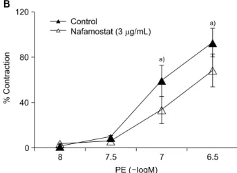
(1) Results of contractile test on papaverine and nafa- mostat mesilate: The normal control group of aorta segments
was pretreated with either 3 μg/mL of papaverine or 3 μg/mL of NM, and then, the results of the contractile re- action of both groups to the PE were studied. Both groups began showing a significant contractile reaction when the PE concentration reached 10
-7M. At 10
-7M of PE, as compared to the control groups, the papaverine-treated group displayed 14.3% and the NM-treated group exhibited a 40.1% con- tractile effect (p<0.05). At 3×10
-7M of PE, as compared to the control groups, the papaverine-treated group showed a 26.9% contractile effect and the NM-treated group exhibited a 68.6% contractile effect (p<0.05). Although the papaverine group seemed to have a better diminishing effect on the con- tractile ability than the NM group, it did not show any sig- nificant difference, from a statistical perspective, at the two different concentrations of PE (p>0.05) (Fig. 1).
The normal control group of aorta segments was pretreated with either 10 μg/mL of papaverine or 10 μg/mL of NM, and then, the results of the contractile reaction of both groups to PE were studied. The NM-treated group began showing a definite contraction at 3×10
-8M of PE, whereas the papaver- ine-treated group started displaying a contraction at 3×10
-7M of PE. At 3×10
-8M of PE, as compared to the control groups, the papaverine-treated group exhibited a 12.5% con- tractile effect and the NM-treated group presented an 83.9%
contractile effect (p<0.05). At 10
-7M of PE, as compared to the control groups, the papaverine-treated group showed a 2.7% contractile effect and the NM-treated group displayed a 39.2% contractile effect (p<0.05). At 3×10
-7M of PE, as compared to the control groups, the papaverine-treated group exhibited a 7.0% contractile effect and the NM-treated group presented a 58.4% contractile effect (p<0.05). At every point in the concentration range of PE from 3×10
-8M to 3×10
-7M, papaverine showed, from a statistical point of view, a sig- nificantly different diminishing effect in the contractile ability from that of NM (p<0.05) (Fig. 2).
(2) Results of relaxation test on papaverine and nafamo-
stat mesilate: The normal control group of aorta segments
was pretreated with either 3 μg/mL of papaverine or 3
μg/mL of NM, and then, the degree of contractile reaction to
the PE was increased to that of the concentration of 3×10
-7M of PE to induce the submaximal contraction of the blood
vessel. Next, ACh was applied gradationally starting from
Fig. 1. Cumulative contraction–response curves for contractile re- sponses to phenylephrine (PE) in the control solution, (A) 3 μg/mL of papaverine, and (B) 3 μg/mL of nafamostat mesilate.
Contractile responses to PE 10
-7M and 3×10
-7M in (A) 3 μg/mL of papaverine and (B) 3 μg/mL of nafamostat mesilate were statistically decreased as compared to those of the control.
(C) The results of contraction with papaverine and nafamostat mesilate are expressed as a percentage as compared to that of the control at the same PE concentration. The percentage con- traction observed in the cases of papaverine and nafamostat mesilate was not significantly different in 10
-7M and 3×10
-7M of PE.
a)p<0.05.
10
-9M to 10
-5M to both the experimental groups, and the results of the relaxation reaction of the aorta were studied.
Both groups started showing significant relaxation results at 10
-8M of ACh. At 10
-8M of ACh, the papaverine-treated group showed 60.9% and the NM-treated group displayed a 76.5% relaxation effect. At 10
-7M of ACh, the papaver- ine-treated group exhibited a 23.4% relaxation effect and the NM-treated group displayed a 32.0% relaxation effect. At 10
-6M of ACh, the papaverine-treated group showed a 24.9% re- laxation effect and the NM-treated group displayed a 22.5%
relaxation effect. At 10
-5M of ACh, the papaverine-treated group exhibited an 18.0% relaxation effect and the NM-treat- ed group presented a 17.5% relaxation effect (Table 1).
Although the papaverine group seemed to have better relaxa- tion ability than the NM group, it did not show any statisti- cally significant difference at any of the four different con-
centrations of ACh (p>0.05) (Fig. 3).
The normal control group of aorta segments was pretreated with either 10 μg/mL of papaverine or 10 μg/mL of NM, and then the blood vessel was treated with PE to induce its submaximal contraction level. However, as compared to the normal control group, the papaverine-treated group showed an insignificant contraction of only 2.7%. Therefore, the relaxa- tion reaction change in the case of ACh with respect to the submaximal contraction of the papaverine-treated group did not show any meaningful results. These results were not analyzed.
2) Results of viability test on human umbilical vein endothelial cells
HUVECs were administered at different concentration lev-
els of NM, ranging from 100 ng/mL to 100 μg/mL in incre-
Fig. 2. Cumulative contraction–response curves for the con- tractile responses to phenylephrine (PE) in the control, (A) 10- μg/mL papaverine, and (B) 10-μg/mL nafamostat mesilate. (A) Contractile responses to 3×10
-8-M, 10
-7-M, and 3×10
-7-M PE in 10- μg/mL papaverine showed a statistically significant decrease as compared to those of the control. (B) Contractile responses to 10
-7-M and 3×10
-7-M PE in 10- μg/mL nafamostat mesilate al- so showed a statistically significant decrease as compared to those of the control. (C) The results of contraction with papaver- ine and nafamostat mesilate are expressed as a percentage as compared to those of the control at the same PE concentration.
There are significant differences between the contraction percent- age of papaverine and nafamostat mesilate in 10
-7-M and 3×10
-7-M PE.
a)p<0.05.
Table 1. Results of relaxation test with acetylcholine were obtained after preincubation with papaverine and nafamostat mesilate
Drug Mole
10
-910
-810
-710
-610
-5Papaverine (%)
Nafamostat mesilate (%)
100 100
60.9 76.5
23.4 32.0
24.9 22.5
18.0 17.5
ments of ten-fold. The results after cultivating the cells for 24 hours showed that the control group displayed 100% viability in the case of 100 ng/mL to 10 μg/mL of NM and 97% via- bility in the case of 1 μg/mL to 100 μg/mL of NM (Table 2, Fig. 4).
HUVECs were administered at different concentration lev- els of papaverine ranging from 100 ng/mL to 100 μg/mL in increments of ten-fold. The results after cultivating the cells for 24 h showed that the control group showed 98% viability
in 100 ng/mL of papaverine, 96% viability in 1 μg/mL of pa- paverine, and 95% and 91% viability in 10 ng/mL and 100 μg/mL of papaverine, respectively (Table 2, Fig. 5).
3) Results of histopathologic and immunohistologic
tests on thoracic aorta of Sprague–Dawley rat
(1) Results of hematoxylin and eosin stain intima and
media membrane thickness comparison: In the H&E stain,
the structural damage and the overall morphology of the nu-
Fig. 3. Cumulative relaxation–response curves for the relaxation responses to acetylcholine (ACh) for the control, (A) 3 μg/mL of papaver- ine, and (B) 3 μg/mL of nafamostat mesilate. Relaxation responses to each ACh concentration in 3 μg/mL of papaverine and 3 μg/mL of nafamostat mesilate were not statistically different compared with those of the control.
Table 2. Results of human umbilical vein endothelial cell culture of the rat thoracic aorta following preincubation with saline, papaverine (0.1, 1, 10, and 100 μg/mL), and nafamostat mesilate (0.1, 1, 10, and 100 μg/mL)
Drug Concentration
Control 0.1 μg/mL 1 μg/mL 10 μg/mL 100 μg/mL
Nafamostat mesilate (%) Papaverine (%)
100 100
100 98
100 96
97 95
97 91
Fig. 4. Results of human umbilical vein endothelial cell viability following treatment with varying concentrations of nafamostat mesi- late (range, 100 ng/mL to 100 μg/mL). The results are expressed as percentage viability.
Fig. 5. Results of human umbilical vein endothelial cell viability following treatment with each papaverine concentration (range, 100 ng/mL to 100 μg/mL). The results are expressed as percentage viability.
clei and the cytoplasm of intima and media membrane cells were studied. Further, thicknesses of the intima and media membranes were measured. The measurements of the intima
membrane thickness were as follows: 5.03±1.08 μm for the
control group, 5.16±1.26 μm for the papaverine-treated group,
and 4.87±1.1.3 μm for the NM-treated group. There was no
Table 3. Quantitative histopathological analysis of the rat thoracic aorta thickness following preincubation with saline, papaverine (10 μg/mL), and nafamostat mesilate (10 μg/mL)
Group (n=48)
p-value
a)Saline Nafamostat mesilate Papaverine
T
intima(μm) T
media(μm) T
intima+media( μm)
5.03±1.08 79.43±4.27 54.46±4.32
4.87±1.13 85.98±2.93
b)90.85±2.69
b)5.16±1.26 72.35±5.46
c)78.86±5.47
c)0.575 0.000 0.000 T
intima, thickness of intima; T
media, thickness of media; T
intima+media, thickness of intima and media.
a)
Statistical significances were tested by one-way analysis of variances among groups.
b)