J Korean Soc Pediatr Nephrol 2014;18:42-46 DOI: http://dx.doi.org/10.3339/jkspn.2014.18.1.42
Copyright © 2014 The Korean Society of Pediatric Nephrology ISSN 1226-5292 (print) ISSN 2234-4209 (online)
급성 신우신염이 재발한 후 불완전 가와사끼병이 발생 한 고도의 방광요관역류가 있는 8개월 남아
CHA의과대학교 분당차병원 소아청소년과 정수진・박성은・이준호
An 8-month-old Male Infant with High Grade Vesicoureteral Reflux who Developed Incomplete Kawasaki disease after Recurrent Pyelonephritis
Kawasaki disease (KD) is a systemic vasculitis that can affect many organ systems.
Renal manifestations include pyuria, hematuria, proteinuria, tubulointerstitial nephritis, acute renal failure, hemolytic uremic syndrome, or renal scarring. Al though its precise pathogenesis remains unknown, it is considered an autoim mune disease.
In the literature, it has been reported that KD may develop in conjunction with urinary tract infections. However, many of these previous studies did not use imaging methods such as renal sonograms, dimercaptosuccinic acid renal scans, and voiding urethrocystograms. We report a case of an 8-month old male infant with high grade vesicoureteral reflux, who developed incomplete KD after recurrent pyelonephritis. Acute pyelonephritis can be an early manifestation of KD. Such cases require the evaluation of urinary tract anomalies according to the guidelines for the management of urinary tract infections.
Key words: Kawasaki disease, Pyelonephritis, Urinary tract infection, Vesicoureteral reflux
Su Jin Jung, M.D., Sung Eun Park, and Jun Ho Lee, M.D.,
Department of Pediatrics, CHA Bundang Medical Center, CHA University, Seongnam, Korea Corresponding Author: Jun Ho Lee, MD Department of Pediatrics, CHA Bundang Medical Center, CHA University, Seongnam, Korea Tel: +82-31-780-5230, Fax: +82-31-780-5011 E-mail: naesusana@gmail.com
*There was no financial and material support for this research and work.
*The authors declare that they have no conflict of interest.
Received: 28 January 2014 Revised: 11 February 2014 Accepted: 20 February 2014
This is an open-access article distributed under the terms of the Creative Commons Attribu- tion Non-Commercial License (http:// crea- tivecom mons.org/licenses/bync/3.0/) which permits unrestricted non-commercial use, distribution, and reproduction in any medium, provided the original work is properly cited.
Introduction
About 10-15% of patients with Kawasaki disease (KD) present with sterile pyuria in the acute phase due to non-specific vasculitis of the urethra [1]. However, previous reports suggest that pyuria in KD is not always sterile, as it can develop in association with urinary tract infections (UTI) [2, 3]. The limitation of these findings is that the acute pyelone- phritis diagnosis at initial manifestation could not be confirmed in these KD patients due to normal 99mTc-dimercaptosuccinic acid (DMSA) scan
results. KD is an autoimmune disease. It has been re- ported that UTI has a role in the pathogenesis of some autoimmune diseases like autoimmune cholangitis, rhe- umatoid arthritis, autoimmune liver disease, thrombotic thrombocytopenic purpura, and lupus nephritis [4-8].
We report a case of an 8-month-old boy with high grade vesicoureteral reflux (VUR), who developed in- complete KD after recurrent pyelonephritis.
Case report
An 8-month-old boy was admitted to our hospital with a 3-hour history of high fever and no associated symptoms. His past medical history revealed two prior admissions. At 3-month-old, he was first admitted due to acute pyelonephritis, confirmed by abnormal urin- alysis {specific gravity, <1.005; pH, 6.0; occult blood, 2+;
leukocyte esterase, 1+; RBC, 1-4/high power field (HPF);
WBC, 10-30/HPF; and bacteria, some}, urine culture {growth of Klebsiella oxytoca with more than 105 colony forming unit (CFU)/mL} sampled by urine bag, C-reactive protein (CRP) 9.78 mg/dL, and renal sonogram (US) {hydronephrosis of both kidneys; right, 7.8 mm, Society for Fetal Urology (SFU) grade 2; left, 10.6 mm, SFU grade 2-3; otherwise unremarkable findings}. At 6 month-old, he was admitted for a second time due to Kawasaki disease and the following associated symptoms: 5- day history of fever, lip redness, strawberry tongue, bilateral non-purulent conjunctivitis, erythematous induration on BCG site, and swelling of both hands and feet. Urinalysis revealed pyuria, while urine culture sampled by urine bag revealed the growth of Pseudomonas aeruginosa with more than 105 CFU/mL. Echocardiography revealed mild tricuspid regurgitation (TR) and mitral regurgitation (MR), with no dilatation of coronary arteries. Following this second admission, he commenced daily treatment with low dose aspirin. On the third admission, his vital signs were taken: heart rate, 125 beats/minute; respiratory rate, 28/minute; and body temperature, 39℃. On examination, he appeared slightly ill looking; however, there were no remarkable findings. Laboratory findings were: hemo- globin, 11.9 g/dL; white blood cell (WBC) count, 23,830/
mm3 (seg 49.5%, lym 40%, mono 10.5%); platelet count, 520,000/mm3; CRP, 2.29 mg/dL; ESR, 45 mm/hr; protein, 7.8 g/dL; albumin, 4.7 g/dL; glucose, 133 mg/dL; blood urea nitrogen/creatinine, 8.9/0.5 mg/dL; GOT/GPT, 44/23 IU/L; and serum electrolytes, 132-4.5-99-17.6 mEq/L. Urinalysis revealed: specific gravity, <1.005; pH, 6.0; occult blood, 2+; protein, (±); leukocyte esterase, 3+; RBC, 1-4/HPF; WBC, 31-50/HPF; and bacteria, some. Urine culture sampled by urine bag revealed the growth of Pseudomonas aeruginosa with 105 CFU/mL.
Imaging studies consisting of a posterior-anterior chest radiograph were normal. Following the diagnosis of recurrent pyelonephritis, he was treated with antibiotics (ceftriaxone + amikacin). However, his high fever per- sisted for 4 days after admission. On the third and fifth hospital day (HD), follow-up CRP values were 11.6 mg/
dL and 2.7 mg/dL, respectively. Other values that were recorded include: creatinine kinase, 64 U/L (38-160 U/
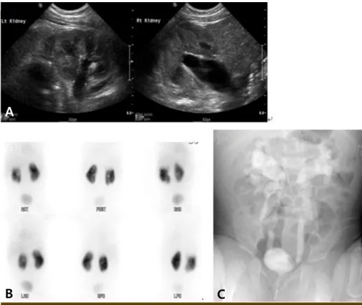
L); troponin-T, 0.006 ng/mL (0-0.1 ng/mL); and proBNP, 376.8 pg/mL (0-320 pg/mL). Imaging studies that were ordered during this admission include US, DMSA scan and voiding urethrocystography (VCUG). Ultrasound findings were as follows: right, 7.7 mm, SFU grade 2 and left, 28 mm, SFU grade 2-3 (Fig. 1A), otherwise no remarkable finding. A DMSA scan revealed cortical defects on the right kidney with normal relative renal uptake ratio (Fig. 1B). A VCUG revealed bilateral grade 4-5 VUR (Fig. 1C). He was discharged on the seventh HD with cefixime. When he visited our clinic one week after discharge, he was doing well overall, although he had developed desquamation of both hands and feet. Two months after the KD diagnosis, a follow- up echocardiogram revealed mild ectasia and luminal irregularity of the left middle coronary artery (1.8–2.6 mm) with mild TR and MR. He was transferred to pe- diatric urologist in our hospital. Then, bilateral uretero- neocystostomy is supposed to be done after the cessation of aspirin.
Discussion
According to the diagnostic criteria of incomplete KD
established by the American Heart Association (AHA), children ≥6 months of age with incomplete presentation might have unexplained fever for ≥5 days associated with 2 or 3 of the principle clinical features in the acute phase [9]. In addition, the AHA recommended a diagnostic algorithm of incomplete KD which comprises of 6 supplemental laboratory and echocardiographic criteria.
More than 3 laboratory criteria (serum albumin ≤3.0 g/
dL, anemia for age, elevation of alanine aminotransferase, platelets after 7 days ≥450,000/mm3, WBC ≥15,000/
mm3, urine WBC ≥10/HPF) support the diagnosis of incomplete KD [9]. For this case, although there was one associated principle clinical feature (desquamation) besides fever, 3 laboratory findings support the diagnosis of incomplete KD.
Although there has been reports about KD in associa- tion with UTI, there was no radiographical evidence of pyelonephritis in these KD patients because their DMSA scans did not be performed or were normal [2, 3].
Meanwhile, Wang et al. [10] reported that among 50
patients with KD, a DMSA renal single-photon emission computed tomography (SPECT) revealed renal inflam- matory foci in 52% of them in the acute phase, 46%
of which had renal scarring on the 6-month follow- up DMSA renal SPECT. This study excluded patients with a previous history of UTI, coincident urinary tract anomaly, UTI between the initial and follow-up scin- tigram, space-occupying lesion at US, and positive urine culture. However, they did not evaluate lower urinary tract anomalies in patients with renal scarring by VCUG, so a possibility that renal scarrings in these KD patients were originated from congenital renal scarring associated with VUR could not be excluded. Furthermore, Oh et al.
[11] reported that no abnormal DMSA renal SPECT were detected among 15 patients in the acute phase of KD.
In this case, the diagnosis of recurrent pyelonephritis in the initial stage was favored by the urine culture results, DMSA scan, the presence of a high grade bilateral VUR, as well as the defervescence with antibiotics as the sole treatment. On the other hand, the diagnosis of incomplete
A
B C
Fig. 1. (A) Bilateral hydronephrosis on renal sonogram. (B) Multiple cortical defects in the right kidney with a normal relative renal uptake ratio on dimercaptosuccinic acid renal scan. (C) Bilateral high grade vesicoureteral reflux on voiding urethrocystogram.
KD at the third admission was favored by a change in fever pattern during admission, an increased CRP on the third HD, a UTI caused by antibiotics-sensitive bacteria, desquamation on both hands and feet, and the follow- up echocardiogram results.
Although the etiology of KD remains unknown, it is currently classified as a systemic vasculitis syndrome, caused primarily by an invasion of medium-sized mus- cular arteries by infiltrating monocytes, macrophages, and lymphocytes [12]. Therefore, the renal involvement seen in some KD cases may be explained by an invasion of medium-sized muscular renal arteries. A hypothetical pathogenesis of KD is proposed under the premise of a protein homeostasis system; where innate and adap tive immune cells control pathogenic proteins that are toxic to host cells at a molecular level [13]. After an infection of unknown KD pathogen, pathogenic proteins produced from unknown focus, spread and bind to endothelial cells of coronary arteries as main target cells [13]. To control the action of pathogenic proteins, immune cells are activated [13]. Pseudomonas aeruginosa causing acute pyelonephritis in this case may be a KD pathogen through immune modulation in renal parenchyma.
However, it can not completely exclude a possibility that recurrent febrile UTI may accidentally accompany with incomplete KD, and urine culture (Pseudomonas aeru- ginosa) was contaminated in this case.
In conclusion, acute pyelonephritis can be an initial manifestation of early KD. In such a case, the evaluation of urinary tract anomaly is needed according to the guideline for the management of UTI. We report a case of an 8-month old boy with high grade VUR who de- veloped incomplete KD after recurrent pyelonephritis.
요약
가와사끼병은 전신성 혈관염을 일으키는 질환중의 하나 로 여러 장기들을 침범할 수 있다. 신장증세로는 농뇨, 혈 뇨, 단백뇨, 간질성 신염, 급성 신부전증, 용혈성 요독 증후 군, 신반흔 등이 있다. 가와사끼병의 신장침범에 대한 병리 기전은 아직 알려져 있지 않지만, 자가면역질환으로 인한 것으로 사려된다. 가와사끼병이 요로감염 이 후에 발병한
다는 몇몇 보고들이 있었다. 하지만, 이미 보고된 논문들에 포함된 많은 요로감염 환자들은 신장방광 초음파, DMSA 스캔이나 배뇨중 요도방광조영술 등을 모두 받은 경우는 없었다. 이에 저자들은 급성 신우신염이 재발한 후 불완전 가와사끼병이 발생한 고도의 방광요관역류가 있는 8개월 남아를 보고하는 바이다. 급성 신우신염은 가와사끼병의 초기 증세일 수 있다. 그런 경우, 환아가 가와사끼병으로 확 진되더라도 요로감염 진료지침에 따라 요로기형에 대한 이 미지 검사를 시행할 필요가 있다고 생각한다.
References
1) Barone SR, Pontrelli LR, Krilov LR. The differentiation of classic Kawasaki disease, atypical Kawasaki disease, and acute ad- enoviral infection: use of clinical features and a rapid direct fluorescent antigen test. Arch Pediatr Adolesc Med 2000;
154:453-6.
2) Husain EH, AI-Rashid M. Kawasaki disease in association with urinary tract infection. Indian Pediatr 2011;48:808-9.
3) Jan SL, Wu MC, Lin MC, Fu YC, Chan SC, Lin SJ. Pyuria is not always sterile in children with Kawasaki disease. Pediatr Int 2010;52:113-7.
4) Wang J, Yang GX, Zhang W, Lu L, Tsuneyama K, Kronenberg M, et al. Echerichia coli infection induces autoimmune cho- langitis and antimitochondrial antibodies in NOD B6 (Idd10/
Idd18) mice : Clin Exp Immunol-used Patrick’s E. Coli 9-9- 13.enl. Clin Exp Immunol 2013 Oct 15. doi : 10.1111/cei.12224 [Epub ahead of print]
5) Puntis D, Malik S, Saravanan V, Rynne M, Heycock C, Hamilton J, el al. Urinary tract infections in patients with rheumatoid arthritis. Clin Rheumatol 2013;32:355-60.
6) Smyk DS, Bogdanos DP, Kriese S, Billinis C, Burroughs AK, Rigopoulou El. Urinary tract infection as a risk factor for autoimmune liver disease : from bench to bedside. Clin Res Hepatol Gastroenterol 2012;36:110-21.
7) Park YA, Schultz EF, Hay SN, Brecher ME. Thrombotic throm- bocytopenic purpura and urinary tract infecdtions : is there a connection? Am J Clin Pathol 2011;135:85-8.
8) Tsai YC, Hou CL, Yao TC, Chen LC, Jaing TH, Huang JL. Risk factors and bacterial profiles of urinary tract infections in patients with systemic lupus erythematosus. Asian Pac J Allergy Immunol 2007;25:155-61.
9) Newburger JW, Takahashi M, Gerber MA, Gewitz MH, Tani LY, Burns JC, et al. Diagnosis, treatment, and long-term mana- gement of Kawasaki disease : a statement for health professionals from the Committee on Rheumatic Fever, Endocarditis, and Ka- wasaki Disease, Council on Cardiovascular Disease in the Young, American Heart Association. Pediatrics 2004;114:1708-33.
10) Wang JN, Chiou YY, Chiu NT, Chen MJ, Lee BF, Wu JM. Renal scarring sequelae in childhood Kawasaki disease. Pediatr Nephrol 2007;22:684-9.
11) Oh JY, Park SJ, Kim SJ, Jang GC, Kim U, Shin JI, et al. Renal manifestations and imaging studies of Kawasaki disease. J Korean Soc Pediatr Nephrol 2013;17:86-91.
12) Takahashi K, Oharaseki T, Yokouchi Y. Pathogenesis of Kawasaki disease. Clin Exp Immunol 2011;164:20-2.
13) Lee KY, Rhim JW, Kang JH. Kawasaki disease : laboratory findings and an immunopathogenesis on the premise of a prot ein homeostasis sytem. Yonsei Med J 2012;53:262-75.