355
Isolation and Characterization of Blakeslea trispora Isolated from Gut of Grasshopper and Soldier Fly Larva in Korea
Thi Thuong Thuong Nguyen and Hyang Burm Lee*
Division of Food Technology, Biotechnology and Agrochemistry, College of Agriculture and Life Sciences, Chonnam National University, Gwangju 61186, Korea
ABSTRACT : During a survey of fungal diversity in insect guts in Korea, two fungal strains, EML-PGH2 and EML-PUKI88, were isolated from the gut of grasshopper and soldier fly larvae inhabiting the bulrush plants at a pond located in the Chonnam National University Arboretum, Gwangju, Korea. Based on their morphological characteristics and a phylogenetic analysis of the internal transcribed spacer (ITS1 and ITS2) and 5.8S rDNA sequences, the strains were identified as Blakeslea trispora. To our knowledge, the zygomycete species B. trispora has not been previously described in Korea.
KEYWORDS : Blakeslea trispora, Grasshopper, Mucorales, Soildier fly larva, Undiscovered taxa
The genus Blakeslea (Choanephoraceae, Mucorales) was established by Thaxter (1914) with the type species B.
trispora [1]. According to Index Fungorum (www.index- fungorum.org), the genus Blakeslea includes only two spe- cies: B. trispora and B. monospora. The species belonging to this genus are characterized by production of both sporangia and sporangiola on sporangiophores, and appen- dages on the sporangiospores, and formation of zygospo- res with opposed suspensors [2]. They are commonly iso- lated from soil, dung, flower, and cowpeas [1, 3-6]. Several studies have shown that B. trispora is the model fungus useful for study of trisporic acid biosynthesis [7].
The Choanephoraceae family was first described by Schröter for only a genus Chaoanephora. However, later Kirk [2] monographed the family and added two genera, Blakeslea and Poitrasia. In the year 2013, Papp et al. [8]
performed the phylogenetic analysis of Gilbertella, Blake- slea, Choanephora, and Poitrasia in Mucoraceae. This
study suggested that the Gilbertella (Gilbertellaceae) is located between Choanephoraceae and Mucoraceae. Later, Voigt and Olsson [9] revised the family Choanephoraceae based on a multigene (act, ref-1alpha, 18S and 28S rRNA) data set obtained from selected species of 50 genera of the Mucorales and demonstrated that Gilbertella could belong to the Choanephoracea. According to these au- thors, four genera, including Blakeslea, Choanephora, Gil- bertella and Poitrasia, classified under the Choanepho- racea.
In Korea, only one species of Choanephora, C. cucurbi- tarum, causing soft rot disease on eggplant (Solanum mel- ongena) was recorded [10]. During a study on the Muco- rales, a species of Blakeslea was first isolated from grass- hopper and soldier fly (Stratiomyidae) larva samples in Korea. To our knowledge, there were no previously pub- lished records of this genus, which was isolated from the gut of grasshopper and soldier fly larva.
The objective of the present study was to perform the morphological and molecular analyses to characterize an unrecorded zygomycete species − Blakeslea trispora in Korea.
Grasshopper from weedy plant and soldier fly larvae inhabiting the bulrush (Typha orientalis) from an aqua- tic plant, were collected at CNU Arboretum located in Chonnam National University, Gwangju, Korea in 2016.
The insect placed in polyethylene and kept at ambient temperature until being transported to the laboratory. The gut was removed from each insect and placed on a sterile Petri dish, cut into small pieces, and spread onto potato
*Corresponding author E-mail: [email protected] Received October 29, 2016 Revised November 18, 2016 Accepted November 21, 2016
This is an Open Access article distributed under the terms of the Creative Commons Attribution Non-Commercial License (http://
creativecommons.org/licenses/by-nc/3.0/) which permits unrestricted non-commercial use, distribution, and reproduction in any medium, provided the original work is properly cited.
Kor. J. Mycol. 2016 December, 44(4): 355-359 https://doi.org/10.4489/KJM.2016.44.4.355 pISSN 0253-651X • eISSN 2383-5249
© The Korean Society of Mycology
dextrose agar (PDA) amended with streptomycin (50 mg/
L). Plates were incubated at 20oC for 3~7 days. Hyphal tips were transferred with a glass needle to PDA plates amended with the antibiotics mentioned above using a stereomicroscope. Pure isolates were transferred to slant tubes, and stored in 20% glycerol at -80°C at the Environ- mental Microbiology Laboratory Herbarium (EMLH;
Chonnam National University, Gwangju, Korea) as EML- PGH2 and EML-PUKI88. Genomic DNA was directly ex- tracted from mycelia using the HiGene Genomic DNA
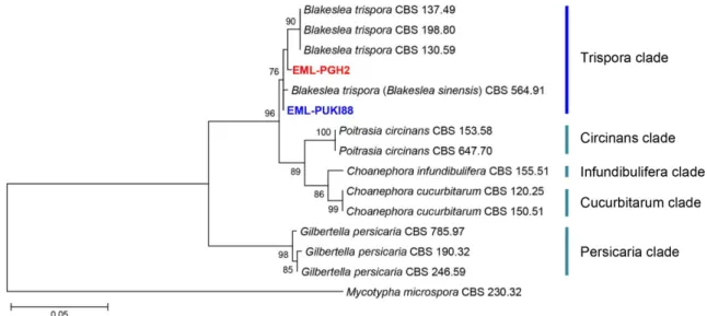
prep kit for fungi (Biopact Corp., Daejeon, Korea). The internal transcribed spacers (ITS1 and ITS2) and 5.8S gene were amplified using primers ITS1 (5'- TCCGTAGGTGA ACCTGCGG-3') and ITS4 (5'-TCCTCCGCTTATTGATA TGC-3') following the method by White et al. [11]. The sequences were initially aligned using CLUSTAL X [12], and edited manually [13]. Phylogenetic analyses were per- formed using MEGA 6 [14] with the default settings. Phy- logenetic trees were constructed from the data using maxi- mum likelihood (ML). The sequences of EML-PGH2 and Fig. 1. Phylogenetic tree based on maximum likelihood analysis of internal transcribed rDNA sequences of Blakeslea trispora EML-PUKI88 and B. trispora EML-PGH2. Mycotypha microspora was used as outgroup. Bootstrap support values of ≥ 50% are indicated at the nodes. The bar indicates the number of substitutions per position.
Table 1. Morphological characteristics of EML-PUKI88 and the reference species, Blakeslea trispora on malt extract agar medium
Character EML-PUKI88 Blakeslea trisporaa
Colony color rapid-growing, first white then yellowish rapid-growing, first white then yellow Sporangiophores bearing sporangia 8.0~12 μm in width, variable in length 11~14 μm in width, pale brown Sporangia 35~50 × 36~60 μm, sub-globose, multispored,
initially white, turning to yellow, dark brown with age
(30)~60~90~(180) μm, spherical, multispored, initially white, turning to yellow and pale brown, dark brown with age Sporangiospores from sporangia 6~9 × 9~15 μm, ellipsoid, reddish-brown,
hyaline appendages at each pole
(6)~8~18~(22) × (3)~4~8~(12) μm, ellipsoid, reddish-brown, hyaline appendages at each pole
Sporangiophores bearing sporangiola 12~26 μm in width, variable in length, bearing 2~26 enlargements
14~28 μm in width,
bearing (1)~8~16~(32) enlargements
Sporangiola 8~16 × 11~18.5 μm, ellipsoidal,
mostly 3 sporangiospores
10~16~(20) × 8~12~(14) μm, ellipsoidal, containing (1)~3~(5) sporangiospores Sporangiospores from sporangiola 11~18.5 × 6~9 μm, ellipsoid, reddish-brown (8)~10~16~(20) × (4)~5~8~(9) μm,
ellipsoid, reddish-brown
Zygospores absent 40~80 μm in diameter
Chlamydospores present present
aFrom the description by Thaxter [1].
EML-PUKI88 strains were deposited in the GenBank data- base with accession numbers, KY047145 and KY047144, respectively. A BLASTn search revealed that in rDNA ITS region EML-PGH2 and EML-PUKI88 revealed sequence similarities of 98.6% (566/574 bp) and 98.7% (567/574 bp) with B. sinensis (current name, B. trispora; GenBank accession no. JN206230) and B. trispora (GenBank acces- sion no. JN943005), respectively. Phylogenetic analysis of ITS region showed that the isolates EML-PGH2 and EML- PUKI88 were placed within Trispora clade along with B.
trispora (Fig. 1).
To confirm the phylogenetic result, the morphology of
the isolate EML-PUKI88 was observed under a light micro- scope (DFC 290; Leica Microsystems, Wetzlar, Germany).
The isolate was cultured on PDA, synthetic mucor agar (SMA; 40 g dextrose, 2 g asparagine, 0.5 g KH2PO4, 0.25 g MgSO4·7H2O, 0.5 g thiamine chloride, and 15 g agar, in 1 L of deionized water), and malt extract agar (33.6 g MEA in 1 L of deionized water; BD, Sparks, MD, USA). Colo- nies were characterized after 5~7 days of cultures at 25°C.
Blakeslea trispora Thaxt., Botanical Gazette Crawfords- ville 58: 353 (1914) (Table 1, Fig. 2).
Description: Colonies grew rapidly at 25°C on MEA, SMA and PDA, filling the petri dish after 2~3 days of
Fig. 2. Morphology of Blakeslea trispora EML-PUKI88. Colonies on synthetic mucor agar (A, D), potato dextrose agar (B, E), and malt extract agar (C, F). A~C, obverse view; D~F, reverse view; G~I, branching sporangiophore with apical vesicles bearing few spored sporangiola (under stereo-microscope); J, sporangiophore with some sporangiola (white arrows) attached on the sur- face of vesicles; K, three-spored sporangiolum; L, terminal spherical vesicle (yellow arrow) after sporangiola detached, showing small spherical pedicels (red arrows) over the surface; M, sporangiophore with a developing sporangium; N, mature sporangium;
O, sporangiospores from sporangium with appendages (purple arrow) (scale bars: J, M, N = 50 μm; K, L, O = 20 μm).
incubation. The initial color of colonies was white, which later turned to yellowish. The colony reverse was also yel- lowish (Fig. 2). Sporangiophores beared sporangia arising from substrate mycelium, non-septate, unbranched, often circinate below the sporangium, 8.0~12 μm in width, vari- able in length. Sporangia measured 35~50 × 36~60 μm, were sub-globose, multispored, initially white and later becoming yellow to dark brown. Sporangiospores from sporangia measured 6~9 × 9~15 μm, were ellipsoid, red- dish-brown, bearing a group of straight fine radiating ap- pendages from either pole. Sporangiophores beared spor- angiola arising from substrate mycelium or aerial hyphae, erect, 12~26 μm in width, variable in length, bearing 2~
26 enlargements. Sporangiola measured 8~16 × 11~18.5 μm, were ellipsoidal, mostly 3 sporangiospores. Sporangi- ospores from sporangiola were ellipsoidal, reddish-brown, and measured 11~18.5 × 6~9 μm, hyaline appendages at each pole. Zygospores were not observed on this medium.
The isolate produces abundant mycelia on PDA agar and the sporulation is excellent on PDA agar. Comparing the colony morphology and culture characteristics of the iso- late on MEA medium, with previous descriptions [1], the present isolate was generally similar to those of B. trispora (Table 1).
To determine the growth rate, EML-PUKI88 was cul- tured on MEA, SMA and PDA media. The plates were incubated at 10°C, 20°C, 25°C, 30°C, and 35°C in the dark for 7 days. The average growth rates of EML-PUKI 88 on MEA, SMA, and PDA were 40.5 mm/day, 51.5 mm/
day, and 59 mm/day at 25°C, respectively. The optimal growth temperature range was 25~30°C. Among the dif- ferent temperatures and culture media, the best mycelial growth was found at 25°C on PDA media. On all media, the isolates grew rapidly at 25~30°C, and stopped growing at a temperature of 10°C.
The genus Blakeslea is considered monotypic. Three species were recognized within genus Blakeslea as B. tris- pora, B. monospora, and B. sinensis. However, recently, Hoffmann et al. [15] and Walther et al. [16] performed phylogenetic analyse including the sequences of B. trisp- ora and B. sinensis which is regarded as synonym of B.
trispora. However, the sequences of B. monospora was not available in GenBank. The present molecular data of this species was consistent with the phylogeny presented by Walther et al. [16]. In the ITS tree, our strains, EML- PUK882 and EML-PGH2, were clustered within Trispora clade containing B. trispora (Fig. 1). Based on the mor- phological, physiological and molecular analyses, the fun-
gus was identified as B. trispora.
Little is known about fungi inhabiting the gut of ins- ects including grasshopper and soldier fly larvae at a pond in Korea. Herein B. trispora is described a as new record of zygomycete fungi belonging to undiscovered taxa in Korea.
Acknowledgements
This work was supported by the Project on Survey and Discovery of Indigenous Species of Korea funded by NIBR of the Ministry of Environment (MOE), Republic of Korea.
REFERENCES
1. Thaxter R. New or peculiar Zygomycetes. 3: Blakeslea, Disso- phora and Haplosporangium, nova genera. Bot Gaz 1914;58:
353-66.
2. Kirk PM. A monograph of the Choanephoraceae. Mycol pap- ers 1984;152:1-61.
3. Benny GL. Methods used by Dr. R. K. Benjamin, and other mycologists, to isolate zygomycetes. Aliso 2008;26:37-61.
4. Ho HM, Chang LL. Notes on Zygomycetes of Taiwan (III):
two Blakeslea species (Choanephoraceae) new to Taiwan. Tai- wania 2003;48:232-8.
5. Li GJ, Hyde KD, Zhao RL, Hongsanan S, Abdel-Aziz FA, Ab- del-Wahab MA, Alvarado P, Alves-Silva G, Ammirati JF, Ari- yawansa HA, et al. Fungal diversity notes 253-366: taxonomic and phylogenetic contributions to fungal taxa. Fungal Divers 2016;78:1-237.
6. Nguyen TT, Lee SH, Bae S, Jeon SJ, Mun HY, Lee HB. Cha- racterization of two new records of zygomycete species belon- ging to undiscovered taxa in Korea. Mycobiology 2016;44:29- 37.
7. Sutter RP, Jelinek BG. Trisporic acid biosynthesis in cultures of Blakeslea trispora with (+) and (-) mycelia separated by a Fig. 3. Effect of temperature and culture medium on mycelial growth of Blakeslea trispora EML-PUKI88. Mycelia were grown on malt extract agar (MEA), synthetic mucor agar (SMA) and potato dextrose agar (PDA), at different temperatures, as indi- cated.
filter. Exp Mycol 1983;7:188-91.
8. Papp T, Acs K, Nyilasi I, Nagy E, Vágvölgyi C. Phylogenetic relationship of the genus Gilbertella and related genera within the order Mucorales based on 5.8S ribosomal DNA sequences.
Acta Biol Hung 2003;54:393-402.
9. Voigt K, Olsson L. Molecular phylogenetic and scanning elec- tron microscopical analysis places the Choanephoraceae and the Gilbertellaceae in a monophyletic group within the Muc- orales (Zygomycetes, Fungi). Acta Biol Hung 2008;59:365-83.
10. Kwon JH, Jee HJ. Soft rot of eggplant (Solanum melongena) caused by Choanephora cucurbitarum in Korea. Mycobiology 2005;33:163-5.
11. White TJ, Bruns TD, Lee SB, Taylor JW. Amplification and direct sequencing of fungal ribosomal RNA genes for phylo- genetics. In: Innis MA, Gelfand DH, Sninsky JJ, editors. PCR protocols: a guide to methods and applications. San Diego:
Academic Press; 1990. p. 315-22.
12. Thompson JD, Gibson TJ, Plewniak F, Jeanmougin F, Higgins
DG. The CLUSTAL_X windows interface: flexible strategies for multiple sequence alignment aided by quality analysis tools.
Nucleic Acids Res 1997;25:4876-82.
13. Hall TA. BioEdit: a user-friendly biological sequence alignment editor and analysis program for Windows 95/98/NT. Nucleic Acids Symp Ser 1999;41:95-8.
14. Tamura K, Stecher G, Peterson D, Filipski A, Kumar S. MEGA 6: Molecular Evolutionary Genetics analysis version 6.0. Mol Biol Evol 2013;30:2725-9.
15. Hoffmann K, Pawłowska J, Walther G, Wrzosek M, de Hoog GS, Benny GL, Kirk PM, Voigt K. The family structure of the Mucorales: a synoptic revision based on comprehensive multi- gene-genealogies. Persoonia 2013;30:57-76.
16. Walther G, Pawłowska J, Alastruey-Izquierdo A, Wrzosek M, Rodriguez-Tudela JL, Dolatabadi S, Chakrabarti A, de Hoog GS. DNA barcoding in Mucorales: an inventory of biodiversity.
Persoonia 2013;30:11-47.