Received: 17 November, 2017 Revised: 27 November, 2017 Accepted: 27 November, 2017
Ⓒ The Korean Society of Mycology
This is an Open Access article distributed under the terms of the Creative Commons Attrib- ution Non-Commercial License (http://creative- commons.org/licenses/by-nc/4.0/) which permits unrestricted non-commercial use, distribution, and reproduction in any medium, provided the original work is properly cited.
Kor. J. Mycol. 2017 December, 45(4): 304-318 https://doi.org/10.4489/KJM.20170033
pISSN : 0253-651X eISSN : 2383-5249
OPEN ACCESS
RESEARCH ARTICLE
Three Unrecorded Fungal Species from Fecal and Freshwater Samples in Korea
Thuong T. T. Nguyen, Monmi Pangging, Hyang Burm Lee
*Division of Food Technology, Biotechnology and Agrochemistry, College of Agriculture and Life Sciences, Chonnam National University, Gwangju 61186, Korea
*Corresponding author: [email protected]
Abstract
While evaluating fungal diversity in fecal and freshwater samples in Korea, three fungal strains, CNUFC-GHD83-1, CNUFC-RD8126, and CNUFC-YR2-1, were isolated from specific habitats including grasshopper and rat feces, and freshwater samples in Korea. On the basis of the morphological characteristics and phylogenetic analysis of the internal transcribed spacer (ITS) and 28S rDNA, the isolates CNUFC-GHD83-1, CNUFC-RD8126, and CNUFC-YR2-1 were identified as Albifimbria terrestris, Cephaliophora tropica, and Mariannaea aquaticola, respectively. These species have not been previously described in Korea.
Keywords: Albifimbria terrestris, Cephaliophora tropica, Mariannaea aquaticola, Specific habitat, Taxonomy
Introduction
Fungi are generally found in aerobic ecosystem, and colonize diverse substrates and play a wide diversity of roles. Many species are cosmopolitan but others are found only in restricted or specific niches or habitats [1]. While evaluating fungal diversity in unusual fungal niches, feces and freshwater have been considered for isolation of rare fungi with specific habitats in Korea [2, 3].
In fece ecosystems, fungi play an important role in the biodegradation of organic materials and the return of nutrients to the environment for reuse [4]. A number of studies on the diversity of fungi have been carried out using different animal dung substrates [2, 4, 5]. However, a few studies have been done using the feces from insect or rat. Stejskal et al.
[6] reported that 35 species belonging to 11 genera such as Alternaria, Arthrinium, Aspergillus, Cladosporium, Epicoccum, Eurotium, Geotrichum, Microascus, Mucor, Penicillium, and Thamnidium were isolated from house mouse using dilution plate method.
Nyberg and Persson [7] observed 24 species of 14 genera in mouse dung. One study of gut
microflora of grasshopper (Melanoplus sanguinipes) has been reported, but there was no
detailed information on the fungal communities [8]. In Korea, two new species (Absidia
stercoraria and Mucor stercorarius) and two new records (Absidia glauca and Paecil- omyces variotii) have been currently reported from rat feces [9-12].
In freshwater ecosystems, fungi play a major role in the decomposition of complex organic compounds thus providing nutrients for other aquatic organisms. Based on a recent literature, there are approximately 622 species (170 genera) of Ascomycetes; more than 531 species of Hyphomycetes (55 genera); and species of Trichomycetes (3 orders, no longer regarded as fungi) among freshwater fungi; however, there is little information on freshwater fungi belonging to Basidiomycetes and Zygomycetes [3]. Intensive studies on the fungal diversity from various habitats including freshwater have been conducted in Korea. Despite intensive studies in freshwater habitats, our knowledge of the diversity of freshwater-derived fungi in general and freshwater Ascomycetes in particular is still lacking compared to that of terrestrial habitats [13].
Pezizomycotina is the largest subphylum of Ascomycetes. In the current classification, Pezizomycotina is divided into 11 classes based on rDNA phylogenies. Especially, the Pezizomycotina contains species which are important to both industry and agriculture. The most well-known order is Hypocreales.
The genus Albifimbria L. Lombard & Crous 2016 belongs to the subphylum Pezizomycotina, order Hypocreales, family Stachybotryaceae. It is characterized by the formation of verrucose setae surrounding the sporodochia and conidia with funnel-shaped, mucoid appendages. Albifimbria species are found in the soil, leaves, cotton, fruit, and air [14]. To date, Albifimbria includes only 4 species, i.e., A. lateralis, A. terrestris, A.
verrucaria, and A. viridis.
The genus Cephaliophora, which belongs to the subphylum Pezizomycotina, order Pezizales, family Ascodesmidaceae, was established by Thaxter in 1903; it includes two coprophilous species, Cephaliophora tropica (type species) and C. irregularis [15].
According to available data from Index Fungorum, 7 species belonging to this genus are known. Members of this genus are characterized by the production of conidia synchronously from a swollen ampulla arising directly from vegetative hyphae and from a short lateral branch [16]. Species in this genus are regarded as nonpathogenic to humans, but can cause infectious keratitis [17, 18]. C. tropica is found as soil saprophytes and dominates subtropical and tropical areas [19]. Published literature has revealed the occurrence of C. tropica primarily in countries of the Asia continent, except South Korea [20].
The genus Mariannaea G. Arnaud ex Samson, which belongs to the subphylum
Pezizomycotina, order Hypocreales, family Nectriaceae, was established by Samson, with
Mariannaea (M.) elegans (Corda) Samson as type species [21]. The first and only
teleomorph connection of Mariannaea to Nectria was introduced by Samuels and Seifert
[22] in 1991. Species belonging to the genus Mariannaea have been frequently isolated
from wood, bark, submerged wood in freshwater streams, insects, soil, and diseased roots
[21-24]. To date, 15 species of Mariannaea have been described according to Index
Fungorum (www.indexfungorum.org). Numerous species have been reported from freshwater habitats, but there are very few reports from Korea [25]. A recent study reported M. elegans and M. samuelsii in beetles [26, 27].
During an inventory of fungal species from feces and freshwater samples, three interesting fungal strains belonging to the subphylum Pezizomycotina were assigned to the genera Albifimbria, Cephaliophora, and Mariannaea.
The objective of the present study was to morphologically and molecularly characterize three unrecorded species in Korea: Albifimbria terrestris, Cephaliophora tropica, and Mariannaea aquaticola.
Materials and Methods
Isolation of fungal strains from fecal samples
In September 2016, grasshoppers were collected at the CNU Arboretum located in Chonnam National University, Gwangju, Korea. Samples were transferred to the laboratory. After 6 hr, feces were collected. Rat fecal samples were collected from a garden at Chonnam National University, Gwangju, Korea using sterile forceps and transferred to the laboratory in plastic bags. The feces of grasshoppers and rats were placed on water agar (20 g of agar, 1 L of deionized water) and incubated at 25°C for 3~7 days. Hyphal tips were transferred to potato dextrose agar (PDA) plates using the tips of heat-stretched capillary tubes under a stereomicroscope.
Freshwater samples were collected from Yeongsangang River located in Gwangju Korea in Feb 2016. These samples were transferred in sterile 50 mL conical tubes, and stored at 4°C until examination. Fungi were isolated by a serial dilution plating method. In this technique, 1 mL water was mixed with 9 mL of sterile distilled water and the solution was shaken for 15 min at room temperature, and serial dilutions were made ranging from 10 -1 to 10 -4 . An aliquot of 0.1 mL from each dilution was transferred to PDA and incubated at 25°C for 3~7 days.
To isolate pure cultures, individual colonies with various morphologies were transferred to PDA plates. Pure isolates were maintained in PDA slant tubes and stored in 20% glycerol at -80°C at the Environmental Microbiology Laboratory Fungarium, Chonnam National University, Gwangju, Korea.
Morphological studies
For detailed morphological analyses, the CNUFC-GHD83-1, CNUFC-RD8126, and
CNUFC-YR2-1 strains were cultured on PDA (Becton Dickinson, Sparks, MD, USA), corn
meal agar (20 g of corn meal extract and 15 g of agar in 1 L of deionized water), oat meal
agar (30 g of oat meal and 15 g of agar in 1 L of deionized water). The plates were incubated
at 25°C in the dark for 7 days. Samples were mounted in lactophenol solution (Junsei Chemical, Tokyo, Japan) and observed under an Olympus BX51 microscope with DIC optics (Olympus, Tokyo, Japan).
DNA extraction, PCR, and sequencing
Genomic DNA was extracted directly from the mycelia of fungal isolates using the Solg Genomic DNA Prep Kit (SolGent, Daejeon, Korea). The internal transcribed spacer (ITS) region and large subunit of 28S rDNA were amplified with the primer pairs ITS4 and ITS5 [28] and LROR and LR5F [29], respectively. The PCR amplification mixture (total volume, 20 µL) contained fungal DNA template, 5 pmol/µL each primer, and Accupower PCR Premix (Taq DNA polymerase, dNTPs, buffer, and a tracking dye; Bioneer, Daejeon, Korea). PCR products were purified using the AccuPrep PCR Purification Kit (Bioneer) according to the manufacturer’s instructions. DNA sequencing was performed using an ABI 3700 Automated DNA sequencer (Applied Biosystems, Foster City, CA, USA).
Phylogenetic analysis
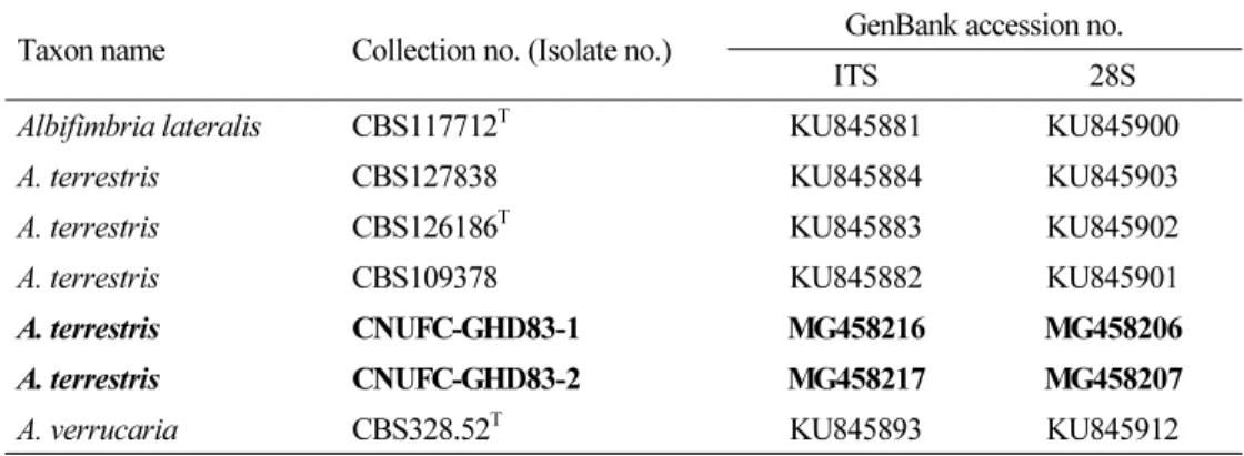
The fungal sequences obtained from the GenBank database were aligned using Clustal_X v.1.83 [30] and edited using BioEdit v.5.0.9.1 [31]. Phylogenetic analyses were performed using MEGA 6 [32] with maximum likelihood (ML) and Kimura’s two-parameter correction method. Myxospora sp., Pseudopithyella minuscula, and Stachybotrys chartarum were used as outgroups. The reliability of internal branches was assessed using the p-distance substitution model with 1,000 bootstrap replications. The CNUFC-GHD83-1, CNUFC-GHD83-2, CNUFC-RD8125, CNUFC-RD8126, CNUFC-YR2-1, and CNUFC- YR2-2 sequences were deposited in the NCBI database under accession numbers as shown in Table 1.
Table 1. Sequences used in this study and GenBank accession numbers
Taxon name Collection no. (Isolate no.) GenBank accession no.
ITS 28S
Albifimbria lateralis CBS117712
TKU845881 KU845900
A. terrestris CBS127838 KU845884 KU845903
A. terrestris CBS126186
TKU845883 KU845902
A. terrestris CBS109378 KU845882 KU845901
A. terrestris CNUFC-GHD83-1 MG458216 MG458206
A. terrestris CNUFC-GHD83-2 MG458217 MG458207
A. verrucaria CBS328.52
TKU845893 KU845912
CBS, Centraalbureau voor Schimmelcultures, Utrecht, The Netherlands; CNUFC, Chonnam National University
Fungal Collection, Gwangju, South Korea; ITS, internal transcribed spacer; T, ex-type strain. Bold letters indicate
isolates and accession numbers determined in our study.
Table 1. (Continued)
Taxon name Collection no. (Isolate no.) GenBank accession no.
ITS 28S
A. verrucaria CBS962.95 KU845895 KU845914
A. viridis CBS244.78 KU845897 KU845916
A. viridis CBS449.71
TKU845898 KU845917
A. viridis CBS127346 KU845899 KU845918
Ascodesmis nigricans CBS428.91 KC012665
Ascodesmis nigricans CBS389.68 DQ168335
Cephaliophora tropica CBS133.33 KC012669
Cephaliophora irregularis CBS218.62 KC012668
Cephaliophora tropica CNUFC-RD8125 MG458219 MG458209
Cephaliophora tropica CNUFC-RD8126 MG458218 MG458208
Coprotus ochraceus JHP-06.121 KC012673
Dimorphiseta terrestris CBS127345
TKU846314 KU846346
Eleutherascus lectardii CBS626.71 DQ168334
Eleutherascus peruvianus CBS101.75 DQ220330
Geopyxis vulcanalis KH.04.37 KC012680
Glaziella aurantiaca PR 6376 KC012681
Inaequalispora prestonii CBS175.73
TKU846316 KU846348
Ilyonectria capensis CBS132815
TJX231151 KM515908
Ilyonectria destructans CBS264.65 AY677273 KM515927
Lasiobolidium orbiculoides CBS344.73 DQ062995
Lasiobolus papillatus KH.08.30 KC012687
Mariannaea aquaticola MFU090225 GQ153838 GQ153837
M. aquaticola MFU090223
TGQ153834 GQ153833
M. aquaticola MFU090224 GQ153836 GQ153835
M. aquaticola CNUFC-YR2-1 MG459018 MG459016
M. aquaticola CNUFC-YR2-2 MG459019 MG459017
M. camptospora CBS209.73
TAY624202 AY554241
M. catenulata CBS491.62
TKM231752 KM232009
M. chlamydospora CGMCC3.17273
TKX986134 KX986141
M. cinerea CGMCC3.17274
TKX986135 KX986142
M. clavispora CBS149.87
TKX986131 KX986138
M. dimorpha HMAS266564
TKF767353 KJ002443
M. elegans CBS217.73A
TKX986132 KX986139
M. elegans DUCC400 JQ690354
M. fusiformis CGMCC3.17272
TKX986133 KX986140
M. humicola CBS740.95
TKM231755 KM231619
M. lignicola CGMCC3.17275
TKX986136 KX986143
CBS, Centraalbureau voor Schimmelcultures, Utrecht, The Netherlands; CNUFC, Chonnam National University
Fungal Collection, Gwangju, South Korea; ITS, internal transcribed spacer; T, ex-type strain. Bold letters indicate
isolates and accession numbers determined in our study.
Table 1. (Continued)
Taxon name Collection no. (Isolate no.) GenBank accession no.
ITS 28S
M. macrochlamydospora FKI-4735
TAB855777 AB855782
M. pinicola CBS745.88
TKM231754 AY554242
M. punicea CBS239.56 AY624201 JF415981
M. samuelsii CBS746.88
TKM231757 KM231621
M. samuelsii DUCC401 JX125048
M. superimposita CBS113472 AB855780 AB855785
Myxospora sp. CBS100347 KU846465 KU846486
Nectria cinnabarina AR4477
THM484548 HM484562
Parvothecium terrestre CBS198.89
TKU846468 KU846489
Peethambara sundara CBS646.77
TKU846471 KU846492
Peethambara sundara CBS521.96 KU846470 KU846491
Pseudombrophila theioleuca DHP3498 KC012696
Pseudopithyella minuscula mh675 AY945849
Pulvinula constellatio KH.03.64 DQ062987
Pulvinula convexella KH.01.020 DQ062986
Pulvinula globifera DHP DR-104 DQ220393
Smaragdiniseta bisetosa CBS459.82 KU847229 KU847255
Stachybotrys chartarum CBS129.13 KM231858 KM231738
Tarzetta catinus KS.94.10A DQ062984
Tarzetta pusilla KH.03.66 DQ062983
Virgatospora echinofibrosa CBS110115 KU847244 KU847270 Virgatospora echinofibrosa MUCL39092 KU847245 KU847271 CBS, Centraalbureau voor Schimmelcultures, Utrecht, The Netherlands; CNUFC, Chonnam National University Fungal Collection, Gwangju, South Korea; ITS, internal transcribed spacer; T, ex-type strain. Bold letters indicate isolates and accession numbers determined in our study.
Results
Phylogenetic analysis
A basic local alignment search tool (BLAST) search of ITS sequences against the NCBI database indicated that CNUFC-GHD83-1, CNUFC-RD8126, and CNUFC-YR2-1 sequences were most similar to sequences from Albifimbria terrestris (GenBank accession no.
KU845884), Cephaliophora tropica (FJ792583), and Mariannaea aquaticola (KT224837)
with 98.4% (570/579 bp), 99.3% (549/553 bp), and 99.2% (518/522 bp) homology,
respectively. On the basis of the 28S rDNA sequence analysis, the three strains had
homologies of 99.1% (819/826 bp), 99.7% (873/876 bp), and 99.9% (839/840 bp) with the
sequences of A. terrestris (KU845902), C. tropica (KC012669), and M. aquaticola
(GQ153837), respectively. On the basis of the ITS and 28S sequence analysis, the isolate
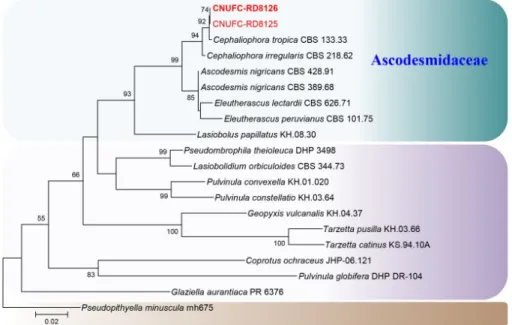
CNUFC-GHD83-1 was identical to Albifimbria terrestris (Fig. 1). CNUFC-RD8126 was identical to Cephaliophora tropica based on 28S sequences analysis (Fig. 2). CNUFC- YR2-1 was identical to Mariannaea aquaticola based on combined ITS and 28S sequences (Fig. 3).
Fig. 1. Phylogenetic trees based on maximum likelihood analysis of the internal transcribed spacer (ITS) and 28S rDNA sequences for Albifimbria terrestris CNUFC-GHD83-1 and A.
terrestris CNUFC-GHD83-2. Myxospora sp. was used as an outgroup. Bootstrap support values
of ≥50% are indicated at the nodes. The bar indicates the number of substitutions per position.
Fig. 2. Phylogenetic tree based on maximum likelihood analysis of 28S rDNA sequences for Cephaliophora tropica CNUFC-RD8125 and C. tropica CNUFC-RD8126. Pseudopithyella minuscula was used as an outgroup. Bootstrap support values of ≥50% are indicated at the nodes. The bar indicates the number of substitutions per position.
Fig. 3. Phylogenetic tree based on maximum likelihood analysis of the combined internal
transcribed spacer and 28S rDNA sequences for Mariannaea aquaticola CNUFC-YR2-1 and
M. aquaticola CNUFC-YR2-2. Stachybotrys chartarum was used as an outgroup. Bootstrap
support values of ≥50% are indicated at the nodes. The bar indicates the number of
substitutions per position.
Taxonomy of CNUFC-GHD83-1
Albifimbria terrestris L. Lombard & Crous, Persoonia 36: 177 (2016) (Table 2, Fig. 4).
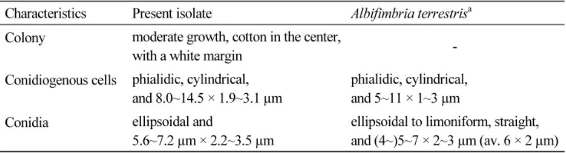
Description: Colonies of the strain grew moderately on PDA, cotton in the center with a white margin, reaching 30~33 mm in diameter at 25°C after 7 days of incubation.
Conidiogenous cells were phialidic, cylindrical, and measured 8.0~14.5 × 1.9~3.1 µm.
Conidia were ellipsoidal and measured 5.6~7.2 µm × 2.2~3.5 µm.
Table 2. Morphological characteristics of CNUFC-GHD83-1 and Albifimbria terrestris on PDA medium at 25°C
Characteristics Present isolate Albifimbria terrestris
aColony moderate growth, cotton in the center,
with a white margin -
Conidiogenous cells phialidic, cylindrical, and 8.0~14.5 × 1.9~3.1 µm
phialidic, cylindrical, and 5~11 × 1~3 µm Conidia ellipsoidal and
5.6~7.2 µm × 2.2~3.5 µm
ellipsoidal to limoniform, straight, and (4~)5~7 × 2~3 µm (av. 6 × 2 µm)
a
From the description by Lombard et al. [14].
PDA, potato dextrose agar.
Fig. 4. Morphology of Albifimbria terrestris CNUFC-GHD83-1. A, Colonies on potato dextrose agar; B, Colonies on oat meal agar; C, Colonies on corn meal agar; D, Scattered aggregated conidiomata (observed under stereo microscope); E, Sporodochial conidiomata; F, Conidia on conidiogenous cells; G, Conidia (scale bars: D = 200 µm, E~G = 20 µm).
Taxonomy of CNUFC-RD8126
Cephaliophora tropica Thaxt., Botanical Gazette Crawfordsville 35: 157 (1903) (Table 3,
Fig. 5).
Table 3. Morphological characteristics of CNUFC-RD8126 and Cephaliophora tropica on PDA medium at 25°C
Characteristics Present isolate Cephaliophora tropica
aColony rapid-growing, golden brown,
reverse reddish brown
rapid-growing, brown
Conidiogenous cells subspherical or clavate and 16.1~27.8 × 13.2~18.3 µm
clavate, 20~30 µm in diameter
Conidia cylindrical, or slightly clavate, 1~4 septate, golden brown, and 27.7~43.8 × 15.5~20.8 µm
subcylindrical, 2~5 septate, pale chocolate-brown, and 50 × 19~20 µm
a
From the description by Thaxter [15].
PDA, potato dextrose agar.
Fig. 5. Morphology of Cephaliophora tropica CNUFC-RD8126. A, Colony on potato dextrose agar; B~H, Subspherical conidiogenous cells and septate or aseptate conidia (scale bars: B~H = 20 µm).
Description: Colonies of the strain grew rapidly on PDA and were golden brown, filling a petri dish in 4 days at 25°C. The reverse colony was reddish-brown. Conidiogenous cells were subspherical to clavate, and measured 16.1~27.8 × 13.2~18.3 µm. Conidia were golden brown, 1~4 septate, cylindrical or slightly clavate, and measured 27.7~43.8 × 15.5~20.8 µm.
Taxonomy of CNUFC-YR2-1
Mariannaea aquaticola Kurniawati, L. Cai & K.D. Hyde, Mycological Progress 9 (3): 338
(2010) (Table 4, Fig. 6).
Table 4. Morphological characteristics of CNUFC-YR2-1 and Mariannaea aquaticola PDA medium at 25°C
Characteristics Present isolate Mariannaea aquaticola
aColony moderate growth, pale yellowish when
young, browning with age; reverse dark brown
moderate growth, pale yellowish, with age brown to dark brown;
reverse brown Phialides flask-shaped, and measured
11.9~21.7 × 2.2~3.5 µm
flask-shaped, and measured 14~25 × 2~3 µm
Conidia ellipsoidal to fusiform,
and measured 4.5~9.5 × 2.5~4.6 µm
ellipsoidal to fusiform,
and measured 5~10 × 2~4.5 µm
Chlamydospore Absent Absent
a