호흡동조방사선 치료 시 종양 치료의 정확도 평가
고신대학교 복음병원 방사선종양학과, 1부산가톨릭대학교 방사선학과
장은성ㆍ강수만ㆍ이철수ㆍ강세식
1목 적: 호흡동조방사선 치료 시 종양의 실제 움직임과 호흡추적장치로 측정한 피부 움직임의 차이를 자체 제작한 구동팬텀 에서 가상의 종양을 이용하여 호흡과 유사하게 움직임에 따른 정적 상태, 동적 상태 및 호흡동조상태에서 표적 위치의 정확 성의 측정치와 실측치를 평가하고 선량분포를 분석, 비교하고자 한다.
대상 및 방법: 호흡에 의해 움직이는 종양 측정을 위해 2차원적으로 움직이는 구동팬텀을 자체 제작하였다. 구동 팬톰의 움 직임은 위. 아래 방향(SI) 각각 1.5 cm 왕복운동, 상ㆍ하 방향 2 cm으로 속도조절(1∼5단계)이 되도록 하였다. 가로 4 cm, 세로 4 cm, 높이 0.5 cm의 아크릴 슬라이스에 직경 0.5 cm 종양을 납으로 표시하고, 위아래로 동일한 아크릴 슬라이스를 2장씩 쌓 은 후 아크릴 슬라이스 세 번째와 네 번째 사이 Dmax 1.5 cm film을 삽입하였다. 가상의 타겟을 구동 팬텀 위에 위치시키고 6 MV X-선이 조사되는 정적인 상태, 호흡동조 및 동적인 상태에서 각각 5 Gy를 조사하였다. 구동팬텀 위에 표식자를 올린 후, 호흡추적장치를 이용하여 사전에 설정한 호흡시간의 변화에 따른 진폭과 위상변화를 분석하였다.
결 과: RPM respiratory gating system을 이용하여 호흡주기를 8단계로 나누어 각각을 12회씩 위상변화를 분석하여 평균과 표 준편차를 구한결과 평균은 3.0 (1.5∼1.5) sec에서 1.7 cm로 가장 크고, 3.0 (1.3∼1.7) sec 5.0 (2.0∼3.0) sec에서 0.2602 cm로 가장 크고 4.0 (2.0∼2.0) sec에서 0.0866 cm로 가장 작았다. 또한 실측치에서 평균 및 표준편차를 구한 결과 t0에서 9.9 (6.6) mm t10에서 10.6 mm (7.3), t20 16.5 mm (10.3), t30 10.2 mm (7.6)으로 나타났으며, 호기나 흡기 시간의 차이에 따른 규칙은 없고 대체로 균일한 평균과 표준편차의 분포를 나타내었다. 또한 정적 상태, 동적 상태 및 호흡동조상태에서 Gafchromic EBT film 의 방사선량을 분석한 결과, ICRU 62에서 권고한 90% 선량분포가 3 mm 이내에 포함되므로 정확성과 정도관리 측면에서 적 합한 것으로 사료된다.
결 론: 구동팬톰을 이용하여 호흡움직임에 따른 정확성 및 선량분포차이를 Gafchromic film을 통하여 확인하였으며 결과를 바 탕으로 호흡에 의해 변화가 생기는 장기에 대한 차이를 고려하여 치료계획을 한다면 종양과 정상조직에 적절한 선량계획을 세울 수 있어 치료효과 향상에 도움을 주게 될 것으로 생각한다.
핵심용어: 호흡동조방사선치료, 구동 팬텀, 4D-CT, 호흡추적장치
이 논문은 2010년 7월 21일 접수하여 2010년 8월 29일 채택되었음.
책임저자:장은성, 고신대학교 복음병원 방사선종양학과 Tel: 051)990-6396, Fax: 051)990-3993 E-mail: [email protected]
서 론
방사선치료 시 환자호흡에 의한 종양의 움직임은 정확한 치료선량 분포에 오차를 발생시킬 수 있다. 이러한 종양 및 주변장기의 움직임 영향을 보정하기 위해 일반적으로 ICRU (International commission on radiation units and measure- ments) report 621)에서는 계획용표적체적(Planning target volume, PTV) 설정 시 움직임 영역을 충분회 포함하여 치료 대상 종양부위가 조사 영역을 벗어나지 않도록 권고하고 있 다. 이와 같은 종양의 움직임 영역을 정확히 설정하고자 4차 원 전산화단층촬영(CT) 모의치료 영상을 활영하는 방법이
적용되고 있는데, 이는 각 호흡주기별 CT 영상에 종양을 설 정한 후 전체 호흡주기에 걸친 종양의 움직임 영향이 모두 고려된 내부표적체적(Internal Target Volume, ITV)을 결정하 여 PTV 설정시 환자 호흡에 의한 움직임 영역이 정확하게 고려되게 하는 방법이다.2-5) 이러한 종양의 움직임 영역을 모 두 고려한 PTV 설정 방법은 종양 내 충분한 치료 선량을 보 장할 수는 있지만 치료방사선 조사 영역이 증가함에 따라 종 양 주변의 주요 장기 및 정상조직의 선량이 증가하는 단점이 있다. 이와 같은 문제점을 해결하고 동시에 방사선 치료 시 종양의 움직임 영향을 줄이기 위해 여러 방법들이 연구, 개 발되어 왔으며, 그 중 환자의 호흡주기 내 특정 영역에서만 치료방사선이 조사되도록 하는 호흡연동 방사선치료 방법이 현재 임상에 많이 적용되고 있다.6-8) 즉, 특정 환자의 호흡주 기에 따른 영상획득이 가능하고 이를 반영한 치료를 수행할
Fig. 1. Real-time position management (RPM). (A) RPM CCD
camera surrounded by infrared LED and display monitor. (B) RPM retro-reflective marker box with added infrared LED. (C) The marker block signal as a function of time from the RPM system.수 있어 호흡에 따른 움직임이 많은 폐암, 상복부 종양, 간암 의 방사선 치료 중 호흡과 같은 이유로 종괴의 위치가 변할 경우 움직임을 고려한 방사선 치료법으로 정상조직의 방사 선 피폭을 최소화할 수 있다. 흉부 또는 복부부위에 종양이 위치할 때 장기의 움직임은 폐의 하엽에 위치할 경우 0.9∼
3.2 cm, 간의 경우 1∼4 cm 가량의 움직임이 보고되고 있
다.9-16) 이러한 호흡연동 방사선치료에서 표식자의 운동주기
중 안정된 특정 위상영역을 기반으로 방사선 조사 구간을 설 정하는 방법은 진폭변위를 기반으로 하는 방법보다 안정적 으로 치료를 수행할 수 있는 장점이 있으나, 치료 시 방사선 조사 구간 내 표식자 진폭의 변화가 발생해도 설정 위상 기 반영역에만 포함 되면 방사선이 조사되는 경우가 발생할 수 있어, 실제 호흡량의 변위에 따른 내부 장기의 움직임 정도 를 정확히 반영하기에는 한계가 있다.16)
본 연구에서는 이와 같은 위상기반 호흡동조 방사선치료 시 발생할 수 있는 표식자 진폭의 변화와 이로 인한 치료 부 위의 움직임에 의한 오차의 분석을 목적으로 호흡으로 인한 장기의 움직임을 모사하기 위해 동체 모형 팬텀과 구동 모터 를 제작하고, IGRT를 사용한 호흡운동 모의실험을 통해 호 흡으로 인한 움직이는 타겟내에 삽입된 Gafchomic EBT film 과 함께 정적, 동적상태 및 호흡동조상태에서 종양의 표적위 치의 정확성과의 측정치와 실측치를 평가하고 선량분포 분 석, 비교하고자 한다.17-20)
대상 및 방법 1. 실험 기자재
IGRT (CLINAC IX, varian USA) The Manufactured Moving phantom .Gafchromic EBT film
.Scanmaker 9800XL
.Virtual Tumor & Marker block .Eclips (varian Medical system, USA) Acuity simulator
Matlab 7.01
2. 실험 방법
1) 호흡연동 감시 시스템(real-time position manage- ment, RPM) respiratory gating system
환자의 흡기나 호기 시 움직임이 크게 나타나는 흉부나 복 부 부위의 피부움직임을 적외선 카메라로 관찰하는 장치이 다. 적외선카메라 주위에는 적외선 발광다이오드가 있어 적 외선을 방출하며 이 적외선은 환자의 흉부나 복부 위에 위치
한 적외선 반사체에 의해 호흡에 따라 움직이며 적외선을 반 사 시킨다. 적외선카메라는 가시광선과 적외선을 동시에 통 과시켜 영상처리를 통해 적외선 반사체의 움직임을 분석하 게 된다(Fig. 1A). 적외선 반사체는 위, 아래 두개의 점으로 구성되어 있는데 이 두 점의 거리는 항상 3 cm로 일정하기 때문에 카메라에서 촬영한 반사체 두 점 사이의 화소 수에 의하여 카메라와 반사체의 거리를 정확히 알 수 있으며 반사 체 움직임의 정량적 측정이 가능하다(Fig. 1B). RPM은 두 가 지 모드로 호흡측정이 가능한데 하나는 위상모드(phase mode)이고 또 하나는 진폭모드(amplitude mode)이다.21,22) 진 폭모드는 환자의 흡기 또는 호기의 위치를 기준으로 측정하 는 것이기 때문에 환자의 호흡량이 치료간(interfraction)에 일정해야 재현성이 좋다. 위상모드는 환자 호흡의 한 주기의 시간을 백분율로 측정하기 때문에 위상모드는 환자의 호흡 주기가 치료 중(intrafraction) 일정해야 한다(Fig. 1C).
A B C D E F G H I
0 100 Gy 200 Gy 300 Gy 400 Gy 500 Gy 600 Gy 700 Gy 800 Gy
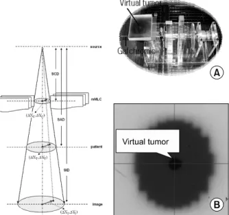
Table 1. Dose values for each segment of the calibration film Fig. 3. 4D gating RT architecture and evaluation setting in
clinac. (A) Gafchromic EBT film insert with a virtual tumor inside for the moving phantom. (B) MV fluence collection by the image Detector with a circular aperture in the 4DRT the adaptive fuction was developed.Fig. 2. Experimental set-up showing ving moving phantom.
Fig. 4. The Calibration was performed to cover the dose range
from 0 to 8 Gy, corresponding to the suitable dose range for external beam therapy. (A) The analysing pixels are marked with blue boxes. (B) The calibration film is displayed in three dimensions.2) 자체 제작한 구동팬텀
호흡운동으로 인한 내부 장기중 횡격막의 움직임을 측정하 여 방사선조사시 그 움직임을 대신할 수 있는 구동 팬톰 시 스템을 제작하였다. 이 시스템은 필름을 놓을 수 있는 구동 팬톰과 이를 제어할 수 있는 제어기로 구성되어 있다(Fig.
2). 구동 팬톰의 움직임은 위, 아래 방향(SI) 각각 1.5 cm 왕 복운동, 상, 하 방향 2 cm으로 속도조절(1∼5단계)이 되도록 하였다. 호흡에 따른 움직임을 자체 제작한 구동팬톰에 적용 하여 방사선을 조사하였다(Fig. 3).
3) Gafchromic EBT film의 선량교정
Gafchromic EBT film의 선형성을 알아보기 위해서 한 장 의 Gafchromic EBT film을 4×4 cm 크기로 9장을 잘라 6 MV 광자선, 조사야 6×6 cm, 1.5 cm (dmax)깊이로 하여 각각 0, 1, 2, 3, 4, 5, 6, 7, 8 Gy를 조사하여(Table 1), 모든 film은 24시간 후 film을 스캔하여 광학 농도(OD; Optical Density) 값을 얻어 film의 선량-광학 농도의 관계를 알아보았다(Fig.
4).
Fig. 5. A diagram showing a 4D CT acquisition process. Images
are acquired and then sorted by the patient’s respiratory phase at the time the image was acquired to create 3D CT images at discrete phases of the respiratory cycle.Fig. 6. Elliptic model of the thoracic and abdominal cross
section during breathing (De Groote et al 2000). The principal axes are dorsoventral and transverse diameter, The spinal cord is fixed. (B) is mathematically equivalent model to (A) with the center of the ellipse considered fixed. (C) is the simulation of respiratory movements, f respiratory frequenly, x and y the mean amplitude and Xm and Ym the mean positions.Fig. 7. Diagram for phase-based 4D CT image reconstruction at
the exhale phase.4) 4차원 CT모의치료 영상획득
4D-CT2)란 호흡으로 인한 종양 및 장기의 움직임을 CT스 캔을 통해 획득하는 방법으로 특정 호흡 상태에서 CT영상을 획득하는 gated-CT와 구별된다.23-25) 두 방법 모두 CT스캔을 시행하는 환자의 호흡을 모니터링 하는 장치가 필요하며 gated-CT의 경우 호흡모니터링장치로부터 특정호흡위상에서 트리거신호를 CT에 보내 CT가 스캔을 하도록 지시하는 방 식이며, 4D-CT의 경우는 CT스캔과 동시에 호흡모니터링신 호를 저장하여 스캔시간과 호흡신호를 이용하여 CT영상을 후처리함으로써 호흡위상별 CT데이터세트를 재구성하는 방 식이다. 대표적인 호흡모니터링장치로는 흉복부의 오르내림 을 CCD카메라를 사용하여 추적하는 Real-Time Position Management System (Varian Medical Systems, Palo Alto, CA;
RPM)26-28)과 호흡에 따른 복부의 팽창도를 스트레인게이지
를 사용하여 측정하는 Respiratory Gating System (Anzai
Medical Company, Japan)28)이 소개되고 있다. 4D-CT 스캔의 경우 동일 위치에서 전체 호흡주기에 걸쳐 연속촬영모드를 이용하여 반복적으로 영상을 얻게 된다.29) 통상적으로 호흡 주기를 10개의 위상으로 나누어 촬영하며, 이 때 10개의 서 로 다른 호흡위상에 대응하는 3D CT데이터를 얻게 된다 (Table 3). 4D CT 영상 획득 과정을 개략적으로 Fig. 5에 도 시하였다.
5) 호흡주기에 따른 위상변화 분석
Gafchromic EBT film에 대한 정적인 상태(static), 호흡동 조 상태(Gating) 및 동적인 상태(moving)에서의 선량 분석 실험을 하기에 앞서, 팬텀을 이용한 종양의 호흡주기에 따른 위상변화 분석을 실시하였다(Fig. 6).
자체 제작한 구동팬텀 위에 표식자를 올린 뒤, 적외선 카메 라를 비롯한 RPM system을 이용하여 사전에 설정한 호흡시 간 변화에 따른 호흡주기를 10단계로 나누어(Fig. 7) 각각 12
Inspiration (sec) Expiration (sec) Total (time)
1.5 1.5
3.0
1.3 1.7
1.0 2.0
2.0 2.0
4.0
1.5 2.5
1.0 3.0
2.0 3.0
5.0
1.5 3.5
1.0 4.0
2.0 4.0
1.5 4.5 6.0
1.0 5.0
Table 2. Set up of the various breathing velocity period
Peak-to-Peak motion SD of tumor positions with in one 4D-CT Mean (mm) SD (mm) Mean (mm) SD (mm) t0
t10
t20
t30
9.9 10.6 16.5 10.2
6.6 7.3 10.3 7.6
2.5 3.8 4.0 3.7
2.8 3.1 2.7 2.9
Table 3. Variability of tumor breathing motion
Fig. 8. Solid line shows the relationship between absorbed dose
and optical density. Dotted line is fitting line. The calibration curve could be fitted to the function of 2nd order. The linearity of GafChromic EBT film response was corrected and between absorbed dose and optical density. Dotted line is fitting line.The calibration curve could be fitted to the function of 2nd order. The linearity of GafChromic EBT film response was corrected and the optical density could be converted into the absolute dose by using this function.
회씩 측정하여 진폭과 위상변화의 평균값을 나타내었다 (Table 2).
6) 정적인 상태(static)와 호흡동조상태(gating) 및 동 적인 상태(moving)에서의 Gafchromic EBT film의 선량분포 분석비교
가로 4 cm, 세로 4 cm, 높이 0.5 cm의 아크릴 슬라이스에 직경 0.5 cm 종양을 납으로 표시하고, 위아래로 동일한 아크 릴 슬라이스를 2장씩 쌓은 후 아크릴 슬라이스 세 번째와 네 번째 사이 Dmax 1.5 cm film을 삽입하였다. 가상의 타겟을 구동 팬텀 위에 위치시키고 6 MV X-선이 조사되는 정적인 상태, 호흡동조 및 동적인 상태에서 각각 5 Gy를 조사하였다 (Fig. 8). 그리고 정적인 상태 호흡동조 및 동적인 상태에서 획득한 Gafchromic EBT film을 스캔하고, 선량분포를 분석 하였다(Fig. 9).
결 과 1 .Gafchromic EBT film의 선량교정
6 MV 광자선에 의한 Gafchromic EBT 필름은 0∼8 Gy 영 역에서 우수한 선형성을 보여주었다(Fig. 8).
2. 호흡주기에 따른 위상변화 분석
RPM respiratory gating system을 이용하여 호흡주기를 10 단계로 나누어 각각 12회씩 위상변화를 분석하였다. 평균과 표준편차를 구한결과 평균은 3.0 (1.5∼1.5) sec에서 1.7 cm 로 가장 크고, 3.0 (1.3∼1.7) sec 5.0 (2.0∼3.0) sec에서 0.2602 cm로 가장 크고 4.0 (2.0∼2.0) sec에서 0.0866 cm로 가장 작았다. 분석결과 호기나 흡기 시간의 차이에 따른 규 칙은 없고 대체로 균일한 평균과 표준편차의 분포를 나타내 었다(Table 4).
3. 정적인 상태와 동적인 상태에서의 Gafchromic EBT film의 방사선량 분석
구동 팬텀 내의 Gafchromic EBT film에 조사한 선량분포 는 Gafchromic EBT film을 이용한 구동팬텀 내 필름으로 측 정한 선량과 계산된 선량분포와 비교하면 90%가 포함되는 선량분포는 오차범위 3 mm 이내에 분포됨을 알 수 있었다 (Fig. 9).
고안 및 결론
호흡조절방사선치료는 환자 피폭을 최소로 하면서 정상조 직을 최대한 보호할 수 있는 최신 치료기법이다. 일반적으로
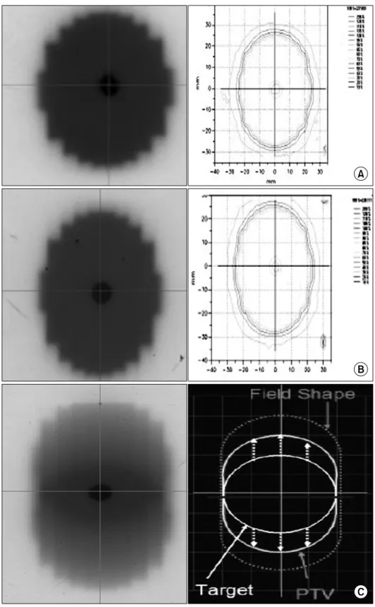
Fig. 9. Gafchromic films of the
three patterns. (A) Static pattern, (B) Gating pattern, (C) Moving pattern.방사선치료계획 시 정상조직 부작용확률(Normal Tissue Com- plication Probability, NTCP)을 평가할 때 정상폐 용적에서 Gross Target Volume (GTV)을 제외한 체적에 부여되는 방사
선부피곡선을 적용하기 때문에 Planning Target Volume (PTV) margin를 줄이는 것이 정상조직손상확률을 최소화하 기 위한 방법이라 하겠다.30-35) 그러나 이 치료법의 대전제는
Total period (inspiration-expiration)
(sec)
Mean (cm) S·D
① 3.0 (1.5∼1.5) 1.7 0.1732
② 3.0 (1.3∼1.7) 1.375 0.1090
③ 3.0 (1.0∼2.0) 1.4333 0.1826
④ 4.0 (2.0∼2.0) 1.45 0.0866
⑤ 4.0 (1.5∼2.5) 1.3882 0.1811
⑥ 4.0 (1.0∼3.0) 1.6667 0.2573
⑦ 5.0 (2.0∼3.0) 1.5421 0.2602
⑧ 5.0 (1.5∼3.5) 1.46 0.2026
⑨ 5.0 (1.0∼4.0) 1.5294 0.1963
⑩ 6.0 (2.0∼4.0) 1.4313 0.1927
⑪ 6.0 (1.5∼4.5) 1.5267 0.1436
⑫ 6.0 (1.0∼5.0) 1.4875 0.1364
Table 4. Mean and standard deviation of breathing phase for the
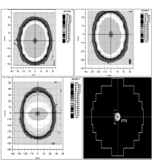
Respiratory cycleFig. 10. After the dose conversion
from the absorbed spectrum of Gafchromic EBT films, comparions of isodose distributions among static, gating, moving patterns were analyzed.호흡추적장치가 관찰하는 움직임의 정도가 종양의 움직임을 정확하게, 또는 용인 가능한 오차범위 이내에서 예측할 경우
에만 적용 가능하다. 호흡조절방사선치료를 시행하기 위해서 는 치료 시 종양 움직임의 정확한 예측은 무엇보다 중요하 다.
필름을 이용한 선량측정방법은 에너지 의존성 이외에 기 존의 필름이 실내 빛에 민감하거나, 그 이외의 몇 가지 단점 이 있으나, 이러한 단점을 보완한 Gafchromic EBT film은 높 은 공간분해능을 가지고 있으며, 다른 필름과 다르게 흑화도 가 우수한 선형성이 있다. 이러한 Gafchromic EBT film의 장 점을 이용하여 호흡에 따른 선량분포의 정확성 평가는 정상 조직의 장해를 최소화하고 종양조직의 치사를 최대화한다는 방사선치료 목적과 부합된다.
이를 위해 본 논문에서는 Gafchromic EBT film의 선량과 광학농도 관계를 실험한 결과, 0∼8 Gy사이에서 우수한 선 형성을 가진다는 것을 확인하였고, 5 Gy를 조사하여 정적인 상태, 동적인 상태 및 호흡동조상태에서의 선량분포를 분석 하였다.
분석한 결과, 호흡에 따른 선량분포 변화는 ICRU 62에서
권고한(Fig. 10) 90%선량분포가 3 mm 이내에 포함되므로 정 확성과 정도관리 측면에서 적합한 것으로 사료된다. 또한 구 동팬톰을 이용하여 호흡움직임에 따른 정확성 및 선량분포 차이를 Gafchromic film을 통하여 확인하였으며 결과를 바탕 으로 호흡에 의해 변화가 생기는 장기에 대한 차이를 고려하 여 치료계획을 한다면 종양과 정상조직에 적절한 선량계획 을 세울 수 있어 치료효과 향상에 도움을 주게 될 것으로 생 각한다.
참고문헌
1. ICRU-62: Prescribing, Recording and Reporting Photon Beam Therapy (supplement to ICRU report 50): International Com- mission on Radiation Units and Measurements, Bethesda, MD 1999
2. Rietzel E, Chen GTY, Choi NC, Willet CG: Four-dimensional image-based treatment planning: target volume segmentation and dose calculation in the presence of respiratory motion. Int J Radiat Oncol Biol Phys 2005;61:1535-1550
3. Rietzel E, Liu AK, Doppke KP, et al.: Design of 4D treatment planning target volumes. Int J Radiat Oncol Biol Phys 2006;
66:287-295
4. Guckenberger M, Wilbert J, Krieger T, et al.: Four- dimensional treatment planning for stereotactic body radio- therapy. Int J Radiat Oncol Biol Phys 2007;67:276-285 5. Colgan R, McClelland J, McQuaid D, et al.: Planning lung
radiotherapy using 4D CT data and a motion model. Phys Med Biol 2008;53:5815-5830
6. Vedam SS, Keall PJ, Kini VR, Mohan R: Determining parameters for respirtion-gted radiotherapy. Med Phys 2001;28:2139-2146
7. Wink NM, Chao M, Antony J, Xing L: Individualized gating windows based on four-dimensional CT information for respiration-gated radiotherapy. Phys Med Biol 2008;53:165- 175
8. Ahmed RS, Shen S, Ove R, Duan J, Fiveash JB, Russo SM:
Intensity modulation with respiratory gating for radiotherapy of the pleural space. Med Dosim 2007;32:16-22
9. Elizabeth AB, Brad RM, Donald MR, et al.: Dosimetric evalution of lung tumor immobilization using breath hold at deep inspiration. Int J Radiat Oncol Phys 2006;50:1091-1098 10. Rosenz weig KE, Hanley J, Mah D, et al.: The deep inspriation breath-hold technique in the treatmemt of inoperable ono- small-cell lung cancer. Int J Radiat Oncol Biol Phys 2000;48:
81-87
11. Shimizu S, Shirato Ogura S, et al.: Detection of lung tumor movement in real-time tumor-tracking radiotherapy. Int J Radiat Oncol Biol Phys 2001;51:304-310
12. Korin HW, Ehman RL: RiedereRespiratory kinematics of the
upper abdominal organs: a quantitative study. Magn Reson Med 1992;23:172-178
13. Davies SC, Hill AL, Holmes RB, Halliwell M, Jackson PC:
Ultrasound quantitation of respiratory organ motion in the upper abdomen. Br J Radiol 1994;67:1096-1102
14. Wagman R, Yorke E, Ford E, et al.: Respiratory Gating for live tumors: use in dose escalation. Int J Radiat Oncol Biol Phys 2003;55:659-668
15. Guckenberger M, Wilbert J, Meyer J, et al.: Is a single respiratory correlated 4D-CT study sufficient for evaluation of bresthing motion. Int J Radiat Oncol Biol Phys 2007;67:1352- 1359
16. Ahn S, Yi B, Suh Y, et al.: A feasibility study on the predication of tumor location in the lung from skin motion.
Br J Radiol 2004;77:588-596
17. 신은혁, 박희철, 한영이, 주상규, 안용찬: 호흡연습장치를 적 용한 호흡교정법의 영향 평가. 대한방사선종양학회지 2008;
26:181-188
18. 김명남, 원철호: 호흡연동에 의한 EBT 단면영상에서의 폐실 질 윤곽선 검출 1999;8:154-162
19. 장은성, 이철수: Gafchromic EBT film을 이용한 뇌정위방사 선치료의 선량분석 가능성 평가. 대한방사선치료학회지 2007;19:27-33
20. G. Li, D. et al.: Advances in 4D medical imaging and 4D radiation therapy. Technol Cancer Res Treat 2008;7:67-82 21. Lujan AE, Larsen EW, Balter JM, et al.: A method for
incorporating organ motion breathing into 3D dose calcula- tions. Med Phys 1999;26:715-720
22. Vedam SS, Keall PJ, Kini VR, et al.: Determining parameters for respiration-gated radiotherapy. Med Phys 2001;28:2139- 2146
23. Vedam SS, Keall PJ, Kini VR, et al.: Acquiring a four dimensional computed tomography dataset using an external respiratory signal. Phys Med Biol 2003;48:45-62
24. Ford EC, Mageras GS, Yorke E, et al.: Respiration-correlated spiral CT: a method of measuring respiratory-induced anato- mic motion for radiation treatment planning. Med Phys 2003;30:88-97
25. Kubo HD, Hill BC: Respiration gated radiotherapy treatment:
a technical study. Phys Med Biol 1996;41:83-91
26. Minohara S, Kanai T, Endo M, et al.: Respiratory gated irradiation system for heavy-ion radiotherapy. Int J Radiat Oncol Biol Phys 2000;47:1097-1103
27. Pan T, Lee TY, Rietzel E, Chen GTY: 4D-CT imaging of a volume influenced by respiratory motion on multi-slice CT.
Med Phys 2004;21:334-340
28. Vedam SS, Keall PJ, Kini VR, et al.: Acquiring a fourdi- mensional computed tomography dataset using an external respiratory signal. Phys Med Biol 2003;48:45-62
29. Mageras GS, Yorke E, Rosenzweig K, et al.: Fluoroscopic evaluation of diaphragmatic motion reduction with a respiratory gated radiotherapy system. J Appl Clin Med Phys
2001;2:191-200
30. Ramsy CR, Daniel Scaperoth D, Arwood D, et al.: Clinical efficacy of respiratory gated conformal radiation therapy.
Medical Dosimetry 2006;24:115-119
31. Giraud P, Yorke E, Ford EC. et al.: Reduction of organ motion in lung tumors with respiratory gating. Int J Radiat Oncol Biol Phys 2007;68:259-266
32. Giraud P, Yorke E, jiang S, Simon L, Rosenzweig K, Mageras G: Reduction of organ motion effects in IMRT and conformal 3D radiation delivery by using gating and tracking techniques.
Cancer Radiotherapy 2006;10:269-282
33. Huntzinger C, Munro P, Lohson S, et al.: Dynamic targeting image-guided radiotherapy. Medical Dosimetry 2006;31:113- 125
34. Keall P, Vedam S, George R, et al.: The clinical imple- mentation of respiratory-gated intensity-modulated radio- therapy. Medical Dosimetry 2006;31:152-162
35. Underberg RWM, Lagerwaard FJ, Slotman BJ, et al.: Use of maximum intensity projections (MIP) for target volume gene- ration in 4DCT scans for lung cancer. Int J Radiat Oncol Biol Phys 2005;63:253-260