C A S E R E P O R T Open Access
An unusual presentation of non-specific cystic degeneration of craniofacial fibrous dysplasia: a case report and review of
literature
Inseok Hong 1,2 , Dong Cheol Kang 1,2 , Dae-Ho Leem 1,2 , Jin-A Baek 1,2 and Seung-O Ko 1,2*
Abstract
Background: Fibrous dysplasia (FD) is a rare, sporadic, and benign congenital condition in which normal cancellous bone is replaced by fibro-osseous tissue with immature osteogenesis. FD localized in the cranial and facial bones is called craniofacial fibrous dysplasia (CFD). Cystic degeneration in CFD cases is rare; cystic degeneration appearing in both the maxilla and the mandible FD lesion is even rarer. The aim of this article was to report a case of fibrous dysplasia of the mandible and maxilla complicated by nonspecific cystic degeneration.
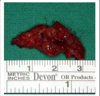
Case presentation: A 30-year-old woman presented with a rare case of non-specific cystic degeneration in a mandible and maxilla FD lesion that occurred 11 years after surgery. She was diagnosed with polyostotic CFD and underwent maxillary and mandibular bone contouring. Cyst enucleation under general anesthesia was performed in the mandibular region due to pain and discomfort.
Conclusions: In cases involving non-aggressive and non-invasive FD cystic degeneration in focal areas, conservative treatment is recommended. However, if cystic degeneration of FD develops rapidly and causes discomfort, pain, or dysfunction, surgical treatment should be considered.
Keywords: Craniofacial fibrous dysplasia, Cystic degeneration, Cyst enucleation, Fibrous dysplasia, Mandible, Maxilla, Polyostotic fibrous dysplasia
Background
Fibrous dysplasia (FD) is a benign disorder characterized by the replacement of normal bone tissue with prolifera- tive fibrous connective tissues [1]. Somatic mutations in the Gs-alpha gene on chromosome 20 can lead to endo- crine tumors, FD, and McCune–Albright syndrome (MAS) [2]. FD occurs in two forms: the monostotic form affecting one bone (approximately 70% of cases), and the polyostotic form affecting at least two bones
(approximately 30% of cases) [3]. FD that appears in the cranial and facial bones is called craniofacial fibrous dys- plasia (CFD). The prevalence of polyostotic and mono- stotic CFD is 71–91% and 10–29%, respectively [4, 5]. In the jaw bone, FD is approximately twofold more preva- lent in the maxilla and usually occurs unilaterally [1].
Non-epithelial-lined cysts sometimes occur in associ- ation with various benign and malignant bone lesions, including FD, giant cell tumors, chondroblastoma, ossi- fying fibroma, benign osteoblastoma, cemento-osseous dysplasia, fibrous histiocytoma, fibrosarcoma, and osteo- sarcoma. These cysts show various patterns and can ap- pear as aneurysmal bone cysts, simple bone cysts, or non-specific cystic degenerations [6].
© The Author(s). 2020 Open Access This article is licensed under a Creative Commons Attribution 4.0 International License, which permits use, sharing, adaptation, distribution and reproduction in any medium or format, as long as you give appropriate credit to the original author(s) and the source, provide a link to the Creative Commons licence, and indicate if changes were made. The images or other third party material in this article are included in the article's Creative Commons licence, unless indicated otherwise in a credit line to the material. If material is not included in the article's Creative Commons licence and your intended use is not permitted by statutory regulation or exceeds the permitted use, you will need to obtain permission directly from the copyright holder. To view a copy of this licence, visit http://creativecommons.org/licenses/by/4.0/.
* Correspondence: [email protected]
1
Department of Oral and Maxillofacial Surgery, School of Dentistry, Chonbuk National University Dental Hospital, 20, Geonji-ro, Deokjin-gu, Jeonju-si, Jeollabuk-do, Republic of Korea
2