Received November 14, 2019 Revised January 17, 2020 Accepted February 24, 2020
Corresponding author: Seungyeup Han Department of Internal Medicine, Keimyung University School of Medicine and Keimyung University Kidney Institute, 1095 Dalgubeol-daero, Dalseo- gu, Daegu 42601, Korea
Tel: +82-53-258-7712 Fax: +82-53-258-4739 E-mail: [email protected]
Effectiveness of valacyclovir prophylaxis against the occurrence of cytomegalovirus infection in kidney transplant recipients
Woo Yeong Park
1,2, Yaerim Kim
1,2, Jin Hyuk Paek
1,2, Kyubok Jin
1,2, Sung Bae Park
1,2, Seungyeup Han
1,21Department of Internal Medicine, Keimyung University School of Medicine, Daegu, Korea
2Keimyung University Kidney Institute, Daegu, Korea
Background: Cytomegalovirus (CMV) infection is a crucial infection in kidney transplant recipients (KTRs) despite advancements in diagnostic and treatment methods. There are still many controversies about the ways to prevent CMV infection.
Methods: We retrospectively analyzed 153 KTRs who underwent kidney transplantation (KT) between September 2013 and January 2016. We classified KTRs into two groups:
valacyclovir prophylaxis group (intravenous ganciclovir for 2 weeks after KT, followed by oral valacyclovir for 3 months) and historical control group (only intravenous ganci- clovir for 2 weeks after KT). We evaluated the incidence of CMV infection, clinical out- comes, CMV-free survival rate between the two groups and risk factors for the develop- ment of CMV infection.
Results: Mean time between KT and diagnosis of CMV infection was 4.5±3.3 months.
The valacyclovir prophylaxis group showed lower incidence of CMV infection than the historical control group (21.7% vs. 43.9%, P=0.011). The valacyclovir prophylaxis group showed higher CMV-free survival rate than the control group (P=0.011). In multivari- able-adjusted analysis, independent risk factors for the development of CMV infection were no valacyclovir prophylaxis, older age at KT, thymoglobulin induction, and delayed graft function.
Conclusions: Valacyclovir prophylaxis for 3 months showed significant reduction in the incidence of CMV infection in KTRs. Therefore, we suggest valacyclovir prophylaxis for 3 months in KTRs with risk factors such as old age, thymoglobulin induction, and de- layed graft function.
Keywords: Antibiotic prophylaxis; Antithymocyte serum; Cytomegalovirus infections;
Kidney transplantation; Risk factors
pISSN 2671-8790 eISSN 2671-8804
© The Korean Society for Transplantation This is an Open Access article distributed under the terms of the Creative Commons Attribution Non-Commercial License (http://creativecommons.org/licenses/
by-nc/4.0/) which permits unrestricted non-commercial use, distribution, and reproduction in any medium, provided the original work is properly cited.
INTRODUCTION
Cytomegalovirus (CMV) infection is a crucial infection in kidney transplant recipients (KTRs) [1]. CMV infection usually happens within 1 year after kidney transplantation
(KT), which is an unstable period of immunosuppression
and ultimately can lead to early graft failure [1]. Recently,
the prevalence of CMV infection is increasing due to the
use of strong immunosuppressants such as antithymo-
cyte globulin (ATG), rituximab, or bortezomib [2]. If proper
treatment is not provided in this condition, CMV that has invaded multiple organs can lead to death [3]. In addition, bacterial infections, other viral infections, and fungal in- fections can often occur, and acute rejection can occur simultaneously [4,5]. Thus, it is important to detect and treat CMV infection early and prevent it, if possible.
Even though the diagnosis and treatment for CMV infection have been developed, many controversies exist about the ways to prevent CMV infection, and each cen- ter’s policies vary. The Kidney Disease: Improving Global Outcomes guidelines and the US guidelines recommend- ed CMV prophylaxis in KTRs with a high risk of CMV infection, that is, kidney donors whose CMV serostatus was positive and recipients whose CMV serostatus was negative before KT, for 3 months after KT, and for 6 weeks after anti-rejection therapy such as steroid pulse therapy or the use of ATG [3,6-8]. In our center, we used intrave- nous ganciclovir for 2 weeks for CMV prophylaxis during the recovery period after KT, but we could not decrease the incidence of CMV infection. Most guidelines recom- mended oral valganciclovir for CMV prophylaxis, but oral valganciclovir was very expensive and has some adverse effects. Recent research reported that the effect of oral valacyclovir is not inferior to that of oral valganciclovir [9], but its efficacy remains unclear. Therefore, we evalu- ated the effectiveness of oral valacyclovir prophylaxis for 3 months in comparison with the use of an intravenous ganciclovir for 2 weeks to prevent CMV infection in KTRs.
METHODS
This study was approved by the Institutional Review Board of Keimyung University Dongsan Medical Center (IRB No. 2017-10-026). All clinical investigations were conducted in accordance with the guidelines of the 2013 Declaration of Helsinki. The clinical and imaging data were obtained with patients’ consent for the purpose of scientific research publication.
Study Design
We retrospectively investigated 153 KTRs between September 2013 and January 2016. In this study, we classified the participants into two groups as follows:
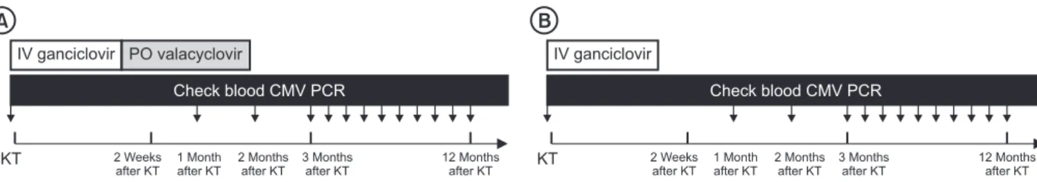
valacyclovir prophylaxis group (intravenous ganciclovir for 2 weeks after KT, followed by oral valacyclovir for 3 months from July 2015) and historical control group (only intravenous ganciclovir for 2 weeks after KT from study starting time to June 2015), as seen in Fig. 1. Blood CMV polymerase chain reaction (PCR) was checked at KT, at 1 month after KT, 3 months after KT, and once a month from 3 to 12 months after KT and once a year after 12 months after KT. Whole blood samples were tested by us- ing QIAcube (Qiagen, Hilden, Germany) and real-time PCR assays for CMV. The result of CMV PCR was expressed to log
10copies/mL. CMV infection is defined as virus iso- lation or detection of viral proteins or nucleic acid in any body fluid or tissue specimen [10]. Several specific defini- tions for CMV detection in blood are recommended such as the detection of virus, antigen, or DNA. Therefore, CMV infection was diagnosed when the blood CMV PCR was more than 100 log
10copies/mL as cutoff values of posi- tive blood CMV PCR, irrespective of the symptoms. In ad- dition, CMV disease was diagnosed as the CMV infection with accompanying symptoms. CMV disease could be classified as viral syndromes with fever, malaise, leuko- penia, thrombocytopenia, or as a tissue invasive disease [8]. We evaluated the incidence of CMV infection, clinical outcomes, and CMV-free survival rate between the two groups and risk factors for the development of CMV infec- tion.
Immunosuppression Protocols
We administered basiliximab (20 mg at days 0 and 4, respectively; Simulect, Novartis, Basel, Switzerland) as the immunosuppressant for induction in KTRs with low immunologic risks. We also administered ATG (Thymo- globulin, Genzyme, Cambridge, MA, USA; 1.5 mg/kg at day HIGHLIGHTS
• Cytomegalovirus (CMV) infection is very important for the prognosis of kidney transplant recipients.
• CMV prophylaxis is necessary to prevent the occur- rence of CMV infection.
• Valacyclovir prophylaxis for 3 months shows significant reduction in the incidence of CMV infection in kidney transplant recipients.
• The independent risk factors for the development of
CMV infection were older age at kidney transplantation,
use of antithymocyte globulin, delayed graft function,
and no valacyclovir prophylaxis.
0 and 1.0 mg/kg between days 1 and 3) in KTRs with high immunologic risks such as 2nd KT, high panel reactive antibody, and fully mismatched human leukocyte antigen (HLA). We used tacrolimus (Prograf; Astellas Pharma Inc., Toyama, Japan), prednisolone, and mycophenolate mofetil (MMF; Cellcept, Hoffmann-La Roche Inc., Nutley, NJ, USA) as the immunosuppressive regimen for maintenance.
We maintained trough levels of tacrolimus at 5–10 ng/mL for 1 month after KT and at 3–8 ng/mL after that.
Prednisolone (30 mg/day) was administered for 2 weeks and was reduced by 10 mg/day. MMF was administered at 1,000 mg/day beginning a month after KT.
In ABO-incompatible KT recipients and highly sen- sitized recipients, rituximab (200 and 375 mg/m
2, re- spectively; Roche Pharma AG, Reinach, Switzerland) was administered once at 2 weeks before KT. Plasmapheresis with 5% albumin and fresh frozen plasma just before KT was performed, and administration of intravenous immu- noglobulin (100 mg/kg) was performed every other day for desensitization.
Patients received intravenous ganciclovir (Cymevene, Roche Pharma, Basel, Switzerland; 2.5–5.0 mg/kg, twice a day according to estimated glomerular filtration rate [eGFR]) against CMV infection for 14 days after KT. Ganci- clovir was administered intravenously in the valacyclovir prophylaxis group for 2 weeks after KT, followed by va- lacyclovir (Valtrex; Glaxo Wellcome, Dartford, UK; a total of 8.0 g, 2.0 g four times a day for normal renal function) orally for 3 months. The doses of antiviral drugs were ta- pered on the basis of renal function [11]. All KTRs received trimethoprim-sulfamethoxazole (80 mg/400 mg, twice a day) against Pneumocystis jiroveci pneumonia and oral fluconazole (5 mL, once a day) against fungal infection for 1 month.
Demographics of KTRs
We investigated donor and recipient age at KT, sex, donor
type, the number of KT, dialysis type before KT, etiologies of end-stage renal disease, the number of HLA mismatch- es, immunosuppressant for induction and maintenance, the rate of biopsy-proven acute rejection, delayed graft function, panel reactive antibody >50%, positive donor specific antibody, CMV infection or disease, BK virus as- sociated nephropathy, allograft function at diagnosis, the proportion of allograft failure, patient death, and time be- tween KT and CMV infection.
Statistical Analysis
Student t-test was performed for continuous variables with a normal distribution, and the variables were ex- pressed as the mean±standard deviation. Chi-square or Fisher’s exact test was performed for categorical vari- ables, and the variables were expressed as the numbers and percentages. CMV-free graft survival rate was ob- tained by the Kaplan-Meier analysis with log-rank test.
Risk factors for CMV infection were analyzed by logistic regression analysis. The P-values <0.05 were statistically significant. Statistical analysis was performed by SPSS ver. 18.0 (SPSS Inc., Chicago, IL, USA).
RESULTS Baseline Characteristics of KTRs
Among 153 KTRs enrolled during the study period, the valacyclovir prophylaxis and control groups included 46 and 107 patients, respectively. The mean follow-up peri- od was 19.9±9.5 months. Eighty-seven patients (56.9%) were males. Sixty-three patients (41.2%) underwent living donor KTs, and 135 patients (88.2%) had the first KT. One hundred-fifteen patients (75.2%) received hemodialysis before KT, and 104 (68.0%) had chronic glomerulonephri- tis as an etiology of end-stage renal disease. Basiliximab
KT
IV ganciclovir PO valacyclovir
Check blood CMV PCR
12 onths after KT
M 2 Weeks
after KT 1 Month after KT
2 onths after KT
M 3 onths
after KT M
A
KT
IV ganciclovir
Check blood CMV PCR
2 Weeks after KT
1 onth after KT
M 2 onths
after KT
M 3 onths
after KT
M 12 onths
after KT M
B
Fig. 1. Study protocol. We classified into the two groups as follows: valacyclovir prophylaxis group (IV ganciclovir for 2 weeks after KT, followed by oral
valacyclovir for 3 months) (A) and control group (only intravenous ganciclovir for 2 weeks after KT) (B). KT, kidney transplantation; IV, intravenous; PO,
per oral; CMV, cytomegalovirus; PCR, polymerase chain reaction.
was administered to 101 patients (66.0%) and ATG to 52 patients (34.0%) as induction immunosuppressants.
Tacrolimus was administered to all patients (100%) as a maintenance immunosuppressant. The proportion of pan- el reactive antibody >50% was 37 (24.2%), and the propor- tion of positive donor specific antibody was 17 (13.4%).
Comparison of Clinical Parameters between Valacyclovir Prophylaxis and Control Groups
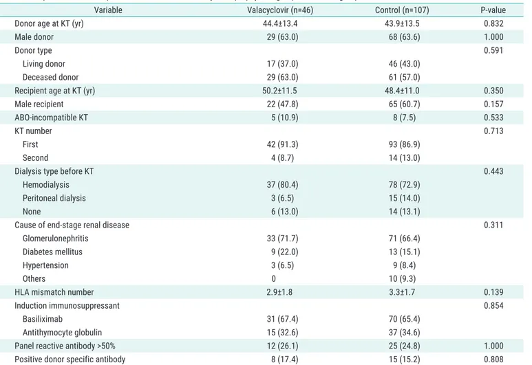
The clinical parameters of the valacyclovir prophylaxis and control groups are demonstrated in Table 1. Regard- ing the donors, there were no significant differences in age at KT, the proportion of male sex, and donor type between the two groups. Regarding the recipients, there were no significant between-group differences in age at KT, the proportion of male sex and ABO-incompatible KT,
KT number, dialysis type before KT, and etiologies of end- stage renal disease. The number of HLA mismatches, the proportion of panel reactive antibody >50%, and positive donor specific antibody were not significantly different between the two groups.
Comparison of Clinical Outcomes between Valacyclovir Prophylaxis and Control Groups
The clinical outcomes between the valacyclovir pro- phylaxis and control groups are presented in Table 2.
Median time (interquartile range) from KT to diagnosis of CMV infection was significantly longer in the valacy- clovir group compared to the control group (6.2 [5.1–7.7]
vs. 3.1 [2.4–4.5] months, P=0.025). There was no sig- nificant between-group difference in the mean recipient age at diagnosis of CMV infection. In Fig. 2A, the rate of
Table 1. Comparison of clinical parameters between the valacyclovir prophylaxis group and control group for CMV infection
Variable Valacyclovir (n=46) Control (n=107) P-value
Donor age at KT (yr) 44.4±13.4 43.9±13.5 0.832
Male donor 29 (63.0) 68 (63.6) 1.000
Donor type 0.591
Living donor 17 (37.0) 46 (43.0)
Deceased donor 29 (63.0) 61 (57.0)
Recipient age at KT (yr) 50.2±11.5 48.4±11.0 0.350
Male recipient 22 (47.8) 65 (60.7) 0.157
ABO-incompatible KT 5 (10.9) 8 (7.5) 0.533
KT number 0.713
First 42 (91.3) 93 (86.9)
Second 4 (8.7) 14 (13.0)
Dialysis type before KT 0.443
Hemodialysis 37 (80.4) 78 (72.9)
Peritoneal dialysis 3 (6.5) 15 (14.0)
None 6 (13.0) 14 (13.1)
Cause of end-stage renal disease 0.311
Glomerulonephritis 33 (71.7) 71 (66.4)
Diabetes mellitus 9 (22.0) 13 (15.1)
Hypertension 3 (6.5) 9 (8.4)
Others 0 10 (9.3)
HLA mismatch number 2.9±1.8 3.3±1.7 0.139
Induction immunosuppressant 0.854
Basiliximab 31 (67.4) 70 (65.4)
Antithymocyte globulin 15 (32.6) 37 (34.6)
Panel reactive antibody >50% 12 (26.1) 25 (24.8) 1.000
Positive donor specific antibody 8 (17.4) 15 (15.2) 0.808
Values are presented as mean±standard deviation or number (%).
CMV, cytomegalovirus; KT, kidney transplantation; HLA, human leukocyte antigen.
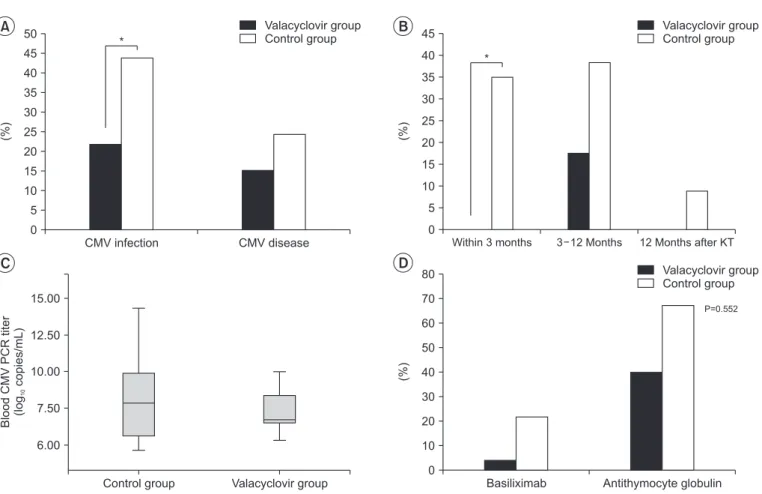
CMV infection was 10 (21.7%) in the valacyclovir pro- phylaxis group and 47 (43.9%) in the control group. The rate of CMV disease was 7 (15.2%) in the valacyclovir prophylaxis group and 26 (24.3%) in the control group.
The valacyclovir prophylaxis group showed significantly lower incidence of CMV infection than the control group (P=0.011), but not CMV disease. In Fig. 2B, when the fol- low-up period was divided into less than 3 months, 3 to 12 months and 12 months after KT, the incidence of CMV infection was the highest between 3 and 12 months after KT (P=0.008). The incidence of CMV infection was signifi- cantly lower in the valacyclovir prophylaxis group than the control group within 3 months after KT (P=0.022). Other periods also showed a low incidence of CMV infection in the valacyclovir prophylaxis group than the control group, but there was no significant difference between the two groups in each period. In addition, the valacyclovir pro- phylaxis group showed a lower median value in the blood CMV PCR titer converted to log values compared to the control group, but there was no significant difference be- tween the two groups in Fig. 2C. In Fig. 2D, the proportion of CMV infection was significantly higher in the ATG in- duction than basiliximab indunction (P<0.001). KTRs with ATG showed the lower incidence of CMV infection in the valacyclovir prophylaxis group (40.0%) compared to the control group (67.6%), but there was no significant differ- ence between the two groups. The allograft function at diagnosis of CMV infection was significantly lower in the valacyclovir prophylaxis group than in the control group,
but the allograft function at 6 and 12 months after diag- nosis of CMV infection did not show significant difference between the two groups. The proportion of delayed graft function, biopsy-proven acute rejection within 1 year, and BK virus-associated nephropathy within 1 year also did not show significant differences between the two groups.
In the valacyclovir group, nine (19.6%) KTRs had leukope- nia and five (10.9%) had thrombocytopenia as the compli- cation of valacyclovir, but leukopenia and thrombocyto- penia improved in all patients after dose reduction. There were no significant differences in the graft loss rate (2.8%
vs. 4.3%, P=0.637) and patient death rate (0.9% vs. 6.5%, P=0.081).
Comparison of Clinical Parameters According to CMV Infection
The clinical parameters between the groups with and without CMV infection are presented in Table 3. The pro- portion of deceased donors was significantly higher in the CMV infection group (P=0.017). Recipient age at KT was significantly higher in the CMV infection group (P=0.001).
The proportion of ATG induction was significantly higher in the CMV infection group (P<0.001). The proportion of panel reactive antibody >50% and biopsy-proven acute rejection tended to be higher in the CMV infection group.
The proportion of delayed graft function and BK virus-as- sociated nephropathy were significantly higher in the CMV infection group (P=0.031 and P=0.020, respectively).
However, donor age at KT, the proportion of donor and re-
Table 2. Comparison of clinical outcomes according to valacyclovir prophylaxisVariable Valacyclovir (n=46) Control (n=107) P-value
Time from KT to diagnosis of CMV infection (mo) 6.2 (5.1–7.7) 3.1 (2.4–4.5) 0.025
Recipient age at diagnosis of CMV infection (yr) 57±10 51±10 0.131
Graft function (eGFR, mL/min/1.73m
2)
At diagnosis of CMV infection 52.0±21.2 67.0±22.7 0.040
6 Months after diagnosis 61.2±16.5 66.5±23.0 0.507
12 Months after diagnosis 64.9±17.4 64.5±21.2 0.960
Delayed recovery of graft function 8 (17.4) 14 (13.1) 0.465
Biopsy-proven acute rejection within 1 year 2 (11.8) 9 (52.9) 0.515
BK virus associated nephropathy within 1 year 1 (100) 3 (100) NS
Leukopenia 9 (19.6) NA
Thrombocytopenia 5 (10.9) NA
Graft loss 2 (4.3) 3 (2.8) 0.637
Patient death 3 (6.5) 1 (0.9) 0.081
Values are presented as median (interquartile range), mean±standard deviation, or number (%).
KT, kidney transplantation; CMV, cytomegalovirus; eGFR, estimated glomerular filtration rate; NS, not significant; NA, not applicable.
cipient sex, ABO-incompatible KT, KT number, the number of HLA mismatches and the proportion of positive donor specific antibody did not show significant differences be- tween the two groups.
Comparison of CMV-Free Survivals between Valacyclovir Prophylaxis Group and Control Group, Risk Factors Related with CMV Infection, and Complications of Valacyclovir
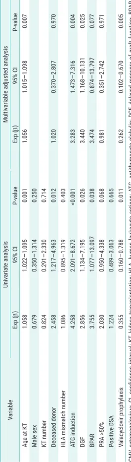
In the Kaplan-Meier analysis, the valacyclovir prophylaxis group showed significantly higher CMV-free survival rate than the control group in Fig. 3 (P=0.011). In multivari- able-adjusted analysis by logistic regression analysis, the independent risk factors for the development of CMV infection were older age at KT, use of ATG, delayed graft function, and no valacyclovir prophylaxis (Table 4). There were some complications when valacyclovir was used.
Leukopenia in nine patients (19.6%) and thrombocytope- nia in five patients (10.9%) occurred despite the control of valacyclovir dosage according to the eGFR. However, all patients improved after reduction of valacyclovir dosage.
DISCUSSION
Prophylaxis for CMV infection after KT is very important because of early graft failure due to CMV infection [1]. We used intravenous ganciclovir to prevent CMV infection for only 2 weeks of hospitalization for postoperative care after KT since 2009, but we could not effectively prevent the occurrence of CMV infection. Therefore, we used oral valacyclovir for 3 months after intravenous administration of ganciclovir for 2 weeks from July 2015 to prevent CMV
Basiliximab Antithymocyte globulin 80
70 60 50 40 30 20 10
(%)
Valacyclovir group Control group
0
D
Within 3 months 45
40 35 30 25 20 15 10 5
(%)
0
Valacyclovir group Control group
3 12 Months 12 Months after KT 50
B
45 40 35 30 25 20 15 10 5
(%)
0
Valacyclovir group Control group
CMV disease CMV infection
*
A
15.00
12.50
10.00
7.50
6.00 BloodCMVPCRtiter (logcopies/mL)10
Control group Valacyclovir group
C
*
P=0.552
Fig. 2. (A) Incidence of cytomegalovirus (CMV) infection and disease between valacyclovir prophylaxis and control groups, *P<0.05. (B) Incidence of
CMV infection between valacyclovir prophylaxis and control groups according to the follow-up period. *P<0.05. (C) Median value in the blood CMV poly-
merase chain reaction (PCR) titer converted to log values between valacyclovir prophylaxis and control groups. (D) Incidence of CMV infection between
valacyclovir prophylaxis and control groups according to the induction immunosuppressant.
0 1 2 3 4 5 6 7 8 9 10 11 12 100
80
60
40
CMV-freesurvivalrate(%) 20
Follow-up (mo)
+
+ + + ++ + + + +
+ +
+
Log-rank, P=0.011 Valacyclovir group Control group
Fig. 3. Cytomegalovirus (CMV)-free survival between the valacyclovir pro-
phylaxis group and the control group.
Table 3. Comparison of clinical parameters according to CMV infection
Variable CMV
infection (+) (n=57)
infection (–) CMV (n=96) P-value Donor age at KT (yr) 45.6±13.3 43.1±13.5 0.275
Male donor 41 (71.9) 56 (58.3) 0.118
Donor type 0.017
Living donor 16 (28.1) 47 (49.0)
Deceased donor 41 (71.9) 49 (51.0)
Recipient age at KT (yr) 52.9±10.4 46.6±11.0 0.001
Male recipient 29 (50.9) 58 (60.4) 0.311
ABO-incompatible KT 5 (8.8) 8 (8.3) 0.999
KT number 0.800
First 51 (89.5) 84 (87.5)
Second 6 (10.5) 12 (12.5)
HLA mismatch number 3.3±1.8 3.1±1.7 0.405
Induction immunosuppressant <0.001
Basiliximab 26 (45.6) 75 (78.1)
Antithymocyte globulin 31 (54.4) 21 (21.9) Panel reactive antibody >50% 18 (34.0) 19 (20.2) 0.077 Positive donor specific antibody 9 (17.6) 14 (14.9) 0.644 Biopsy-proven acute rejection 8 (14.0) 4 (4.2) 0.057 Delayed recovery of
graft function 13 (22.8) 9 (9.4) 0.031
BK virus associated
nephropathy 8 (14.0) 3 (3.1) 0.020
Values are presented as mean±standard deviation or number (%).
CMV, cytomegalovirus; KT, kidney transplantation; HLA, human leukocyte antigen.
Table 4.