http://dx.doi.org/10.5763/kjsm.2013.31.2.85
Received: September 16, 2013 Revised: November 26, 2013 Accepted: November 26, 2013 Correspondence: Chang-Hyuk Choi
Department of Orthopaedics, Catholic University of Daegu and College of Medicine, 33 Duryugongwon-ro 17-gil, Nam-gu, Daegu 705-718, Korea
Tel: +82-53-650-4276, Fax: +82-53-650-4270, E-mail: [email protected]
Copyright ©2013 The Korean Society of Sports Medicine
CC
This is an Open Access article distributed under the terms of the Creative Commons Attribution Non-Commercial License (http://creativecommons.org/
licenses/by-nc/3.0) which permits unrestricted non-commercial use, distribution, and reproduction in any medium, provided the original work is properly cited.
고교 역도선수에서 주관절의 외반 불안정성:
초음파의 유용성
대구가톨릭대학교 의과대학 정형외과학교실
최창혁ㆍ김세식ㆍ박창민ㆍ채승범ㆍ장호진
Valgus Laxity of Elbow Joint in High School Weight Lifters:
Ultrasonographic Assessment
Chang-Hyuk Choi, Se-Sik Kim, Chang-Min Park, Seung-Bum Chae, Ho-Jin Chang Department of Orthopaedics, Catholic University of Daegu and College of Medicine, Daegu, Korea
We evaluated abnormalities in medial portion of elbow in high-school weightlifter compared with the non weightlifter using a stress radiography and ultrsonography. The experimental group(G1) was 26 high school weightlifters with an average age of 17 years old (range, 16—18 years). The control group (G2) were comprised of 25 age matched general students. Both groups received physical examination, simple and valgus stress radiography and ultrasonography on both side of elbow. Physical examination showed 26.9% (14/52 elbows) tenderness and 19.2%
(10/52 elbows) valgus laxity in G1, no tenderness and laxity in G2. There were no differences in medial joint gaps on simple radiography (G1, 3.3 mm, G2, 2.7 mm; p>0.05), but the valgus stress view showed 5.6±0.8 mm medial joint gap in G1 and 3.8±0.8 mm in G2 (p<0.001). Ultrasonography in G1, angular deformity was found in 67.3%
(36/52) and G2 all in normal (p<0.01). The horizontal distance was an average 4.9±1.23 mm for the G1 and 3.1±0.78 mm for the G2 (p<0.001). Vertical distance of the proximal portion of the ulna was average 0.58±0.94 mm for the G1 and 1.59±0.49 mm for the G2 (p<0.001). In G1, angular deformity of male was 50% (15/30 elbows) and female was 95% (21/22 elbows) (p<0.001). Change of horizontal and vertical distance were larger in female (p<0.05). In conclusion, there were increased incidence of medial elbow joint laxity in high school weightlifter, especially in female, regardless of career. Sustained valgus laxity could be prone to ulnar collateral ligament injury and should be evaluated with ultrasonography-assisted dynamic study.
Keywords: Elbow joint, High school weighlifter, Radiography, Ultrasonography
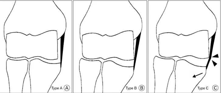
Fig. 1. Three types of angular changes of the medial collateral ligament. (A) Type A: normal stable elbow joint. (B) Type B: increased medial elbow laxity, as manifested by widening of the medial joint space and lateral shift of the proximal part of the ulna. (C) Type C: increased lateral shift of the proximal part of the ulna (arrow) causing impingement of the ulnar collateral ligament on the trochlea (arrowheads).
Introduction
It is well known that chronic valgus stress of the elbow joint often begins in sports activities (such as volleyball spike, the hockey shoot, the American football pass, the javelin throw, the tennis serve, and the baseball pitch), which can lead to ulnar medial collateral ligament injury and other elbow joint overuse injuries 1-5) . These sports have a similar exercise mechanism, which requires fast and strong elbow joint extensibility, accompanied by the valgus stress of the elbow joint and pronation of the supinated forearm 6) . Weight lifting has a similar exercise mechanism and chronic repetitive valgus-stress during exercise, abrupt resisted extension force on lifting can cause injuries at the medial side of the elbow joint. The purpose of this study was to evaluate the problems at the medial portion of the elbow joint in high-school weightlifters using a stress radiographic and ultrsonographic examination.
Methods
1. Demographics
The experimental group (G1) was 26 high school weight lifters,
15 male (right hand dominant in 12), and 11 female (right hand dominant in 9) with an average age of 17 years. Their weight was 63 kg on average (male, 68 kg; female, 60 kg), and height was 170 cm in average (male, 174 cm; female, 161 cm), and the period involved in the sport was 4 years in average. The control group (G2) was comprised of 25 volunteers without elbow problems (right-hand dominant in all) whose weight and height had been controlled; 15 were male and 10 were female, and their average age was 17 years. The average weight was 63 kg (male, 69 kg; female, 61 kg), and height was 171 cm in average (male, 175 cm; female, 160 cm). They did not have any symptoms in the elbow joint area and were not involved in any other sports activities with throwing or weight lifting. This study protocol was approved by the institutional ethics committee of our hospital.
2. Physical examination and radiographic study
After a medical history was taken for general information,
a physical examination was attempted of both elbows on each
athlete about tenderness or laxity, and simple and valgus stress
radiograph of the elbow joint were taken. Valgus stress to the
elbow was given as elbow flexed 30 o in a comfortable sitting
position.
Fig. 2. Ultrasonographic image of the elbow joint in normal and medial laxity. (A) Ultrasonography of normal elbow joint. The medial joint space is shown as a nonechoic space between the subchondral bone of the trochlea (arrowheads) and that of the coronoid process (large ar- row). The ulnar collateral ligament (UCL) is identified as a band-like structure that attaches to the medial epicondyle and the tubercular portion of the coronoid process. The superficial surface of the ligament is seen outlined by a hyperechoic straight line (small arrows). (B) Ultrasonogra- phic findings in ligament injury. The image showing type C angular changes with increased medial elbow laxity, as manifested by widening of the medial joint space (h=8.4 mm) and the lateral shift of the proximal part of the ulna (v=-1.2 mm). UCL tear is shown as a nonechoic gap be- tween torn margin of the ligament (between x and +).
h=horizontal distance of the medial joint space (with the assumption that the outline of the ulnar collateral ligament is a horizontal line), and v=vertical distance of the medial joint space.
3. Ultrasonographic study
Ultrasonographic examination was conducted by using a 7.5 Hz linear array transducer (Philips HD11 XE, Bothell, WA, USA).
For the ultrasonographic examination, the patient was positioned in the supine position, with the arm undergoing examination stretched across the bed. After the arm was 90 o in external rotation, the elbow joint bent at 70 o , and the forearm in a neutral position, gravity bearing stress was put on the forearm in order to cause tension in the medial portion of the elbow joint. The transducer was positioned on the medial portion of the elbow joint to make both the top of the medial epicondyle and the medial tubercular portion of the coronoid process appear together in the ultrasonographic image. The ulnohumeral joint between the
trochlea and the coronoid process was presented as echo-free space. The ulnar collateral ligament appeared as a hyperechoic straight-line, band shape attached to the medial tubercular portion of the coronoid process and medial epicondyle. The 3 types of angular change (Fig. 1), the gap of the elbow joint, and the lateral movement of the elbow joint under gravitation were measured (Fig. 2).
4. Statistical analysis
All statistical analyses were performed using the SPSS ver.
12.0 (SPSS Inc., Chicago, IL, USA). Chi-square test, Fischer exact test, Student’s t test, and Mann-Whitney U test were used, and a p<0.05 was considered statistically significant.
Results
1. Physical examination
Experimental group (G1) showed 14 cases of tenderness (5 cases in males, 9 cases in females). Three persons (6 cases) had tenderness in both elbow joints, among them 1 was male and 2 were female. There were 10 cases of valgus laxity (2 cases in males, 8 cases in females), and 2 females (4 cases) had valgus instability in both elbow joints. There was no instability or tenderness found in the control group (G2).
2. Radiographic examination
There were no remarkable differences in medial joint gaps on simple radiographic examination (G1, 3.3 mm; G2, 2.7 mm;
p>0.05), but the valgus stress view showed that the medial joint gap was 5.6±0.8 mm for the G1 and 3.8±0.7 mm for the G2, showing an average 1.8 mm increase (independent t test, p<0.001).
3. Ultrasonographic examination
1) Angular deformity of the elbow joint
There were significant differences between G1 and G2 in angular
deformity, with 30.8% of normal type A in G1 and 100% in
G2 (p<0.001). In G1, angular deformity of the elbow joint was
found in 69.2% of cases (36/52 cases), comprised of 33 cases
of type B (dominant arm, 20 cases; non-dominant, 13 cases)
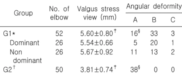
Table 1. Valgus laxity on stress radiograph and angular deformity between weight-lifter and control group
Group No. of elbow
Valgus stress view (mm)
Angular deformity
A B C
G1* 52 5.60±0.80
‡16
§33 3
Dominant 26 5.54±0.66 5 20 1 Non
dominant
26 5.67±0.92 11 13 2
G2
†50 3.81±0.74
‡38
§0 0
*High school weightlifter;
†High school general students;
‡
Independent t test, p=0.000;
§Dependent t test, p=0.000.
Table 2. Horizontal and vertical distances of medial elbow joint on ultrasonographic image between weight-lifter and control group
Group No. of elbow
Horizontal distance (mm)
Vertical distance (mm)
G1* 52 4.92±1.23
‡0.58±0.94
§Dominant 26 5.06±1.12 0.48±0.86 Non
dominant
26 4.75±1.28 0.67±1.01
G2
†50 3.11±0.78
‡1.59±0.49
§*High school weightlifter;
†High school general students;
‡
Independent t test, p=0.000;
§Dependent t test, p=0.000.
Table 3. Valgus laxity on stress radiograph and angular deformity between male and female in weight-lifter group
Gender No. of elbow
Angular deformity
A* B
†C
Male 30 15 15 0
Female 22 1 18 3
*Independent t test p=0.000;
†p=0.782.
Table 4. Horizontal and vertical distances of medial elbow joint on ultrasonographic image between male and female in weight-lifter group
Gender No. of elbow
Horizontal distance (mm)*
Vertical distance (mm)
†Male 30 4.47±1.13 0.85±1.06
Female 22 5.40±1.22 0.41±0.82
*Independent t test, p=0.04;
†p=0.042.
and 3 cases of type C (dominant arm, 1 cases; non-dominant, 2 case). There were no significant differences in valgus laxity and angular deformities between the dominant and non-dominant elbow (Table 1).
2) Horizontal and vertical distance of the elbow joint The horizontal distance (width of the medial elbow) was an average 4.9 mm (range, 3.7–6.2 mm) for the G1, and 3.1 mm (range, 2.3–3.9 mm) for the G2, showing an about 1.8 mm increase (independent t-test, p<0.001). There was no statistical difference between the dominant arm and the non-dominant arm in G1 (Table 2). Vertical distance (lateral movement) of the proximal portion of the ulna was on average 0.58 mm (range, -0.36–1.02 mm) for the G1, and 1.59 mm (range, 1.10–2.08 mm) for the G2, making it a remarkable 0.94 mm decrease (independent t-test, p<0.001) (Table 2). There was no statistically significant difference between the dominant and non-dominant arm in G1 (Table 2).
3) Male and female differences in experimental group For the angular deformity, male athletes had 15 cases of type A, 15 cases of type B, while the female athletes had 1 case of type A, 18 cases of type B, and 3 cases of type C, which revealed a remarkable disparity (p=0.001) (Table 3). In addition, 1 case of partial tear and 1 case of total tear were confirmed in female athletes, and the case of the total tear medial collateral ligament reconstruction surgery was performed.
For the ultrasonographic distances, the horizontal distance of male athletes was on average 4.47 mm (range, 3.34–5.60 mm), whereas the female athletes’ average of 5.40 mm (range, 4.18–6.62 mm) with more remarkable widening of the medial joint gap (p=0.04). For the vertical distances in the experimental group, male athletes had an average of 0.85 mm (range, -0.21–1.91 mm), while the females athletes had an average of 0.41 mm (range, -0.41–1.23 mm) showing more lateral movement (p=0.042) (Table 4).
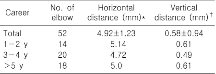
4) Differences according to sports related period in experimental group
In G1, the sports-related career length were divided into 3
group time periods, group A (14 elbows) with one to 2 years
career, group B (20 elbows) with 3 to 4 years career, group
C (18 elbows) with over 5 years career. The horizontal distance,
Table 5. Horizontal and vertical distances of medial elbow joint on ultrasonographic image according to career
Career No. of elbow
Horizontal distance (mm)*
Vertical distance (mm)
†Total 52 4.92±1.23 0.58±0.94
1−2 y 14 5.14 0.61
3−4 y 20 4.72 0.49
>5 y 18 5.0 0.61
*Independent t test, p=0.747;
†p=0.865.
Table 6. Correlation between angular deformity and ultra- sonographic findings (horizontal and vertical distance)
Angular deformity
No. of elbow
Horizontal distance (mm)
Vertical distance (mm) Normal
(type A)
16 2.02±2.21* 5.09±0.83
†Abnormal (type B, C)
36 5.34±1.01
‡0.17±0.79
§*Mann-Whitney U test, p=0.000;
†p=0.000;
‡p=0.000;
§