64
Corresponding to: Ho-Kyung Chun, Department of Surgery, Samsung Medical Center, Sungkyunkwan University School of Medicine, 50, Ilwon-dong, Gangnam-gu, Seoul 135-710, Korea. Tel: 02- 3410-3469, Fax: 02-3410-0040, E-mail: [email protected] Received January 16, 2008, Accepted March 18, 2008
This article was presented in part at the 59th annual meeting of the Korean Surgical Society, November 7-9, Seoul, Korea.
The First Transcolonic Cholecystectomy in Korea: A NOTES Survival Study in an Animal Model
1
Center for Colorectal Cancer, National Cancer Center, Goyang,
2
Department of Surgery, Samsung Medical Center, Sungkyunkwan University School of Medicine, Seoul,
3
Department of Surgery, Kyungpook National University School of Medicine, Daegu, Korea
Dae Kyung Sohn, M.D.1, Seung-Yong Jeong, M.D.1, Yong Beom Cho, M.D.2,Ji Won Park, M.D.1, Woo Yong Lee, M.D.2, Gyu-Seog Choi, M.D.3, Ho-Kyung Chun, M.D.2 and Korean NOTES Study Group
Natural orifice translumenal endoscopic surgery (NOTES) is a new and rapidly evolving hybrid procedure in which the endoscope is introduced into the peritoneal cavity through the stomach, colon, vagina or urethra. A trans- colonic approach is applicable for upper abdomen surgery. The present report describes a successful transcolonic cholecystectomy performed in a pig. The total operation time was 180 minutes. The pig survived without any complications for 7 days post-operatively. Necropsy after euthanasia showed no evidence of organ injury, bleeding or suppurative peritonitis, and it revealed that the colonic wall that had been incised and then closed using multiple endoscopic clippings was water-tight and air-tight. This is the first transcolonic cholecystectomy performed in Korea.
This approach may represent a promising new method for performing NOTES. (J Korean Surg Soc 2008;75:64-69)
Key Words: NOTES, Cholecystectomy, Endoscopy, Transcolonic surgeryINTRODUCTION
Natural orifice translumenal endoscopic surgery (NOTES) is a new and rapidly evolving hybrid procedure that utilizes flexible endoscopy technology to perform intra-abdominal laparoscopic surgical procedures.(1-4) This novel approach provides adequate visualization of the peritoneal cavity and the facility to perform surgical maneuvers. A variety of dia- gnostic and therapeutic procedures including liver biopsy, tubal ligation, oophorectomy, hysterectomy, cholecystectomy and gastrojejunostomy have already been demonstrated in experimental animal models.(5-9)
Using a transcolonic approach to access the peritoneal cavity was first reported in 2006.(10) This approach appeared beneficial for upper abdominal surgery as it allowed more consistent identification of structures and provided better en face orientation and scope stability despite the risks of infection and the difficulty of colonic closure.(6,11)
The present study in a pig model is the first to describe the use of NOTES for transcolonic chole- cystectomy in Korea.
CASE REPORT
1) AnimalThe study protocol was approved by the Association for
Assessment and Accreditation of Laboratory Animal Care
(protocol No. 001113) at the Laboratory Animal Research
Center of the Samsung Biomedical Research Institute,
Seoul. The study used a female pig weighing 38 kg. The
Fig. 1. A guidewire was placed into the peritoneal cavity through the colonic incision.
pig was housed at the Laboratory Animal Research Center of the Samsung Biomedical Research Institute, Seoul.
2) Preoperative preparation
The pig was deprived of food except for 4 liters of Colonlyte
Ⓡ(Meditech Korea Pharm, Seoul, Korea) for 24 hours before the procedure. Bowel preparation involved two enemas with a 1 liter-soap saline solution. Cefazolin (22 mg/kg intravenous (IV)) was administered the day before surgery. Anesthesia was induced with a bolus IV injection of 20 mg/kg ketamine and 2 mg/kg xylazin.
General anesthesia was maintained using enflurane on a semi-closed inhalation circuit after endotracheal intubation.
Cefazolin (22 mg/kg) was IV administered immediately before the procedure.
The colon was prepared by endoscopic irrigation with normal saline and a non-sterile endoscope. A cefazolin suspension (1 g in 1 L normal saline) was then instilled through the endoscope. After aspiration, the rectum was irrigated with 0.5 L diluted povidone-iodine solution, and the anus and gluteal surfaces were scrubbed with 10%
povidone-iodine. The peri-umbilical areas were similarly scrubbed.
A double-channel upper endoscope (GIF-2TQ260M;
Olympus Optical Co, Ltd, Tokyo, Japan) was treated with 2.4% glutaraldehyde solution and then air-dried for high-level disinfection before the procedure. All reusable instruments including endoscopic accessories and laparoscopic devices were sterile as provided by the manufacturer or due to gas sterilization.
3) Surgical techniques
Under aseptic conditions, a 5-mm-sized port was created in the peritoneal cavity via a small incision in the supr- aumbilical skin. Continuous CO2 insufflation through the port was used to create a pneumoperitoneum with a pressure of less than 10 mm Hg. This abdominal port was also used for observing endoscopic procedures using a laparoscopic camera (5 mm diameter), or for grasping and retracting organs using a laparoscopic clamp.
A double-channel endoscope was advanced to a distance
of 25 cm from the anus. After the endoscope light at the anterior wall of the colon was identified laparoscopically, a 2-mm incision was made in the colon wall using a needle knife. A 17-mm dilating balloon (CRE
TMWireguided Balloon Dilator, Boston Scientific, Natick, USA) was passed over a guidewire (Jagwire
ⓇHigh Performance Guidewire, Boston Scientific, Natick, USA) to enlarge the initial incision up to 17 mm (Fig. 1). The endoscope was passed through the opening and advanced into the peri- toneal cavity.
The stomach, hepatic lobes, spleen, small intestine,
colon and other pelvic organs were clearly observed. The
endoscope was advanced en face to the liver, and then the
gall bladder (GB) was exposed and evaluated (Fig. 2). A
laparoscopic clamp was introduced into the peritoneal
cavity through the abdominal port. The GB showed signs
of inflammation and was carefully retracted using the
clamp. The cystic duct and artery were clearly identified,
ligated and cut using clips (Easyclip
ⓇHX-610-090L,
Olympus Medical Systems Corp., Tokyo, Japan) and a
needle knife (KD-1L-1, Olympus Medical Systems Corp.,
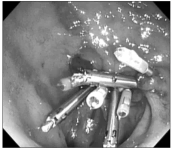
Tokyo, Japan) with coagulating currents (Fig. 3). The GB
bed was then dissected using a needle knife, flex-knife
Ⓡ(Olympus Medical Systems Corp., Tokyo, Japan) and
hook-knife
Ⓡ(Olympus Medical Systems Corp., Tokyo,
Fig. 2. Endoscopic view of the gall bladder. Fig. 4. Dissection of the gall bladder using a flex-knifeⓇ.
Fig. 5. Closure of the colonic incision using endoclips.
Fig. 3. The cystic duct was identified and ligated using endoclips.
Japan) using standard endoscopic submucosal dissection (ESD) methods (Fig. 4). After all attachments to the liver were severed, the GB was withdrawn into the colonic lumen using grasping forceps (FG-49L-1, Olympus Medical Systems Corp., Tokyo, Japan), and removed via the anus.
The colonic incision was then closed using multiple endoscopic clips (Resolution clip
Ⓡ, Boston Scientific, Natick, USA and Easyclip
ⓇHX-610-090L, Olympus Medical Systems Corp., Tokyo, Japan) (Fig. 5). Complete closure of the colonic wall was confirmed using laparoscopic observa- tion through the abdominal port. The abdominal incision
was then closed. The total time for the procedure was 180 minutes. There were no complications other than minor bleeding from the liver bed which was easily controlled by clipping and coagulation.
4) Postoperative care and necropsy
The pig was fed a standard diet from post- operative day
2. Cefazolin (22 mg/kg) was IV administered once per day
for 5 days. The pig recovered rapidly without complications
for 7 days postoperatively. On day 7, the pig weighing 38
kg was electively euthanized and a necropsy performed. The
Fig. 6. Necropsy results. (A) Sparse adhesions between the liver bed and omentum. (B) Serosal aspect of the well-healed colonic incision site. (C) Air-inflation test showing complete closure of the colonic incision site. (D) Mucosal aspect of the colonic incision site with endoclips.
necropsy findings indicated no evidence of organ injury, bleeding or suppurative peritonitis. Sparse adhesions between liver bed and omentum were observed (Fig. 6A). The colonic incision site appeared to have healed well, with no evidence of gross inflammatory changes or defects (Fig. 6B).
The colon was segmentally resected to determine the state of incision site closure. Both air-inflation and water- filled tests indicated complete closure of the site (Fig. 6C, D).
DISCUSSION
The evolution of minimally invasive techniques for abdo- minal surgery has resulted in improved surgical outcomes, including faster wound healing, shorter hospital stays and
increased cosmetic benefits.(12,13) NOTES may further minimize abdominal surgery invasiveness by eliminating abdominal incisions and multiple trocar puncture sites.
This in turn may reduce the incidence of postoperative
pain, wound infection, hernias and adhesions.(2,14)
Compared with transgastric or transvaginal approaches,
the transcolonic approach described here may have some
limitations in terms of preparation and sterilization.(10,11,15)
However, the approach provides for safe surgery with a
good visual field and ease of scope handling, and it can
be applied regardless of gender. While further investi-
gations into standardized colonic preparation, sterilization
and colonic wall incision closure are required before this
technique can be used clinically, the transcolonic approach
may represent a promising new method for NOTES of the upper abdomen.
Advanced therapeutic endoscopic skills as well as surgical experience is essential for performing NOTES safely. Our experience indicates that a range of procedures is required, including scope insertion through the colonic lumen, ballooning at the colonic wall incision site and other standard manipulation skills including torque, rotation, retroflexion, tip deflection for accessing and processing in the abdominal cavity. In addition, highly advanced techni- ques such as ESD are needed for dissecting the GB from the liver bed. Ideally the procedure requires an experienced surgeon with advanced endoscopic skills, or alternatively it requires a combination of a surgeon and an endoscopist for NOTES.
In July 2005, leaders from the American Society of Gastrointestinal Endoscopy (ASGE) and the Society of American Gastrointestinal and Endoscopic Surgeons (SAGES) formed a working group called the Natural Orifice Surgery Consortium for Assessment and Research (NOSCAR). The primary responsibility of this group is the safe and responsible development of NOTES for clinical practice.
The group has identified challenges that need to be overcome before NOTES can become widespread in clinical practice (2). In August 2007, the Korean NOTES Study Group (KNSG) was organized for conducting and guiding NOTES research. We anticipate the KNSG will play an important role in the research and clinical application of NOTES.
ACKNOWLEDGMENT
We thank all members of the KNSG for assistance and support.
DISCLOSURE
Endoscopic equipment and disposable endoscopic acce- ssories were supplied by Olympus Korea (Seoul, Korea) and the Olympus Corporation (Tokyo, Japan).
All laparoscopic instruments were supplied by Johnson
and Johnson, Korea Co., LTD. (Seoul, Korea).
Disposable balloons and guidewires were supplied by Boston Scientific Korea (Seoul, Korea).
REFERENCES
1) Kalloo AN, Singh VK, Jagannath SB, Niiyama H, Hill SL, Vaughn CA, et al. Flexible transgastric peritoneoscopy: a novel approach to diagnostic and therapeutic interventions in the peritoneal cavity. Gastrointest Endosc 2004;60:114-7.
2) Rattner D, Kalloo A. ASGE/SAGES Working Group on Natural Orifice Translumenal Endoscopic Surgery. October 2005. Surg Endosc 2006;20:329-33.
3) Jagannath SB, Kantsevoy SV, Vaughn CA, Chung SS, Cotton PB, Gostout CJ, et al. Peroral transgastric endoscopic ligation of fallopian tubes with long-term survival in a porcine model.
Gastrointest Endosc 2005;61:449-53.
4) Feretis C, Kalantzopoulos D, Koulouris P, Kolettas C, Archon- tovasilis F, Chandakas S, et al. Endoscopic transgastric procedures in anesthetized pigs: technical challenges, compli- cations, and survival. Endoscopy 2007;39:394-400.
5) Park PO, Bergstrom M, Ikeda K, Fritscher-Ravens A, Swain P.
Experimental studies of transgastric gallbladder surgery:
cholecystectomy and cholecystogastric anastomosis (videos).
Gastrointest Endosc 2005;61:601-6.
6) Bergstrom M, Ikeda K, Swain P, Park PO. Transgastric anastomosis by using flexible endoscopy in a porcine model (with video). Gastrointest Endosc 2006;63:307-12.
7) Lima E, Rolanda C, Pego JM, Henriques-Coelho T, Silva D, Carvalho JL, et al. Transvesical endoscopic peritoneoscopy: a novel 5 mm port for intra-abdominal scarless surgery. J Urol 2006;176:802-5.
8) Wagh MS, Merrifield BF, Thompson CC. Survival studies after endoscopic transgastric oophorectomy and tubectomy in a porcine model. Gastrointest Endosc 2006;63:473-8.
9) Kantsevoy SV, Jagannath SB, Niiyama H, Chung SS, Cotton PB, Gostout CJ, et al. Endoscopic gastrojejunostomy with survival in a porcine model. Gastrointest Endosc 2005;62:
287-92.
10) Pai RD, Fong DG, Bundga ME, Odze RD, Rattner DW, Thompson CC. Transcolonic endoscopic cholecystectomy: a NOTES survival study in a porcine model (with video).
Gastrointest Endosc 2006;64:428-34.
11) Fong DG, Pai RD, Thompson CC. Transcolonic endoscopic abdominal exploration: a NOTES survival study in a porcine model. Gastrointest Endosc 2007;65:312-8.
12) Barkun JS, Barkun AN, Sampalis JS, Fried G, Taylor B, Wexler MJ, et al. Randomised controlled trial of laparoscopic versus mini cholecystectomy. The McGill Gallstone Treatment Group.
Lancet 1992;340:1116-9.
13) Fuchs KH. Minimally invasive surgery. Endoscopy 2002;34:
154-9.
14) Swain P. A justification for NOTES--natural orifice translu- menal endosurgery. Gastrointest Endosc 2007;65:514-6.
15) Wilhelm D, Meining A, von Delius S, Fiolka A, Can S, Hann von Weyhern C, et al. An innovative, safe and sterile sigmoid access (ISSA) for NOTES. Endoscopy 2007;39:401-6.