564 CASE REPORT
Korean Circ J 2008;38:564-569
Print ISSN 1738-5520 / On-line ISSN 1738-5555 Copyright ⓒ 2008 The Korean Society of Cardiology
A Fatal Case of Simultaneous, Very Late Thrombosis Involving Three Drug-Eluting Stents in Three Coronary Arteries
Yong Soo Ahn, MD1, Jang Hyun Cho, MD1, Dong Han Kim, MD1, Young June Hwang, MD1, Hong Myong Jung, MD1, Min Seok Choi, MD1, Joon Young Kim, MD1 and Myung Ho Jeong, MD2
1Division of Cardiology, Department of Internal Medicine, St. Carollo Hospital, Suncheon,
2The Heart Center of Chonnam National University Hospital, Gwangju, Korea
ABSTRACT
Late stent thrombosis is one of the most serious complications associated with morbidity and mortality after coronary drug-eluting stent implantation, and is mainly caused by the withdrawal of antiplatelet agents. We report our experience of late stent thrombosis simultaneously involving three different coronary arteries in a young male patient who was treated with three drug-eluting stents two years ago. The patient stopped taking antiplatelet agents for several days. The patient did not recover from cardiogenic shock, even after repeated ballooning with thrombus aspiration, intra-aortic balloon pumping, and temporary pacing during cardiopulmonary resuscitation.
(Korean Circ J 2008;38:564-569)
KEY WORDS: Stents; Thrombosis; Platelets.
Introduction
Since the initial introduction of drug-eluting stents (DES), their use has increased rapidly because the clini- cal and angiographic outcomes have been favorable.1-3) However, random trials with strict inclusion criteria have revealed that the incidence of thrombosis associated with DES is not different from bare-metal stents (BMS).4)5) In the clinical setting, this finding may be relevant because complex lesions, the length or extent of the occluded lesion, the underlying disease, and the compliance of patients has more influence on stent thrombosis than the material from which stents are made.6-8) Late stent thrombosis is an especially remarkable severe complica- tion of DES, which has caused myocardial infarctions or deaths, and these events have been mainly associated with the withdrawal of antiplatelet agents.9)10)
We will report our experience with late stent throm- bosis simultaneously involving three different coronary arteries after cessation of an antiplatelet agent for several days in a young patient treated with DES implantation.
Case
A 35 year-old man with a history of heavy smoking presented to the emergency room with the sudden onset of chest pain of 2 hours duration. The electrocardiogram (ECG) obtained in the emergency room showed ST- segment elevation in the anterior and inferior leads. Two years previously, he was admitted with similar symptoms at another hospital. The ECG at that time showed T wave inversion in the inferior leads (Fig. 1). Coronary angiography (CAG) revealed the presence of three vessel disease with total occlusion of the middle right coro- nary artery (RCA) and critical stenosis in the proximal left ascending coronary artery (LAD) and intermediate branch. Immediately after the CAG, percutaneous coro- nary intervention (PCI) for mid - RCA was performed with a 3.0×28 mm Taxus stent (Boston Scientific, Bos- ton, MA, USA). One week later, the follow-up angio- graphy for the RCA lesion revealed a patent stent and a second stage PCI for the proximal LAD and interme- diate branch lesions was performed with 3.5×24 mm and 3.0×24 mm Taxus stents, respectively. The final CAG showed no residual stenosis with good distal flow (Fig. 2).
Twenty months after the initial procedure, the patient was transferred to our hospital with an acute inferior wall ST elevation myocardial infarction (STEMI) after emergency treatment with a thrombolytic agent {Tenec-
Received: June 17, 2008
Revision Received: August 18, 2008 Accepted: August 21, 2008
Correspondence: Jang Hyun Cho, MD,Division of Cardiology, Department of Internal Medicine, St. Carollo Hospital, 1742 Jorye-dong, Suncheon 540-719, Korea
Tel: 82-61-720-2113, Fax: 82-61-720-6160 E-mail: [email protected]
Yong Soo Ahn, et al.·565
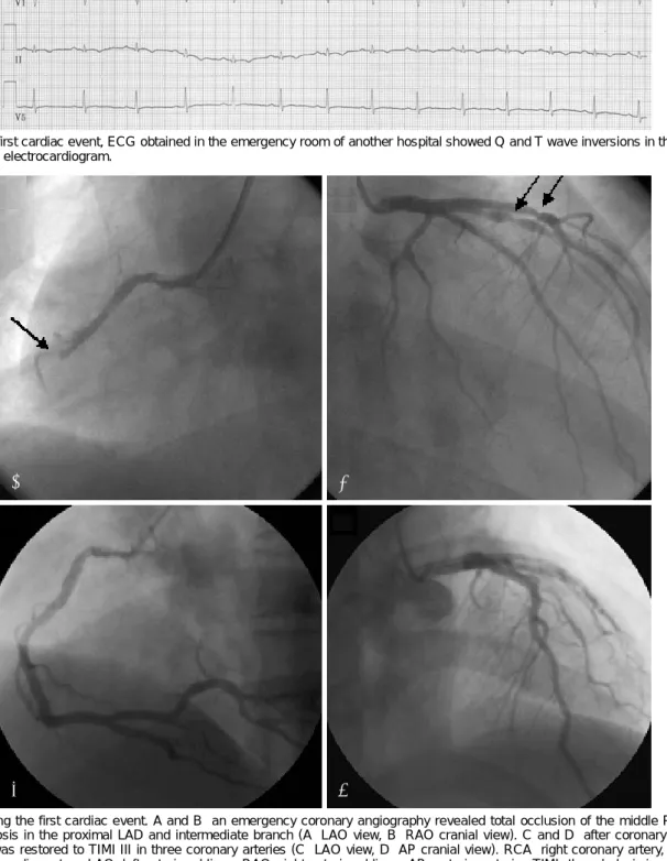
Fig. 1. The first cardiac event, ECG obtained in the emergency room of another hospital showed Q and T wave inversions in the inferior leads. ECG: electrocardiogram.
Fig. 2. During the first cardiac event. A and B: an emergency coronary angiography revealed total occlusion of the middle RCA, and critical stenosis in the proximal LAD and intermediate branch (A: LAO view, B: RAO cranial view). C and D: after coronary stenting, distal flow was restored to TIMI III in three coronary arteries (C: LAO view, D: AP cranial view). RCA: right coronary artery, LAD: left anterior descending artery, LAO: left anterior oblique, RAO: right anterior oblique, AP: anterioposterior, TIMI: thrombolysis in myocardial infarction.
D C
B A
566·Fatal Case of Stent Thrombosis
teplase (40 mg)} at a local hospital.
He complained of chest pain continuously during the 3 hours prior to thrombolytic therapy. The follow-up ECG obtained in our hospital showed persistent ST elevation in the inferior leads (Fig. 3), the cardiac en- zymes as (CK, CK-MB, and troponin-I) were elevated, and echocardiography also showed hypokinetic move- ment of the inferoposterior wall. After loading with 300 mg of aspirin and 300 mg of clopidogrel, he was taken directly to the catheterization room for rescue PCI. The CAG showed patency in the previously stented sites of the middle RCA, proximal LAD, and intermediate branch, with good distal flow (Fig. 4). Gradually, the chest pain and ST elevation on the ECG resolved. The post-treatment clinical course was uneventful and the patient was discharged 2 weeks later on dual antiplatelet agents {aspirin (100 mg) and clopidogrel (75 mg)}.
Four months after the second diagnostic CAG, the patient presented to the emergency room with an in-
ferior and anteroseptal wall STEMI. He complained of severe chest pain and dyspnea, and dull mental status, and became stuporous in the emergency room. He had not taken his medication over several days because of a disregard for his health, including the heavy consump- tion of alcohol. The ECG showed complete AV block, a right bundle branch block, and ST-segment elevation in both the anterior and inferior leads (Fig. 5). The systolic blood pressure decreased to 60 mmHg and the heart rate dropped to 25 beats/min. Several minutes later, the patient’s mental state changed to coma and his blood pressure was not checked. Cardiopulmonary resuscita- tion (CPR) was started promptly and he was transferred to the catheterization room. His condition deteriorated further, so CPR was continued. An emergency CAG revealed simultaneous total occlusion of the middle RCA, proximal LAD, and intermediate branch, the previously paced sites of the deployed sites of the Taxus stents. After insertion of an intra-aortic balloon coun-
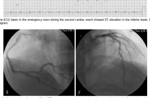
Fig. 3. The ECG taken in the emergency room during the second cardiac event showed ST elevation in the inferior leads. ECG: ele- ctrocardiogram.
Fig. 4. During the second cardiac event (A and B) an emergency coronary angiography revealed patency in the previous stented sites of the middle right coronary artery, the proximal left anterior descending artery, and the intermediate branch with good distal flow (A: LAO view, B: AP cranial view). LAO: left anterior oblique, AP: anterioposterior.
B A
Yong Soo Ahn, et al.·567 terpulsation and a temporary pacemaker, thrombus as-
piration was performed with a Thrombuster catheter.
Later, antegrade flow for the middle RCA was obtained
by thrombectomy and ballooning at the occluded lesion.
However, coronary flow at the occluded lesions of the proximal LAD and intermediate branch and vital status
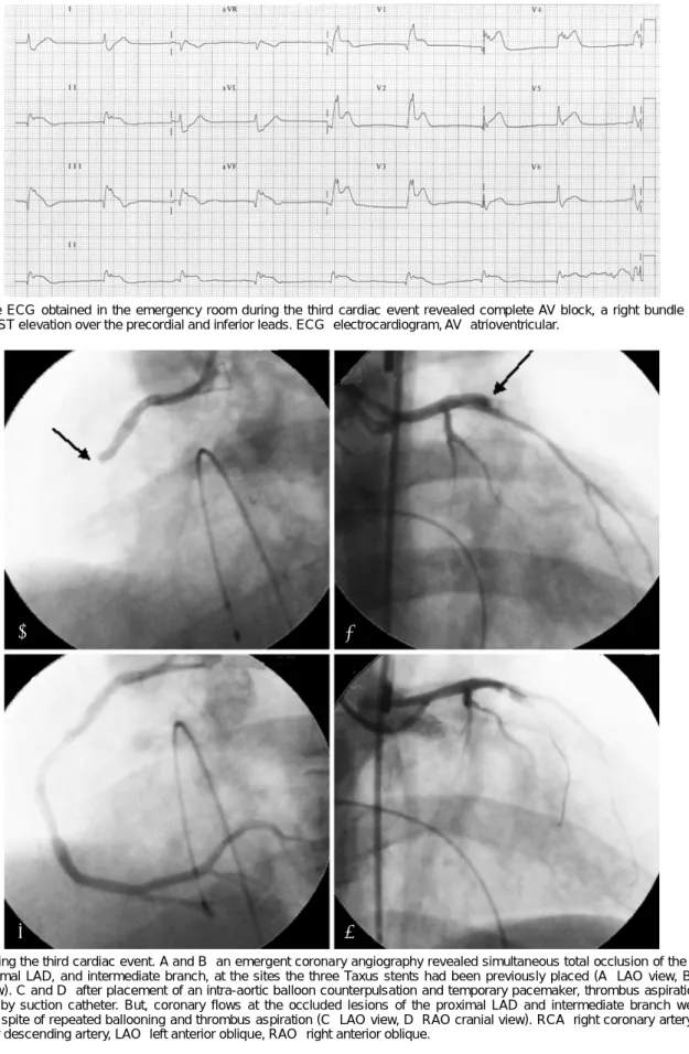
Fig. 5. The ECG obtained in the emergency room during the third cardiac event revealed complete AV block, a right bundle branch block, and ST elevation over the precordial and inferior leads. ECG: electrocardiogram, AV: atrioventricular.
Fig. 6. During the third cardiac event. A and B: an emergent coronary angiography revealed simultaneous total occlusion of the middle RCA, proximal LAD, and intermediate branch, at the sites the three Taxus stents had been previously placed (A: LAO view, B: RAO cranial view). C and D: after placement of an intra-aortic balloon counterpulsation and temporary pacemaker, thrombus aspiration was performed by suction catheter. But, coronary flows at the occluded lesions of the proximal LAD and intermediate branch were not restored in spite of repeated ballooning and thrombus aspiration (C: LAO view, D: RAO cranial view). RCA: right coronary artery, LAD:
left anterior descending artery, LAO: left anterior oblique, RAO: right anterior oblique.
C D
A B
568·Fatal Case of Stent Thrombosis
was not recovered, despite continuous thrombus aspira- tion and repeated ballooning (Fig. 6). Unfortunately, the patient died in the catheterization room.
Discussion
Stent thrombosis is one of the most severe complica- tions associated with morbidity and mortality after DES or BMS implantation.11) By definition, early stent throm- bosis occurs within 30 days after stent implantation, while late stent thrombosis occurs beyond 30 days.12) Additionally, very late stent thrombosis is defined as a thrombus occurring beyond 1 year. Early stent throm- bosis occurs in about 1.0-1.5% of patients with DES;
the probability of occurrence with a BMS is similar. In the largest series of eight cases, late stent thrombosis unexpectedly occurred in 0.35-0.72% of patients with DES. In addition, most cases have been associated with discontinuation of antiplatelet therapy late after stent implantation.13)14)
Delayed re-endothelialization of the stent, because of the antiproliferative nature of the agents, appears to be implicated in the pathophysiology of very late stent thrombosis. On the basis of this theory, DES causes delayed arterial healing and increased inflammatory reactions compared to BMS, so DES seems to be asso- ciated with higher rates of late stent thrombosis than BMS.15) Unlike DES, the overall rate of stent thrombosis which occurs in patients with BMS is the subject of but few reports. However, some recent studies have reported that there are no differences in the incidences of late stent thrombosis between patients treated with DES and BMS.11)
In the clinical setting, acute coronary syndrome, bi- furcation treatment, diabetes, and premature disconti- nuation of antiplatelet therapy are considered strong pre- dictors of stent thrombosis. Effective antiplatelet agents have a critical role in the prevention of stent throm- bosis.6-8) Therefore, in most cardiac centers prolonged dual antiplatelet therapy with aspirin and clopidogrel is recommended for the prevention of stent thrombosis after PCI.
Previously, some studies have reported that aspirin withdrawal after PCI increases the occurrence of a co- ronary event, such as stent thrombosis.6)9)13) A study noted that aspirin withdrawal in patients with coronary artery disease increased morbidity within 1 month after cessation of aspirin, whether treated by BMS or DES,16) and another study reported that eight cases occurred during late stent thrombosis after the withdrawal of aspirin.14) Yet another study noted that the inadvertent interruption of clopidogrel therapy also can induce late stent thrombosis.17) Dual antiplatelet therapy can prevent stent thrombosis, but withdrawal of any one agent could result in the cardiovascular events at any time.
Even if a patient had a coronary stent deployed before elective non-cardiac surgery, aspirin should not be dis- continued, except for intracranial neurosurgery and transureteral prostectomy. A recent review proved that aspirin neither increased the perioperative severity nor the mortality associated with bleeding.18) In addition, whatever non-cardiac surgery is planned, it must be performed later than 6 weeks after stent implantation if possible, because stent thrombosis frequently develops within 3-6 weeks of a cardiac procedure.19) These events have been mainly related to insufficient stent re-endo- thelialization, heightened platelet reactivity, and with- drawal of the antiplatelet agent.
In this case, our patient did not take his prescribed medication continuously in spite of prescription of dual antiplatelet agents after the second attack, and he died due to very late stent thrombosis of three different co- ronary arteries at the same time, two years after DES implantation.
In summary, this case emphasizes that late stent thrombosis should be considered in all patients treated with coronary DES implantation, and may be prevented by using an antiplatelet agent continuously.
REFERENCES
1) Grube E, Silber S, Hauptmann KE, et al. Two year-plus follow up of paclitaxel-eluting stent in de novo coronary narrowing (TAXUS I). Am J Cardiol 2005;96:79-82.
2) Degertekin M, Serruys PW, Foley DP, et al. Persistent inhibition of neointimal hyperplasia after sirolimus-eluting stent implan- tation: long-term (up to 2 years) clinical, angiographic, and in- travascular ultrasound follow-up. Circulation 2002;106:1610-3.
3) Fajadet J, Wijns W, Laarman GJ, et al. Randomized, double-blind, multicenter study of the Endeavor zotarolimus-eluting phosphoryl- choline-encapsulated stent for treatment of native coronary artery lesions: clinical and angiographic results of the ENDEAVOR II trial. Circulation 2006;114:798-806.
4) Moreno R, Fernandez C, Hermandez R, et al. Drug-eluting stent thrombosis: result from a pooled analysis including 10 randomiz- ed studies. J Am Coll Cardiol 2005;45:954-9.
5) Schampaert E, Moses JW, Schofer J, et al. Sirolimus-eluting stents at two years: a pooled analysis of SIRIUS, E-SIRIUS, and C- SIRIUS with emphasis on late revascularizations and stent throm- bosis. Am J Cardiol 2006;98:36-41.
6) Iakovou I, Schmodt T, Bonizzoni E, et al. Incidence, predictors, and outcome of thrombosis after successful implantation of drug- eluting stent. JAMA 2005;293:2126-30.
7) Daemen J, Wenaweser P, Tsuchida K, et al. Early and late coro- nary stent thrombosis of sirolimus-eluting and paclitaxel-elut- ing stents in routine clinical practice: data from a large two- institutional cohort study. Lancet 2007;369:667-78.
8) Park SH, Hong GR, Seo HS, Tahk SJ. Stent thrombosis after successful drug-eluting stent implantation. Korean Circ J 2005;
35:163-71.
9) McFadden EP, Stabile E, Regar E, et al. Late thrombosis in drug- eluting coronary stents after discontinuation of antiplatelet the- rapy. Lancet 2004;364:1519-21.
10) Moon JY, Jeong MH, Jun CH, et al. The frequency, treatment and clinical outcomes of stent thrombosis after use of TAXUS
Yong Soo Ahn, et al.·569
stent. Korean Circ J 2007;37:641-6.
11) Wernick MH, Jeremias A, Carrozza JP. Drug-eluting stents and stent thrombosis: a cause for concern? Coron Artery Dis 2006;
17:661-5.
12) Park DW, Park SW. Stent thrombosis in the era of the drug- eluting stent. Korean Circ J 2005;35:791-4.
13) Ong AT, Hoye A, Aoki J, et al. Thirty-day incidence and six- month clinical outcome of thrombotic stent occlusion after bare- metal, sirolimus, or paclitaxel stent implantation. J Am Coll Cardiol 2005;45:947-53.
14) Ong AT, McFadden EP, Regar E, de Jaegere PP, van Domburg RT, Serruys PW. Late angiograhpic stent thrombosis (LAST) events with drug-eluting stents. J Am Coll Cardiol 2005;45:2088- 92.
15) Joner M, Finn AV, Farb A, et al. Pathology of drug-eluting stents in humans: delayed healing and late thrombotic risk. J Am Coll
Cardiol 2006;48:193-202.
16) Collet JP, Montalescot G, Blanchet B, et al. Impact of prior use or recent withdrawal of oral antiplatelet agents on acute coronary syndromes. Circulation 2004;110:2361-7.
17) Goel PK, Gill GS. Angioplasty for the late stent thrombosis, the new technical challenge: a single centre experience. Int J Car- diol 2008;123:335-7.
18) Burger W, Chemnitius JM, Kneissl GD, Rucker G. Low-dose aspirin for secondary cardiovascular prevention-cardiovascular risks after its perioperative withdrawal versus bleeding risks with its continuation: review and meta-analysis. J Intern Med 2005;
257:399-414.
19) Leibowitz D, Cohen M, Planer D, et al. Comparison of cardio- vascular risk of noncardiac surgery following coronary angio- plasty with versus without stenting. Am J Cardiol 2006;97:1188-91.