https://doi.org/10.20307/nps.2017.23.1.21
21
Diarylbutane-type Lignans from Myristica fragrans (Nutmeg) show the Cytotoxicity against Breast Cancer Cells through Activation
of AMP-activated Protein Kinase
Thi Van Thu Le
1, Phi Hung Nguyen
1, Hong Seok Choi
1, Jun-Li Yang
2, Keon Wook Kang
2, Sang-Gun Ahn
3, and Won Keun Oh
2,*
1
College of Pharmacy, Chosun University, Gwangju 501-759, Republic of Korea
2
Korea Bioactive Natural Material Bank, Research Institute of Pharmaceutical Sciences, College of Pharmacy, Seoul National University, Seoul 151-742, Republic of Korea
3
Department of Pathology, College of Dentistry, Chosun University, Gwangju 501-759 Republic of Korea
Abstract – In our program to search for new AMP-activated protein kinase (AMPK) activators from plants that exert potential anticancer property, we found that an EtOAc extract of Myristica fragrans (nutmeg) activated AMPK enzyme in human breast cancer MCF-7 cells. Two major diarylbutane-type lignans, macelignan and meso-dihydroguaiaretic acid (MDGA), were isolated as active principles from this extract. Treatment of breast cancer cells with two compounds induced cellular apoptosis, evidenced by cleavage of poly-(ADP-ribose) polymerase (PARP) and Ser 15 phosphorylation of p53. Moreover, macelignan and MDGA significantly inhibited the colony formation of MCF-7 breast cancer cells on soft agar. Intraperitoneal injection of macelignan and MDGA (20 mg/kg) suppressed the tumor growth of 4T1 mammary cancer cells. These results indicate that the chemopreventive effects of two major diarylbutane-type lignans from Myristica fragrans (nutmeg) may be associated with induction of apoptosis presumably through AMPK activation.
Keywords − Myristica fragrans, Diarylbutane lignan, MCF-7, AMP-activated protein kinase (AMPK)
Introduction
Breast cancer is the most common malignant tumor affecting women with more than one million new cases each year.
1,2Up to now, treatment of breast cancer remains a challenge. Available therapies including radiation, endocrine, and conventional chemotherapy are often limited by high toxicity, lower efficacy, therapeutic resistance, and therapy-related morbidity.
3Therefore, searching for more effective therapeutic agents with novel mechanisms of action is desired to combat breast cancer. Recent interest has focused on cell signaling pathways controlling both cell metabolism and cell growth.
4,5And also, the association of reduced cancer risk and edible food has also captured the interests of scientists.
6Adenosine monophosphate-activated protein kinase (AMPK), an energy sensing/signalling intracellular protein
found in all eukaryotes, is a heterotrimeric serine/threonine protein kinase that is composed of a catalytic α-subunit and regulatory β-and γ-subunits.
7AMPK has been implicated in carcinogenesis because of the finding that a well-known tumor suppressor LKB1 (liver kinase B1) is an upstream activating kinase for AMPK, and many effects of LKB1 on tumor suppressing are likely to be mediated by AMPK.
8Moreover, it has been reported that AMPK activation could have therapeutic potential in breast cancer. Once activated, AMPK inhibits fatty acid synthesis and the AKT-mTOR pathway, and activates the p53-p21 axis. All these molecular mechanisms are thought to play a key role in breast carcinogenesis. Histological evaluation of AMPK signaling in primary breast cancer also showed the reduced signal of AMPK, compared with the strong expression in normal breast epithelium, in axillary node metastasis of two cohorts of patients.
9Myristica fragrans Houtt. (Myristicaceae), an aromatic evergreen tree cultivated in the India, South Africa, and other tropical countries, is widely used as a spice in many African and Asian countries.
10Lignans, which are known to exert many chemopreventive effects,
11were reported as
*Author for correspondence
Won Keun Oh, Korea Bioactive Natural Material Bank, College of Pharmacy, Seoul National University, Gwanak-gu, Seoul 151-742, Republic of Korea.
Tel: +82-2-880-7872; E-mail: [email protected]
the main chemical constituents of this plant. In this regard and as part of our program to search for new AMPK activators with chemopreventive activity from plants,
12we found that diarylbutane-type lignans from M. fragrans exhibited potential AMPK activation effects and strong growth inhibition activities against MCF-7 breast cancer cells. As major active principles, two diarylbutane-type lignans, macelignan (1)
13and MDGA (2)
14, were isolated using bioassay-guided fractionation method. Herein we will report the isolation and structure elucidation of the two compounds as well as their anti-cancer effects and possible mechanisms of action.
Experimental
General experimental procedures − Optical rotations were determined on a JASCO P-2000 polarimeter using a 100 mm glass microcell. NMR spectra were obtained on a Varian Unity Inova 600 MHz spectrometer with TMS as the internal standard at the Korea Basic Science Institute (KBSI, Gwangju Center, Korea). MS data were recorded on a Micromass QTOF2 (Micromass, Wythenshawe, UK) mass spectrometer. Silica gel (Merck, 63 - 200 μm particle size) and RP-18 (Merck, 40 - 63 μm particle size) were used for column chromatography (CC). TLC was carried out with silica gel 60 F254 and RP-18 F254 plates. All solvents used for extraction and isolation were of analytical grade.
Plant material − The dried seeds of M. fragrans (nutmeg) were purchased at a folk medicine market in Gwangju city, Republic of Korea. The sample was identified by Prof. WK Oh at Seoul National University, and its specimen (No. 2012-0009) has been deposited at the Department of Pharmacy, Seoul National University, Republic of Korea.
Extraction and isolation − The dried seeds of M.
fragrans (5 kg) were extracted with EtOAc (10 L × 3 times) at room temperature. After concentration under reduced vacuum to give a darkness (550 g), this EtOAc-extract was directly subjected to a silica gel open column (20 × 30 cm) using a stepwise gradient of n-hexane/EtOAc (from 50:1, 40:1, 30:1, 20:1, 15:1, 10:1 to 0:1, 5 L for each system) to afford ten fractions (E.1-E.10) according to their TLC profiles. Compound 1 (macelignan, 15 g) was crystallized using a n-hexane: acetone system (1/1) from fraction 3 (E.3). Fraction 5 (E.5) was subjected to a RP-C18 open column (5.0 × 60 cm) using an isocratic of MeOH/H
2O (v/v, 2:1), and resulted in the isolation of compound 2 (MDGA, 5 g).
Macelignan (1) − White crystal (n-hexane/acetone, 1/1);
1
H-NMR (600 MHz, CDCl
3) δ
H: 6.82 (1H, d, J = 7.9 Hz, H-5'), 6.72 (1H, d, J = 7.8 Hz, H-5), 6.65 (lH, d, J = 1.8 Hz, H-2'), 6.64 (lH, dd, J = 1.8, 7.9 Hz, H-6'), 6.61 (lH, d, J = 1.6 Hz, H-2), 6.60 (lH, dd, J = 1.6, 7.8 Hz, H-6), 5.91 (1H, d, J = 2.1 Hz, O-CH
2-O), 5.48 (lH, s, 4-OH), 3.86 (3H, s, OCH
3), 2.72 (2H, dd, J = 4.9, 13.7 Hz, H-7a/7'a), 2.28 (1H, dd, J = 9.2, 13.7 Hz, H-7b/7'b), 2.25 (lH, dd, J = 9.4, 13.7 Hz), 1.67-1.77 (2H, m, H-8/8'), 0.83 (3H, d, J = 6.6 Hz, H-9'), 0.82 (3H, d, J = 6.6 Hz, H-9);
13C NMR (150 MHz, CDCl
3): 133.8 (C-l), 135.7 (C-l'), 111.7 (C-2), 108.1 (C-2'), 146.6 (C-3), 147.7 (C-3'), 143.8 (C-4), 145.6 (C-4'), 114.2 (C-5), 109.4 (C-5'), 121.8 (C-6/6'), 39.2/39.0 (C-7/7'), 39.5/39.4 (C-8/8'), 16.3/16.2 (C-9/9'), 100.6 (OCH
2-O), and 56.0 (-OCH
3).
meso-Dihydroguaiaretic acid (MDGA, 2) − White powder;
1H-NMR (600 MHz, CDCl
3) δ
H: 6.82 (2H, d, J = 8.2 Hz, H-5/5'), 6.65 (2H, dd, J = 1.5, 8.4 Hz, H-6/6'), 6.61 (2H, d, J = 1.5 Hz, H-2/2'), 3.85 (6H, s, OCH
3), 2.73 (2H, dd, J = 5.1, 13.6 Hz, H-7b/7'b), 2.28 (2H, dd, J = 9.3, 13.6 Hz, H-7a/7'a), 1.75 (2H, m, H-8/8'), 0.84 (6H, d, J = 6.6 Hz, H-9/9');
13C-NMR (100 MHz, CDCl
3) δ
C: 133.8 (C-1/1'), 111.4 (C-2/2'), 146.3 (C-3/3'), 143.5 (C-4/
4'), 113.9 (C-5/5'), 121.7 (C-6/6'), 39.0 (C-7/7'), 39.2 (C-8/
8'), 16.3 (C-9/9'), 56.9 (3-/3-OMe).
Reagents and antibodies − McCoy’s 5A medium, Dulbecco’s Modified Eagle medium (DMEM), L-glutamine, gentamicin, and fetal bovine serum (FBS) were purchased from Invitrogen (Carlsbad, CA, USA). Polyvinylidene difluoride (PVDF) membrane was obtained from Millipore (Bedford, MA, USA). 3-[4,5-Dimethylthiazol-2-thiazoyl]- 2,5-diphenyltetrazolium bromide (MTT) was bought from Sigma-Aldrich (St. Lous, MO, USA). Antibodies against phospho-p53 (ser-15), phospho-AMPK, cleaved PARP, and PARP were purchased from Cell Signaling Technology Inc. (Beverly, MA, USA), and goat anti-mouse IgG HRP, goat anti-rabbit IgG HRP and bovine anti-goat IgG HRP were from Santa Crutz Biotechnology (Santa Cruz, CA, USA). Fixation/permeabilization solution (Cytofix/Cyto- perm) was purchased from BD biosciences (San Jose, CA, USA).
Cell culture − The screening cell lines (MCF-7 and MDA-MB-231 human breast carcinoma cells, and the multidrug-resistant cell lines (TAMR/MCF-7 and ADR/
MCF-7) were maintained at 37
oC in a humidified atmosphere containing 5% CO
2. To establish the TAMR- MCF7 cells, a stepwise drug selection was continued until the MCF7 cell population could sustain viability and proliferation when challenged with 3 mM of tamoxifen.
The established TAMR-MCF7 cells were maintained in
DMEM with 10% charcoal/dextran-treated FBS and
3 mM of tamoxifen. All the media used were McCoy’s 5A and DMEM supplemented with 10% heat-inactivated fetal bovine serum, 4.5 g/L D-glucose, 100 mg/L sodium pyruvate and L-glutamine. The cells were subcultured every 3 days using the standard trypsinization procedure.
Cytotoxicity assay − The cell viability was assessed using a 4-[3-(4-iodophenyl)-2-(4-nitrophenyl)-2H-5-tetra- zolio]-1,3-benzene disulfonate (WST-1) based cytotoxi- city assay kit to determine the IC
50of the isolated com- pounds (Daeil lab service Co., Ltd, Korea). In these assays, 1 × 10
4(MCF-7, TAMR/MCF-7, MDA-MB-231) or 1.5 × 10
4(ADR/MCF-7) cells in 100 µL of the culture medium per well were seeded in 96-well plates and allowed to adhere for 24 hr prior to treatment. The final concentration of DMSO in the culture medium was maintained at 0.05% (v/v) to avoid solvent toxicity.
Subsequently, 10 µL of the kit solution was added to each well of the plate and the absorbance was measured at 450 nm using an ELISA reader. The survival percentages are defined as the absorbance in the experiment wells com- pared to that in the control wells. The cytotoxicity results are expressed as the mean standard deviation and represent the concentration inhibiting 50% cell growth (IC
50). Each experiment was carried out three times in triplicate.
Colony forming assay − MCF-7 cells (2 × 10
4) were exposed to different dose of macelignan or MDGA in 1 mL of 0.3% basal medium Eagles (BME) agar containing 10% FBS, 2 mM L-glutamine, and 25 ug/mL gentamicin.
Colonies were scored after 2 weeks of incubation with or without treatment of the lignans at 37
oC in 5% CO
2in air. Each experiment was performed in triplicate.
Western blot analysis − MCF-7 cells were incubated with appropriate concentrations of compounds for indicated time and then lysed in EBC lysis buffer [50 mM Tris-HCl (pH 7.6), 120 mM NaCl, 1 mM EDTA (pH 8.0), 0.5%
NP-40, and 50 mM sodium fluoride]. Cell debris was removed by centrifugation at 12,000 rpm for 15 min, at 4
oC. Protein concentrations in the cell lysates were determined using a Bio-rad protein assay kit. The proteins were resolved by sodium dodecyl sulfate-polyacrylamide gel electrophoresis (SDS-PAGE) and transferred onto polyvinylidene difluoride membrane (PVDF). The mem- branes were blocked and hybridized with the appropriate primary antibody overnight at 4
oC. The immunoreactive antigen was then recognized by using a horseradish peroxidase-labeled anti-rabbit IgG and an enhanced chemiluminescence detection kit.
In vivo tumor growth inhibition assay − Six-week- old female Balb/c mice (Joongang Experimental Animal Co., Seoul, Korea) were anesthesized with 50 mg/kg
pentobarbital and the rudimentary mammary ducts was cleared. 4T1 mammary cancer cells were harvested by trypsinization and centrifuged and resuspended in DMEM at a density of 3 × 10
6/100 μl. 100 μl cell mixtures were injected into the cleared fat pad. Compounds treatment began from 1 week after 4T1 cell transplantation.
Macelignan and MDGA (20 mg/kg) or an equal volume of the vehicle was intraperitoneally injected every two days for a total of 8 times. At the end of the treatment period, animals were sacrificed and solid tumors were excised for further studies. Body weights were recorded daily. All experiments were conducted under protocols approved by the Animal Care and Use Committee at College of Dentistry of Chosun University.
Statistical analysis − All experiments were performed three times. Results are expressed as means ± S.E.M.
Statistical significance was determined by one-way analysis of variance (ANOVA) followed by the Tukey-Kramer multiple comparisons test. A significant value was defined as p < 0.05.
Results
Identification of AMPK activators from M. fragrans as potential anticancer agents − During our searching novel AMPK activators from plants, we found that an EtOAc extract of M. fragrans exhibited a potent effect on AMPK activation. Bioassay-guided fractionation using repeated silica gel and RP-C18 column chromatoghraphy resulted in a wide range of lignans that strongly increased phosphorylation level of the Thr
172residue of AMPK a subunit in MCF-7 breast cancer cells. These compounds were also screened for cytotoxicity against MCF-7 cells.
The results suggested that the diarylbutane-type lignans showed the most potent inhibition effect on cell growth (> 90% at the concentration of 20 μg/mL). By analysis of optical rotation values, 1D- and 2D-NMR spectroscopic and mass spectrometry data as well as comparing these data with literature values, two major bioactive com- pounds were identified as macelignan and MDGA.
13,14Furthermore, HPLC analysis (Fig. 1) showed the high content of macelignan and MDGA at EtOAc layer of Myristica fragrans. Thus, the two compounds were isolated excessively for further studies.
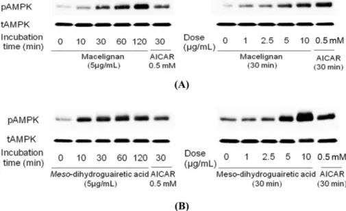
Effects of macelignan and MDGA on AMPK
activation − We utilized MCF-7 cell lines to investigate
the effects of macelignan and MDGA on AMPK
activation, and AICAR was used as positive control. As
shown in Fig. 2, western blot analysis of a time course
assay with MCF-7 cells demonstrated that macelignan
and MDGA increased the phosphorylation of the Thr
172residue of the AMPK α subunit in a time-dependent manner at 5 μg/mL. When the dose-dependent effects of macelignan and MDGA on AMPK activation were checked, the phosphorylation of AMPK at MCF-7 cancer
cells was increased with dose-dependently at the concentrations of 1, 2.5, 5, and 10 μg/mL. These data indicated that both compounds can stimulate the pho- sphorylation and activity of AMPK at dose-dependent and time-dependent manners.
Fig. 1. A representative HPLC profile of major compounds from the total EtOAc layer of Myristica fragrans with detections at 205 and 280 nm. Key to peak identity: (1) macelignan, (2) MDGA. Chromatographic method used: 0 - 40 min (50 - 70% MeOH), 40 - 52 min (70 - 100% MeOH), 52 - 60 min (100% MeOH).
Fig. 2. Phosphorylation of AMPK by macelignan and MDGA in MCF-7 cells. Cells were seeded using DMEM supplemented with 10%
heat-inactivated fetal bovine serum without antibiotic and cultured for 24 hrs at 37 ºC in humidified air condition containing 5% CO
2. The
cells were then starved with serum free DMEM media for 24 hrs and treated with 5 µg/ml of macelignan and MDGA for 10 min, 30 min,
1 hr, and 2 hrs (the left panels of A and B) or treated with 0, 1, 2.5, 5, and 10 µg/ml of macelignan and MDGA (the right panels of A and
B), respectively. The cells were harvested with cold PBS and lysed with 1 × NP40 lysis buffer. Proteins in whole cell lysates were
separated by SDS-PAGE and immunoblotted with antibodies against phospho-AMPK and total AMPK. AICAR, an AMPK activator, was
used as a positive control in this experiment. Data are representative of three independent experiments that gave similar results.
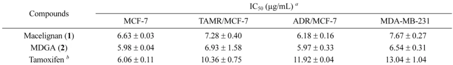
Anti-proliferative effects of macelignan and MDGA − In order to evaluate the effects of macelignan and MDGA on the growth of human breast cancer cells, MCF-7 (estrogen receptor-positive), MDA-MB-231 (estrogen receptor-negative), tamoxifen-resistant MCF-7 (TAMR/
MCF-7), and adriamycin-resistant MCF-7 (MCF-7/ADR) cancer cells were treated with various concentrations of two compounds. As shown in Table 1, both compounds showed significant growth inhibitory effects which are stronger than that of positive control in all tested cell lines at a dose-dependent manner. Macelignan and MDGA showed similar inhibition patterns on different breast cancer lines with IC
50values ranging from 5.97 ± 0.33 to 7.67 ± 0.27 μg/mL.
Inhibitory effects of macelignan and MDGA inhibition on colony formation of MCF-7 cells − Cell
colony formation has been found to be a more sensitive parameter of toxicity than cell viability because colony formation is assessed while the cells are in a state of proliferation, and thus more susceptible to toxic effects.
15Soft agar colony formation assays have been used to determine the growth and drug sensitivity of MCF-7 cells.
16,17In this study, MCF-7 cells were divided into control group and sample group treated with different concentrations of two lignans. The cells were cultured for two weeks in the presence of two lignans. As shown in Fig. 3, both macelignan and MDGA significantly inhibited the colony-formation ability of MCF-7 cells in a dose-dependent manner. The colony formation of MCF-7 cells after treatment of macelignan and MDGA was significantly inhibited compared with the control group.
The inhibition was evident not only in colony number but Table 1. Anti-proliferative effects of diarylbutane lignans from M. fragrans in MCF-7, MDA-MB-231, TAMR/MCF-7, and ADR/MCF-7
Compounds IC
50(µg/mL)
aMCF-7 TAMR/MCF-7 ADR/MCF-7 MDA-MB-231
Macelignan (1) 6.63 ± 0.03 7.28 ± 0.40 6.18 ± 0.16 7.67 ± 0.27
MDGA (2) 5.98 ± 0.04 6.93 ± 1.58 5.97 ± 0.33 6.54 ± 0.31
Tamoxifen
b6.06 ± 0.11 10.36 ± 0.75 11.92 ± 0.04 13.04 ± 1.04
a
Cells were treated with different concentrations of two compounds for 48 hr.
b