281
ABBREVIATIONS: MAPKs, mitogen-activated protein kinase; CASPs, caspase; JNK, Jun NH2-terminal protein kinase; BCL2, B-cell CLL/lymphoma 2; BAX, BCL2-associated X protein; PARP, poly (ADP-ribose) polymerase.
Received May 25, 2009, Revised June 19, 2009, Accepted July 20, 2009
Corresponding to: Joo-Ho Chung, Kohwang Medical Research Institute, Department of Pharmacology, School of Medicine, Kyung Hee University, 1, Hoegi-dong, Dongdaemun-gu, Seoul 130-701, Korea.
(Tel) 82-2-961-0281, (Fax) 82-2-968-0560, (E-mail) [email protected]
*The first two authors contributed equally to this article.
Naringin Protects against Rotenone-induced Apoptosis in Human Neuroblastoma SH-SY5Y Cells
Hak-Jae Kim1,*, Jeong Yoon Song2,*, Hae Jeong Park1, Hyun-Kyung Park2, Dong Hwan Yun3, and Joo-Ho Chung1
1Department of Pharmacology and Kohwang Medical Research Institute, 2East-West Neo Medical Center, 3Department of Physical Medicine and Rehabilitation, School of Medicine, Kyung Hee University, Seoul 130-701, Korea
Rotenone, a mitochondrial complex I inhibitor, can induce the pathological features of Parkinson’s disease (PD). In the present study, naringin, a grapefruit flavonoid, inhibited rotenone-induced cell death in human neuroblastoma SH-SY5Y cells. We assessed cell death and apoptosis by measuring mitogen- activated protein kinase (MAPKs) and caspase (CASPs) activities and by performing 3-(4,5-dimethyl- thiazol-2-yl)-2,5-diphenyltetrazolium bromide (MTT) assay, 4,6-diamidino-2-phenylindole (DAPI) staining, and terminal deoxynucleotidyl transferase-mediated dUTP nick end labeling (TUNEL) staining. Naringin also blocked rotenone-induced phosphorylation of Jun NH2-terminal protein kinase (JNK) and P38, and prevented changes in B-cell CLL/lymphoma 2 (BCL2) and BCL2-associated X protein (BAX) expression levels. In addition, naringin reduced the enzyme activity of caspase 3 and cleavages of caspase 9, poly (ADP-ribose) polymerase (PARP), and caspase 3. These results suggest that naringin has a neuro- protective effect on rotenone-induced cell death in human neuroblastoma SH-SY5Y cells.
Key Words: Apoptosis, Naringin, Parkinson’s disease, Rotenone, SH-SY5Y
INTRODUCTION
Parkinson’s disease (PD) is an age-related progressive neurodegenerative disorder with a prevalence of 1∼2% in people over the age of 50 (Shastry, 2001). PD pathogenesis includes oxidative stress, mitochondrial dysfunction, ex- citotoxicity, calcium overloading, trophic factor deficiency, inflammatory processes, as well as genetic factors (Ramsey and Giasson, 2007; Tansey et al., 2007; Yuan et al., 2007).
PD is characterized by a selective degeneration of dop- aminergic neurons and the presence of Lewy bodies in the neurons of the substantia nigra (Bradshaw et al., 2004).
Although the cause of neuronal death in PD is still un- known, oxidative stress and mitochondrial complex I defi- ciency may play a role in the accumulation of modified pro- teins and degeneration of dopaminergic neurons (Olanow et al., 2004). Complex I inhibitors like rotenone cause de- generation of dopaminergic neurons and motor dysfunction (Greenamyre et al., 2001; Shamoto-Nagai et al., 2003). Dopa- minergic neuron degeneration in PD is mediated through apoptotic pathways by activation of mitogen-activated pro- tein kinases (MAPKs) and caspases (CASPs) (Wang et al., 2002; Pei et al., 2003; Newhouse et al., 2004).
Natural products such as bioflavonoids have antioxidant activity (Haenen et al., 1997; Ng et al., 2000) and inhibit
lipid peroxidation in biological membranes (Maridonneau- Parini et al., 1986). PD-98059, a potent inhibitor of MAP kinase kinases (Dudley et al., 1995), prevents okadaic acid-induced cell death in cultivated rat neurons (Rundén et al., 1998), indicating that bioflavonoids may be cytopro- tective. The flavonoid naringin, for example, has shown an- tiviral (Kaul et al., 1985) and antiallergic (Tsai et al., 1999) activities through regulation of reactive oxygen species.
Naringin also inhibits H2O2-induced cytotoxicity, apoptosis, and genotoxicity in mouse P388 cells (Kanno et al., 2003).
Moreover, naringin can protect rat hepatocytes from tox- in-induced over-phosphorylation, disruption of the keratin cytoskeletal network, and toxin-induced apoptotic cell death (Rundén et al., 1998). Therefore, we tested the anti-apoptotic effects of naringin in rotenone-treated SH-SY5Y cells, a commonly used cellular PD model.
METHODS Cell culture and drug treatment
Human SH-SY5Y cells, obtained from American Type Culture Company (ATCC, MD, USA), were cultivated in Dul- becco’s modified Eagle’s medium (DMEM) supplemented with 10% heat-inactivated fetal bovine serum (GIBCO, MD, USA) and 100 U/ml penicillin/streptomycin. Cultures were main- tained in a humidified incubator at 37oC with 5% CO2, and
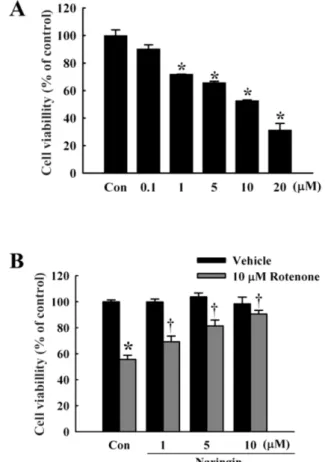
Fig. 1. Effect of naringin on rotenone-induced cell death in SH-SY5Y cells. (A) MTT cell viability assay at 0.1, 1, 5, 10, and 20 μM rotenone for 24 h in SH-SY5Y cells. (B) Naringin inhibits rotenone-induced cytotoxicity in SH-SY5Y cells, treated with 10 μM rotenone for 24 h. Naringin (1, 5, and 10 μM) was added 4 h before rotenone treatment. Independent experiments were repeated three times. Values are presented as mean±SEM. Con, control. *p<0.05 compared to non-treated cells; †p<0.05 compared to rotenone-treated cells.
the medium was changed every 2 days. Rotenone (Sigma, MO, USA) was made fresh in dimethyl sulfoxide prior to each experiment. Naringin (Sigma) was dissolved in distilled water. Naringin was added 4 h prior to rotenone treatment.
MTT assay
Cell viability was determined by the 3-(4,5-dimethylthiazol- 2-yl)-2,5-diphenyltetrazolium bromide (MTT, Sigma) assay as previously described (Park et al., 2006). Viability was measured with a microplate reader (Molecular Devices, CA, USA) at 595 nm.
DAPI staining
4,6-Diamidino-2-phenylindole (DAPI, Sigma) staining was performed, as described previously (Park et al., 2006). Cells were fixed in methanol and incubated in 1 μg/ml DAPI solution for 30 min in the dark. Stained cells were observed with a fluorescence microscope (Zeiss, Germany).
TUNEL assay
The terminal deoxynucleotidyl transferase-mediated dUTP nick end labeling (TUNEL) assay (Roche, IN, USA) was per- formed using a commercial kit according to the manu- facturer’s protocol. Cells were fixed in acetic acid at −20oC, and then incubated with TUNEL reaction mixture for 1 h at 37oC, followed by addition of peroxidase-conjugated de- tection antibody. DNA fragments were stained using dia- minobenzidine (DAB, Sigma) as a substrate for the pero- xidase.
Western blot
Cells were lysed in RIPA buffer (150 mM NaCl, 1%
Nonidet P-40, 1 mM EDTA, 0.5% deoxycholic acid, 2 μg/ml aprotinin, 1 mM phenylmethylsulfonyl fluoride, 5 mM ben- zamidine, 1 mM sodium orthovanadate containing 1×pro- tease inhibitor cocktail (Roche). Protein content was meas- ured using the Bio-Rad colorimetric protein assay kit (Bio-Rad, CA, USA). Equal amounts of protein (60 μg) were separated by 10% SDS-PAGE and transferred onto a PVDF membrane (Millipore, Germany). After blocking with 5%
non-fat milk, membranes were probed with primary anti- bodies against phospho-c-Jun NH2-terminal protein kinase (p-JNK. 1:1,000), phospho-P38 MAPK (p-P38, 1:1,000, Santa Cruz, CA, USA), B-cell CLL/lymphoma 2 (BCL2, 1:1,000, Santa Cruz), BCL2-associated X protein (BAX, 1:1,000, Santa Cruz), cleaved poly (ADP-ribose) polymer- ase-1 (PAR, 1:1,000, Santa Cruz), cleaved CASP3 (1:
1,000, Santa Cruz), cleaved CASP9 (1:1,000, Santa Cruz), and β-Actin (1:5,000, Cell Signaling Technology, MA, USA) overnight at 4oC. Horseradish peroxidase-conjugated antimouse or antirabbit IgG (Serotec, UK) were used as sec- ondary antibodies. An Enhanced Chemiluminescence (ECL) detection system (Amersham Biosciences, Sweden) was used to detect the protein bands on the membrane.
CASP3 activity
CASP3 activity was measured using an assay kit (Sigma) according to the manufacturer’s protocol. SH-SY5Y cells were lysed, CASP3 substrate (Ac-DVED-p-NA) was added, and the mixture was incubated overnight in a humidified
environment at 37oC. The concentration of p-NA released from the CASP3 substrate was measured using a micro- plate reader (Molecular Devices) at 405 nm and calculated from a calibration curve of p-NA standards.
Statistical analysis
Data are expressed as mean±SEM and were analyzed by one-way ANOVA, followed by Tukey’s HSD post-hoc test, using SPSS software (version 17.0; SPSS Inc., IL, USA).
p<0.05 was considered statistically significant.
RESULTS
Effect of naringin on rotenone-induced cell death Rotenone treatment (0.1, 1, 5, 10, and 20 μM for 24 h) of SH-SY5Y cells induced a dose-dependent cytotoxicity, with approximately 50% viability at 10 μM (Fig. 1A).
Naringin dose-dependently (2, 5, and 10 μM) protected against death induced by 10 μM rotenone (Fig. 1B), with
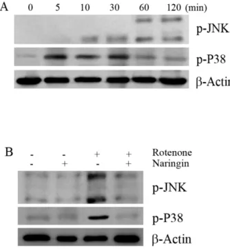
Fig. 3. The effect of rotenone on the phosphorylation of JNK and P38 in SH-SY5Y cells. (A) The effect of rotenone on the phosphory- lation of JNK and P38 by Western blot analysis. Cells were treated with 10 μM rotenone for 120 min. (B) Western blot to determine the effect of naringin on rotenone-induced phosphorylation of JNK and P38 in SH-SY5Y cells. The cells were pretreated with 10 μM rotenone for 120 min, with 10 μM naringin added 4 h prior to rotenone treatment. The cells were harvested for the phosphory- lation assay at 30 and 120 min after rotenone treatment. β-Actin was used as an internal standard protein. Three independent experiments were performed for this assay.
Fig. 2. Effect of naringin on rotenone-induced apoptotic features in SH-SY5Y cells pretreated with 10 μM rotenone for 24 h. Naringin (10 μM) was treated 4 h prior to rotenone treatment. Condensed chromatin was stained dark brown (lower). Scale bar, 100 μm.
Three independent experiments were performed for this study.
about 90% protection at 10 μM naringin.
Naringin inhibits rotenone-induced apoptosis Phase-contrast microscopy revealed that naringin de- creased rotenone-induced cell shrinkage, irregularity in shape, and cellular detachment (Fig. 2, upper). Naringin at 10 μM significantly inhibited apoptotic body formation in SH-SY5Y cells treated with 10 μM rotenone for 24 h and protected against nuclear condensation, DNA fragmen- tation, and perinuclear apoptotic bodies (Fig. 2). Further- more, naringin significantly reduced the number of TUNEL-positive cells (Fig. 2, lower).
Naringin affects P38 and JNK phosphorylation The effect of naringin on phosphorylation of JNK and P38 was investigated in SH-SY5Y cells treated with 10 μM ro- tenone for 2 h using Western blot analysis. Rotenone-in- duced phosphorylation of JNK and P38 reached maximum at 30 min and 2 h, respectively (Fig. 3A). Naringin sig- nificantly blocked rotenone induced phosphorylation of JNK and P38 (Fig. 3B).
Naringin affects apoptotic proteins
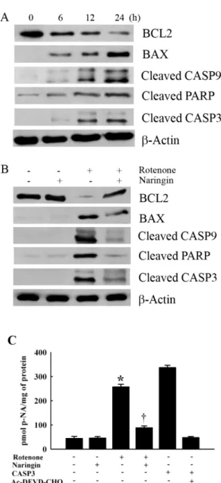
Rotenone reduced BCL2 expression and increased BAX expression in a time-dependent manner (Fig. 4A). Naringin treatment significantly changed rotenone-induced BCL2 and BAX expression profiles (Fig. 4B). Rotenone time-de- pendently increased the intracellular levels of cleaved CASP9, PARP, and CASP3 (Fig. 4A). Naringin treatment significantly suppressed the cleavages of CASP9, PARP, and CASP3 in rotenone-treated cells (Fig. 4B).
CASP3 activity
CASP3 enzyme activity was analyzed by measuring the hydrolysis of a peptide substrate, Ac-DEVD-p-NA, in rote- none-treated SH-SY5Y cells. Naringin treatment signifi- cantly inhibited rotenone-mediated hydrolysis of Ac-DEVD- pNA (Fig. 4C).
DISCUSSION
We measured the ability of naringin to protect rotenone- treated human SH-SY5Y cells, a PD cell model. Naringin dose-dependently reduced rotenone-induced cell death. DAPI staining and TUNEL assay results showed that naringin significantly inhibited chromatin condensation and DNA strand breaks in rotenone-treated cells. Naringin treatment significantly inhibited rotenone-induced phosphorylation of JNK and P38. Naringin also prevented decrease of BCL2 expression and increase of BAX expression, caused by rote- none treatment. These results indicate that naringin pre- vents rotenone-induced apoptosis through inhibiting JNK and P38 activity.
JNK and P38 are members of the MAPK subfamily and regulate neuronal survival, death, and differentiation (Chang and Karin, 2000; Davis, 2000; June and Mouradian, 2001).
As confirmed here, rotenone treatment induces apoptosis via phosphorylation of P38 and JNK but not extracellular signal-regulated kinase (ERK) in human neuroblastoma cells (Davis, 2000; Newhouse et al., 2004). The activation of JNK and P38 is required for inhibiting the anti-apoptotic protein, BCL2, regulating the release of cytochrome c from the mitochondria to the cytoplasm, and inducing the activa- tion of CASP 9 (Davis, 2000; Junn and Mouradian, 2001;
Li et al., 2004). Klintworth et al. (2007) reported that JNK activation is a common mechanism underlying dopamin-
Fig. 4. The effect of rotenone on the expression of apoptotic proteins BCL2, BAX, CASP9, PARP, and cleaved CASP3 in SH-SY5Y cells.
(A) The effect of rotenone on the expression of BCL2, BAX, CASP9, PARP, and cleaved CASP3 by Western blot analysis. Cells were treated with 10 μM rotenone for 24 h. (B) Western blot assay to determine the effect of naringin on rotenone-induced expression of the apoptotic proteins, BCL2, BAX, cleaved CASP9, cleaved PARP, and cleaved CASP3 in SH-SY5Y cells. Cells were pretreated with 10 μM rotenone for 24 h, with 10 μM naringin added 4 h prior to rotenone treatment. β-Actin was detected as an internal standard protein. (C) Effect of naringin on rotenone-induced activity of CASP3 in SH-SY5Y cells. Cells were pretreated with 100 μM rotenone for 24 h and 10 μM naringin was then added 4 h prior to rotenone treatment. The cleaved CASP3 substrate acetyl-Asp- Glu-Val-Asp-p-Nitroanilide (Ac-DEVD-p-NA) was measured at 405 nm. CASP3 was used as a positive control. The CASP3 inhibitor (Ac-DEVD-CHO) added with CASP3 was used as negative control.
Values are presented as mean±SEM. Three independent experiments were performed for this assay. *p<0.05 compared to non-treated cells; †p<0.05 compared to rotenone-treated cells.
ergic cell death induced by both paraquat and rotenone in cell lines and primary cultures.
Rotenone also induces apoptosis in dopaminergic SH- SY5Y cells through activation of CASPs (Newhouse et al., 2004). CASPs, the primary mediator of apoptosis, cleave a number of cellular proteins including PARP, which is an important step in several types of apoptosis. Treatment with CASP inhibitors such as benzyloxycarbonyl-Val-Ala- Asp (Z-VAD) and N-acetyl-Asp-Glu-Val-Asp (Ac-DEVD), in- hibited rotenone-induced apoptosis in SH-SY5Y cells, sug- gesting a general role of CASPs in rotenone-induced apopto- sis (Wang et al., 2002; Pei et al., 2003). In this study, nar- ingin pretreatment significantly inhibited CASP3 enzyme activity as well as CASP9, PARP, and CASP3 cleavage in rotenone-treated neuroblastoma cells.
In conclusion, naringin showed significant antiapoptotic effects in rotenone-treated human SH-SY5Y cells by in- hibiting phosphorylation of JNK and P38, as well as the activation of CASP9, PARP, and CASP3. These findings in- dicate that naringin may have therapeutic potential for PD.
ACKNOWLEDGEMENTS
This study was supported by a grant of the Small and Medium Business Administration (S5107A11901).
REFERENCES
Bradshaw J, Saling M, Hopwood M, Anderson V, Brodtmann A.
Fluctuating cognition in dementia with Lewy bodies and Alzhei- mer's disease is qualitatively distinct. J Neurol Neurosurg Psychiatry 75: 382−387, 2004.
Chang L, Karin M. Mammalian MAP kinase signaling cascades.
Nature 410: 37−40, 2000.
Davis RJ. Signal transduction by the JNK group of MAP kinases.
Cell 103: 239−252, 2000.
Dudley DT, Pang L, Decker SJ, Bridged AJ, Saltiel AR. A synthetic inhibitor of the mitogen-activated protein kinase cascade. Proc Natl Acad Sci USA 92: 7686−7689, 1995.
Greenamyre JT, Sherer TB, Betarbet R, Panov AV. Complex I and Parkinson’s disease. IUBMB Life 52: 135−141, 2001.
Haenen GR, Paquay J, Korthouwer R, Bast A. Peroxynitrite scavenging by flavonoids. Biochem Biophys Res Commun 236:
591−593, 1997.
Junn E, Mouradian MM. Apoptotic signaling in dopamine induced cell death: the role of oxidative stress, P38 mitogen-activated protein kinase, cytochrome c and caspases. J Neurochem 78: 374−
383, 2001.
Kanno S, Shouji A, Asou K, Ishikawa M. Effects of naringin on hydrogen peroxide-induced cytotoxicity and apoptosis in P388 cells. J Pharmacol Sci 92: 166−170, 2003.
Kaul TN, Middlenton E Jr, Ogra PL. Antiviral effect of flavonoids on human viruses. J Med Virol 15: 71−79, 1985.
Klintworth H, Newhouse K, Li T, Choi WS, Faigle R, Xia Z.
Activation of c-Jun N-terminal protein kinase is a common mechanism underlying paraquat- and rotenone-induced dopa- minergic cell apoptosis. Toxicol Sci 97: 149−162, 2007.
Li P, Nijhawan D, Budihardjo I, Srinivasula SM, Ahmad M, Alnemri ES, Wang X. Cytochrome c and dATP dependent formation of Apaf-1/caspase-9 complex initiates an apoptotic protease cascade. Cell 91: 479−489, 2004.
Maridonneau-Parini I, Braquet P, Garay RP. Heterogeneous effect of flavonoids on K+-loss and lipid peroxidation induced by oxygen free radicals in human red cells. Pharmacol Res Commun 18: 61−72, 1986.
Newhouse K, Hsuan SL, Chang SH, Cai B, Wang Y, Xia Z.
Rotenone-induced apoptosis is mediated by p38 and JNK MAP
kinases in human dopaminergic SH-SY5Y cells. Toxicol Sci 79:
137−146, 2004.
Ng TB, Liu F, Wang ZT. Antioxidative activity of natural products from plants. Life Sci 66: 709−723, 2000.
Olanow CW, Perl DP, DeMartino GN, McNaught, KS. Lewy-body formation is an aggresome-related process: a hypothesis. Lancet Neurol 3: 496−503, 2004.
Park HJ, Shin DH, Chung WJ, Leem K, Yoon SH, Hong MS, Chung JH, Bae JH, Hwang JS. Epigallocatechin gallate reduces hypoxia-induced apoptosis in human hepatoma cells. Life Sci 78:
2826−2832, 2006.
Pei W, Liou AK, Chen J. Two caspase-mediated apoptotic pathways induced by rotenone toxicity in cortical neuronal cells. FASEB J 17: 520−522, 2003.
Ramsey CP, Giasson BI. Role of mitochondrial dysfunction in Parkinson’s disease-implications for treatment. Drugs Aging 24:
95−105, 2007.
Rundén E, Seglen PO, Haug FM, Ottersen OP, Wieloch T, Shamloo M, Laake JH. Regional selective neuronal degeneration after protein phosphatase inhibition in hippocampal slice cultures:
evidence for a MAP kinase-dependent mechanism. J Neurosci 18: 7296−7305, 1998.
Saporito MS, Brown EM, Miller M S, Carswell S. CEP-1347/KT-7515, an inhibitor of c-jun N-terminal kinase activation, attenuates the 1-methyl-4-phenyl tetrahydropyridine-mediated loss of nigrostriatal dopaminergic neurons in vivo. J Pharmacol Exp Ther 288: 421−
427, 1999.
Saporito MS, Thomas BA, Scott RW. MPTP activates c-Jun NH(2)- terminal kinase (JNK) and its upstream regulatory kinase MKK4 in nigrostriatal neurons in vivo. J Neurochem 75: 1200−1208, 2000.
Shamoto-Nagai M, Maruyama W, Kato Y, Isobe K, Tanaka M, Naoi M, Osawa T. An inhibitor of mitochondrial complex I, rotenone, inactivates proteasome by oxidative modification and induces aggregation of oxidized proteins in SH-SY5Y cells. J Neurosci Res 74: 589−597, 2003.
Shastry BS. Parkinson disease: etiology, pathogenesis and future of gene therapy. Neurosci Res 41: 5−12, 2001.
Tansey MG, McCoy MK, Frank-Cannon TC. Neuroinflammatory mechanisms in Parkinson’s disease: potential environmental triggers, pathways, and targets for early therapeutic intervention.
Exp Neurol 208: 1−25, 2007.
Tsai SH, Lin-Shiau SY, Lin JK. Suppression of nitric oxide synthase and the down-regulation of the activation of NfkappaB in macropahages by resveratrol. Br J Pharmacol 126: 673−680, 1999.
Wang X, Qin ZH, Leng, Y, Wang Y, Jin X, Chase TN, Bennett MC.
Prostaglandin A1 inhibits rotenone-induced apoptosis in SH- SY5Y cells. J Neurochem 83: 1094−1102, 2002.
Yuan H, Zheng JC, Liu P, Zhang SF, Xu JY, Bai LM. Pathogenesis of Parkinson’s disease: oxidative stress, environmental impact factors and inflammatory processes. Neurosci Bull 23: 125−130, 2007.