Introduction
Among musculoskeletal injuries, ankle sprains are common [1]. In addition, the recurrence rate of ankle sprain is high, and symptoms often persist [2]. Similarly, frequent sprains after the index injury cause chronic ankle instability (CAI) [3, 4]. Symptoms caused by CAI are characterized by the recurrence of pain and swelling
during activity and weakening of the muscles around the ankle joint [4].
CAI, which has a high recurrence rate and long-lasting discomfort, can be noted as a characteristic sensorimotor defect. This is a decreased conscious perception, indicating a defect in afferent somatosensory information and efferent motor control [5]. In the study of cortical excitability, there was a difference in cortical excitability
Received: Mar 3, 2021 Revised: Mar 14, 2021 Accepted: Mar 15, 2021
Corresponding author: Seungwon Lee (ORCID https://orcid.org/0000-0002-0413-0510)
Department of Physical Therapy, College of Health Science and Social Welfare, Sahmyook University 815 Hwarang-ro, Nowon-gu, Seoul 01795, Republic of Korea
Tel: (82 for International)-02- 3399-1630 Fax: 02- 3399-1639 E-mail: [email protected]
This is an Open-Access article distributed under the terms of the Creative Commons Attribution Non-Commercial License (http://creativecommons.org/licenses/
by-nc/4.0) which permits unrestricted non-commercial use, distribution, and reproduction in any medium, provided the original work is properly cited.
Copyright © 2021 Korean Academy of Physical Therapy Rehabilitation Science
Short-term effects of joint mobilization with versus without voluntary movement in patients with chronic ankle instability:
A single-blind randomized controlled trial
Hyunjoong Kim
a,b, Seonghyeok Song
c, Sangbong Lee
c, Seungwon Lee
d,e1)aSports Rehabilitation Center, The Better Hospital, Gwangju, Republic of Korea
bDepartment of Physical Therapy, Gwangju Health University, Gwangju, Republic of Korea
cDepartment of Physical Therapy, Graduate School of Sahmyook University, Seoul, Republic of Korea
dDepartment of Physical Therapy, College of Health Science and Social Welfare, Sahmyook University, Seoul, Republic of Korea eInstitute of SMART Rehabilitation, Sahmyook University, Seoul, Republic of Korea
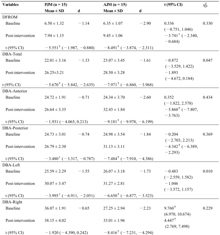
Objective: Joint mobilization for arthrokinematics altered by the positional fault of chronic ankle instability (CAI) is an effective intervention for stabilization. In this study, we compared the effects of ankle dorsi flexion range of motion (DFROM) and dynamic balance ability (DBA) in CAI patients via passive joint mobilization (PJM), a method traditionally performed in previous studies, and active joint mobilization (AJM), a method that can have a greater effect on cortical excitability with spontaneous movements.
Design: Single-blind two-arm randomized controlled trial
Methods: A total of 30 participants were registered: 15 each to the PJM and AJM groups. Each participant received a total of 10 intervention sessions, 10 minutes per session, 5 times a week for 2 weeks. PJM used Maitland's mobilization method to apply joint mobilization with talus in the posterior direction and AJM used an angular joint motion to induce patient's voluntary motion of medial malleolus anterior gliding and lateral malleolus posterior gliding, respectively. DFROM of the ankle was measured by using tape and DBA was evaluated by using the balance system.
Results: Significant improvement was observed after intervention in both the PJM and AJM groups except for the DBA-anterior and DBA-right variables of the PJM group. There were statistically significant differences between the AJM and PJM groups in the DFROM, DBA-anterior, DBA-posterior, and DBA-right variables.
Conclusions: The overall improvement of DFROM and DBA was found to be more effective in joint mobilization including voluntary movement. When it is accompanied by voluntary movement, it further affects the neuromuscular system of the ankle.
Key Words: Joint instability, Ankle injuries, Postural balance, Musculoskeletal manipulations, Joint mobilization pISSN 2287-7576
eISSN 2287-7584
2021, 10(1), 1-9 www.jptrs.org
and joint laxity between CAI patients and healthy adults, and it was reported that cortical excitability was decreased in ankle instability patients subjected to transcranial magnetic stimulation. This means that the chronic state of joint laxity has high cortical excitability, and it means that cortical excitability as well as joint laxity needs to be reduced [6, 7].
In other words, CAI has great influence on the nervous system, which acts as a modulator of body movement. Therefore, the main rehabilitation protocol for patients with CAI is to improve neuromuscular function. This can be explained by a decrease in joint position sense due to damage to the ankle ligament receptors [8] and peroneal nerve [9] resulting from the injury [10].
Among the conventional interventions for CAI, anterior to posterior talocrural joint mobilization effectively improves CAI and may show some neuromuscular mechanisms by increasing fibularis musculotendinous stiffness [11]. However, the effect of passive joint mobilization (PJM) by the therapist is limited to the neuromuscular mechanism as there is no active movement.
Therefore, active joint mobilization (AJM) is judged by the patient’s active repetition accompanied by pain-free motion to sustained mobilization performed by the therapist and can have a greater influence on cortical excitability. This aims at the central nervous system level of motion corrected under the therapist’s guidance, as re-education on pain-free motion patterns is implied in neuroscience theory [12-15].
In CAI patients, joint mobilization combined with voluntary movement will have more influence on the neuromuscular system than PJM, which corresponds to conventional joint mobilization. Therefore, in this study, the range of motion and balance ability are compared and verified to find out the effect according to with or without voluntary movement.
Methods Study design
This single-blind two-arm parallel design randomized controlled trial included two evaluation sessions (baseline and post-intervention) and 10 intervention sessions (PJM and AJM). In the outcome measure, the primary variable was the dorsi flexion range of motion
(DFROM) of the ankle joint, while the secondary variable was dynamic balance ability (DBA).
Participants
Recruitment of potential participants was recruited by staff and students through the bulletin board of Gwangju Health University, Gwangju, Korea. Forty-four potential participants who diagnosed an ankle sprain were recruited; of them, 30 were ultimately enrolled in the study.
The inclusion criteria were: (a) self-reported instability and feeling of “giving way,” (b) a history of sprains in only one ankle, (c) at least two sprains on the same side in the past 2 years, (d) Feeling different in sensation compared to an intact ankle, and (e) not receiving other treatment during the study [16].
The exclusion criteria included: (a) acute ankle sprain within the last 6 months, (b) a history of bilateral ankle injury, (c) bony injury related to ankle sprain, and (d) a history of surgery on the back or lower extremities [16].
Prior to the study, all participants were informed about the purpose and procedure in accordance with the ethical standards of the Declaration of Helsinki. Only those who voluntarily signed an informed consent form were enrolled.
Randomization and blinding
Participants were randomly and blindly assigned to the PJM and AJM groups using random allocation software (ver. 1.0; Isfahan University, Iran).
Intervention
The PJM and AJM interventions were performed on the affected foot for 10 min in each session by two physical therapists who have more than 5 years of clinical experience and have been trained in Orthopedic manual physical therapy. The participants received a total of 10 sessions five times a week for 2 weeks [17].
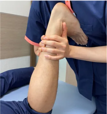
Passive joint mobilization
PJM uses Maitland’s mobilization method, grade III
(high amplitude in the end range of the joint and 1-s
vibration in the middle range through linear motion in
which tissue resistance is felt). The participant assumed
a supine position, and the physical therapist held the
talus with one hand and the tibia with the other hand and
performed joint mobilization in the posterior direction
with the hand holding the talus (Figure 1) [18].
Active joint mobilization
In the AJM, the participant bent their knees in the prone position. The physical therapist held the medial malleolus with one hand and the lateral malleolus with the other. At the same time, the physical therapist put the participant’s soles against the sternum and pressed them in the plantar direction. At this time, the medial malleolus would glide in the anterior direction and the lateral malleolus would glide in the posterior direction.
The first procedure passively recognizes participant’s joint motion, while the second procedure involves the voluntary motion (Figure 2) [19-21].
Outcomes
Participants are enrolled in the study and evaluated for DFROM and DBA prior to intervention (baseline), and immediately after two weeks of intervention (post- intervention).
Dorsiflexion range of motion
The DFROM of the ankle was measured using a weight-bearing lunge. The therapist placed a tape measure on the floor perpendicular to a flat wall without protrusion and affixed it to prevent motion. The second toe and heel of the participant’s foot to be measured were placed parallel to the tape measure, and the non-measured foot was separated from the measuring foot as much as the individual’s foot. The standard for placing the first second toe was 10 cm and bent until the knees touched the wall.
The maximum range of ankle instep flexion was until the
heel reached without falling off the floor [22]. As a precaution, the trunk of the body should not be tilted forward and should be bent toward the wall only at the knee. If the knee could reach the wall during bending, the foot was moved forward, while if the knee easily touched the wall, the foot was moved backward. This measurement was performed three times for each of the left and right feet, and the average value of the measured values was recorded as the ankle angle.
The inter-rater reliability of DFROM has a high intraclass correlation coefficient of 0.99 [23], and minimal detectable change is from 1.1 to 1.5 cm [24].
Dynamic balance ability
DBA was evaluated with the participant in a standing position on both feet using the Biodex Balance System (Biodex Balance System; Biodex Medical Systems Inc., USA). The balance measurement equipment consisted of a fixed circular scaffold with a sensor that detects anterior/posterior, left/right motion, a monitor that can visually check and match a target, a computer for data transmission and analysis, and a computer for analysis data output. It consisted of four main types, including printers. The higher the score, the better the balance ability. At the time of the measurement, both arms were collected on the chest, the center of gravity of the body was moved to maintain it according to the target, and the
Figure 1. Passive joint mobilization Figure 2. Active joint mobilization
foot position was carefully measured to prevent movement [25].
In the evaluation of the DBA, the scaffold was not fixed, and scaffold instability could be adjusted in various stages from 1 to 12 (least stable to most stable).
In this study, DBA was measured by setting the level of stool movement to one of eight stages. As in the static balance ability evaluation, the foot position was not changed, but the task time and route were recorded by setting the target point and moving the body center to reach the point. Measurements were made twice for 20 s with a 10-s break between them. Measurement of DBA using the Biodex Balance System was reported to have an ICC of 0.42 to 0.80 with appropriate intratester reliability [26].
Sample size
In the study by Cruz-Díaz, Lomas Vega (16), the sample size was calculated by changing the range of motion before and immediately after joint mobilization in CAI patients. The calculated Cohen’s d was 1.36, and when two groups and power were set to 0.95, using G*power 3.1 (G-power 3.1; Heinrich-Heine-Universität Düsseldorf, Germany), 26 samples were required. A total of 30 participants were enrolled in the study according to expected dropout and the central limit theorem.
Statistical analysis
All statistical analyses were performed using IBM SPSS Statistics version 25.0 (SPSS 25.0; IBM Corp., USA). The homogeneity test was performed using the chi-squared test (categorical variables) and independent t-test (continuous variables). An independent t-test was used to verify the effect of the difference between groups, while a paired t-test was used to determine the
difference before versus after the interventions. Cohen’s d and partial eta squared (
) were used to determine the effect size of the treatment.
was calculated using repeated measures analysis of variance (RM ANOVA).
All statistical significance levels (α) were set at 0.05.
Results
This study was conducted between April and June 2019. A total of 30 participants were recruited, and there were no dropouts (Figure 3). Table 1 presents the participants’ general characteristics. There were no significant intergroup differences.
Dorsiflexion range of motion
There was statistically significant improvement in the PJM (d = −1.14, P < 0.001) and AJM (d = −2.90, P <
0.001) groups before versus after the intervention. More significant improvement was noted in the AJM group than in the PJM group (
= 0.330, P < 0.01; Table 2).
Dynamic balance ability
In DBA-total, the difference before versus after the intervention was significantly improved in the PJM (d =
−1.33, P < 0.001) and AJM (d = −1.61, P < 0.001) groups. There was no significant intergroup difference (
= 0.047, P > 0.05; Table 2).
In DBA-anterior, the PJM group showed no significant improvement (d = −0.71, P > 0.05), whereas the AJM group showed significant improvement (d = −2.60, P < 0.001).
Therefore, the AJM group showed significantly greater improvement than the PJM group (
= 0.434, P < 0.001;
Table 2).
PJM (n = 15) AJM (n = 15)
χ
2/tSex (male/female) 9/6 7/8 0.536
Age (years) 24.60 ± 2.72 22.87 ± 2.39 1.855
Height (cm) 166.27 ± 8.76 167.47 ± 10.41 − 0.342
Weight (kg) 64.20 ± 14.29 68.20 ± 16.64 − 0.706
BMI (kg/m
2) 22.96 ± 3.16 23.96 ± 3.50 − 0.821
Values are presented as mean ± standard deviation or number.
AJM: active joint mobilization, BMI: body mass index, PJM: passive joint mobilization.
Table 1. Participants’ general characteristics (n = 30)
In DBA-posterior, significant improvement was seen in both the PJM group (d = −0.74, P < 0.01) and the AJM group (d = −1.84, P < 0.001). The AJM group showed greater improvement than the PJM group (
= 0.369, P < 0.001; Table 2).
In DBA-left, significant improvement was seen in both the PJM group (d = −1.55, P < 0.01) and the AJM group (d = −1.73, P < 0.001). There was no significant intergroup difference (
= 0.010, P > 0.05; Table 2).
In DBA-right, the PJM group did not show any significant improvement (d = −0.65, P > 0.05), whereas the AJM group showed significant improvement (d = − 2.23, P < 0.001). Significant differences were found before versus after treatment, but a separate sub-analysis (repeated-measures analysis of variance) showed more significant improvement in the AJM group than in the PJM group (F = 8.321,
= 0.229, P < 0.01; Table 2).
Discussion
In this study, we compared the effects on ankle DFROM and DBA in CAI patients via PJM, a method traditionally performed in previous studies, and AJM, a method that can have a greater effect on neuromuscular system with voluntary movements.
After PJM and AJM were administered a total of 10 times to CAI patients, significant improvement was observed after the intervention in both groups except DBA-anterior and DBA-right in the PJM group. In addition, significantly greater improvement was noted in AJM than in PJM in DFROM, DBA-anterior, DBA- posterior, and DBA-right. Joint mobilization had a large effect on each dependent variable (
> 0.14) [27] of DFROM, DBA-anterior, DBA-posterior, and DBA-right.
Significant improvement in ankle DFROM showed a large effect (d > 0.80) [28] in the PJM and AJM groups.
These results are similar to those of previous studies that
showed significant improvement when performing
Figure 3. CONSORT (Consolidated Standards for Reporting of Trials) study flow diagram
anterior to posterior talocrural joint mobilization in CAI patients [18, 29]. In addition, in the case of voluntary movements, the same results as those of previous studies
were obtained [30, 31]. The minimal detectable change of DFROM for CAI patients reported in previous studies was reportedly 0.85 cm [32], which supports the results
Variables PJM (n = 15) AJM (n = 15) t (95% CI)
Mean ± SD d Mean ± SD d
DFROM
Baseline 6.50 ± 1.32 −1.14 6.35 ± 1.07 −2.90 0.336
(−0.751, 1.046)
0.330
Post-intervention 7.94 ± 1.15 9.45 ± 1.06 −3.741
†(−2.340,
−0.684) t (95% CI) −5.551
‡(−1.987, −0.880) −8.491
‡(−3.874, −2.311)
DBA-Total
Baseline 22.01 ± 3.16 −1.33 23.07 ± 3.45 −1.61 −0.872
(−3.529, 1.422)
0.047
Post-intervention 26.25±3.21 28.50 ± 3.28 −1.893
(−4.672, 0.184) t (95% CI) −5.670
‡(−5.842, −2.635) −7.971
‡(−6.860, −3.968)
DBA-Anterior
Baseline 24.72 ± 1.91 −0.71 24.34 ± 3.70 −2.60 0.352
(−1.822, 2.578)
0.434
Post-intervention 26.64 ± 3.35 32.43 ± 1.84 −5.860
‡(−7.807,
−3.763) t (95% CI) −1.931 (−4.065, 0.213) −9.181
‡(−9.978, −6.199)
DBA-Posterior
Baseline 24.73 ± 3.01 −0.74 24.98 ± 3.54 −1.84 −0.204
(−2.703, 2.213)
0.369
Post-intervention 26.79 ± 2.30 31.13 ± 3.11 −4.342
‡(−6.389,
−2.293) t (95% CI) −3.480
†(−3.317, −0.787) −7.484
‡(−7.910, −4.386)
DBA-Left
Baseline 25.59 ± 2.29 −1.55 26.07 ± 3.18 −1.73 −0.483
(−2.559, 1.582)
0.010
Post-intervention 30.07 ± 3.47 31.27 ± 2.81 −1.046
(−3.572, 1.157) t (95% CI) −3.995
†(−6.911, −2.051) −6.650
‡(−6.877, −3.523)
DBA-Right
Baseline 36.07 ± 1.91 −0.65 27.25 ± 2.94 −2.23 9.760
‡(6.970, 10.674)
0.229
Post-intervention 38.15 ± 4.02 33.01 ± 1.96 4.447
‡(2.769, 7.498) t (95% CI) −1.920 (−4.390, 0.242) −8.416
‡(−7.231, −4.294)
Values are presented as mean ± standard deviation.
AJM: active joint mobilization, CI: confidence interval, DBA: dynamic balance ability, DFROM: dorsiflexion range of motion, PJM: passive joint mobilization.
*