The Structural and Functional Role of p53 as a Cancer Therapeutic Target
Chang Woo Han†, So Young Park†, Mi Suk Jeong and Se Bok Jang*
Department of Molecular Biology, College of Natural Sciences, Pusan National University, Busan 46241, Korea Received March 14, 2018 /Revised April 17, 2018 /Accepted April 18, 2018
The p53 gene plays a critical role in the transcriptional regulation of cellular response to stress, DNA damage, hypoxia, and tumor development. Keeping in mind the recently discovered manifold physio- logical functions of p53, its involvement in the regulation of cancer is not surprising. In about 50%
of all human cancers, inactivation of p53’s protein function occurs either through mutations in the gene itself or defects in the mechanisms that activate it. This disorder plays a crucial role in tumor evolution by allowing the evasion of a p53-dependent response. Many recent studies have focused on directly targeting p53 mutants by identifying selective, small molecular compounds to deplete them or to restore their tumor-suppressive function. These small molecules should effectively regulate vari- ous interactions while maintaining good drug-like properties. Among them, the discovery of the key p53-negative regulator, MDM2, has led to the design of new small molecule inhibitors that block the interaction between p53 and MDM2. Some of these small molecule compounds have now moved from proof-of-concept studies into clinical trials, with prospects for further, more personalized anti-carcino- genic medicines. Here, we review the structural and functional consequences of wild type and mutant p53 as well as the development of therapeutic agents that directly target this gene, and compounds that inhibit the interaction between it and MDM2.
Key words : MDM2 interaction, p53, p53 mutant, tumor suppression
†Authors contributed equally.
*Corresponding author
*Tel : +82-51-510-2523, Fax : +82-51-581-2544
*E-mail : [email protected]
This is an Open-Access article distributed under the terms of the Creative Commons Attribution Non-Commercial License (http://creativecommons.org/licenses/by-nc/3.0) which permits unrestricted non-commercial use, distribution, and reproduction in any medium, provided the original work is properly cited.
Introduction
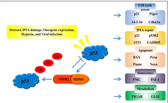
Numerous biological functions of the p53 gene, which has been identified as a tumor suppressor gene [51], have been discovered, including modulation of apoptosis, cell cycle ar- rest, senescence, metabolism, and DNA repair in response to various cellular stresses such as DNA damage, oncogene expression, hypoxia, and viral infection (Fig. 1) [30, 41].
Furthermore, p53 plays an essential role in preventing the development of cancer [18, 49]. Early on, p53 was classified as an oncogene and identified as the most frequently mu- tated gene in a variety of human cancers [21]. Indeed, p53 is inactivated by mutations or loss in about 50% of all human cancers. Unlike other tumor suppressor genes, p53 is primar- ily altered in human cancers by missense mutations, which are located in the DNA-binding domain (DBD) and caused by a single amino acid change at various sites. Missense mu-
tations in hot-spot sites (Arg175, Tyr220, Gly245, Arg248, Arg249, Arg273, and Arg282) are most frequently observed in human cancers. Additionally, nonsense or frame-shift mutations, which are less frequent than missense mutations, constitute 10% of the p53 gene mutations in cancers [36, 40]. Mutations can result in loss of DNA binding, disruption of structural stability, and loss of transcriptional transactivation function [4, 20, 52, 56]. Several recent studies have shown that p53 reactivation leads to tumor regression, which is considered a very promising anticancer strategy. Therefore, many re- search groups are searching for small molecules or peptides that can reactivate mutant p53 [9].
Under normal conditions, the expression of p53 protein is tightly controlled to remain low by the E3 ubiquitin pro- tein ligase MDM2 (Mouse double minute 2), which binds to the N-terminus transactivation domain of p53 [34]. Mouse double minute 2 regulates p53 stability through mono- or poly-ubiquitination and proteasome-mediated degradation [6]. Mouse double minute X (also known as MDM4) is a structural homologue of MDM2 that plays a critical role in down-regulation of p53 activity [22]. The presence of cellular stress inhibits the interaction between p53 and MDM2 by the post-translational modification of both proteins, leading to p53 accumulation and activation [45]. Thus, the inter- action of p53 with MDM2 is an attractive target for develop- - Review -
Fig. 1. The p53 signaling pathway. The p53 tumor suppressor gene acts as the center of a complex network of numerous biological functions that translates in response to various cellular stresses, DNA damage, oncogene expression, hypoxia, and viral infection. Activation of p53 acts as a transcription factor that induces the transcription of target genes involved in regulating cell cycle arrest, DNA repair, apoptosis, senescence, and metabolism.
ment of anticancer therapy, and various small molecules have been developed as protein-protein interaction in- hibitors based the structure of the p53-MDM2 complex.
Several compounds, including nutlins, are known to prevent MDM2/MDM4 from binding to wild-type p53 and thereby block its degradation. Nutlins derived from imidazole scaf- fold were first reported, and Nutlin3a, the most potent nutlin inhibitor, selectively activates p53 and induces apoptosis in selected cancer cell lines. However, most drug discovery ef- forts have focused on small molecule inhibitors that interfere with the p53-MDM2/MDM4 interaction [55].
In this review, we summarize the structures of wild-type and mutant p53 and their diverse structural and functional consequences. In addition, we review therapeutic agents that reactivate mutant p53 and the compounds that directly in- hibit the interaction between p53 and MDM2, and we look into the prospect of future development of therapeutic agents targeting p53.
Structure and function of p53
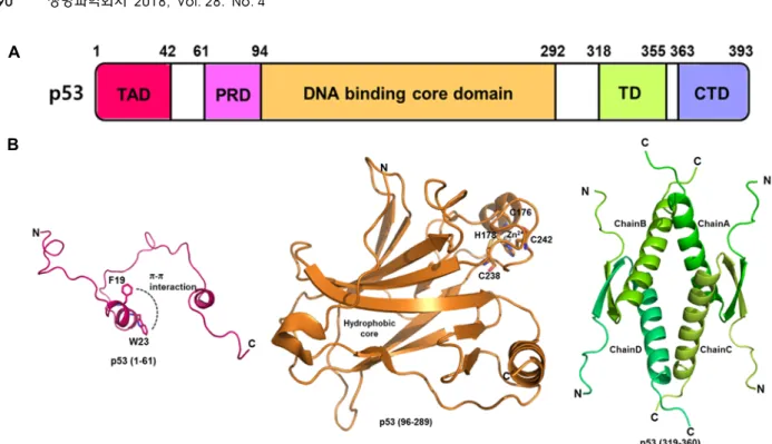
Human p53, a nuclear phosphoprotein of MW of 53 kDa, contains 393 amino acids and is composed of structural and functional main domains: an amino-terminal transactivation domain (TAD1; amino acids 1-40, TAD2; aa 41-61), a pro-
line-rich domain with multiple copies of the PXXP sequence (PRD; aa 61-94, where X is any amino acid), the central DNA-binding domain (DBD; aa 94-292), a tetramerization domain (TD; aa 318-355) and a carboxyl terminal regulatory domain (CTD; aa 363-393) (Fig. 2). N-terminal TAD, which is required for p53 transcriptional activity, interacts with many proteins including CREB-binding protein (CBP)/
p300, p300/CBP-associated factor (PCAF), MDM2, tran- scription factor II D (TFIID), transcription factor IIH (TFIIH), and TATA-binding protein associated factors (TAFs) [27, 48].
These partner proteins overlap with the binding site of TAD.
Few studies of the structure and function of the N-terminal domain of p53 have been conducted, and NMR studies have shown this region was natively unstructured. In 1996, the structure of the transactivation domain peptide of p53 bound to the N-terminal domain of MDM2 was first solved by X-ray crystallography. Amino acids 17-28 of TAD form a random coil to helix that interacts with a hydrophobic cleft in the N-terminal domain of MDM2 [32]. In NMR structural studies of the Taz2/p532-39 complex, the region of p53 has been shown to be highly flexible, forming a short α-helical conformation within amino acids 15-27 of TAD in complex with CBP Taz2 [14]. The TAD1 region corresponds to con- served hydrophobic residues essential for the p53 trans- activation function, while residues 18-25 form an amphi-
A
B
Fig. 2. Overall structure of the p53 gene. (A) Structure of the p53 domain (TAD: Transactivation Domain, PRD: Proline-Rich Domain, TD: Tetramerization Domain, CTD: C-Terminal Domain). (B) Schematic ribbon diagrams of the human p53 TAD domain (PDB ID, 5HOU), p53 DBD domain in the absence of DNA (PDB ID, 2OCJ), and p53 TD domain (PDB ID, 1SAL).
pathic helix when coupled with the partner proteins [26].
Similar to TAD1, the TAD2 region folds into an amphipathic alpha-helix when it binds to replication protein A and TFIIH [1, 10]. Investigations of the helical characteristics of the N-terminal region using molecular dynamics (MD) simu- lations have shown that the region of p53 (aa 17-29) contains a stable helical form and depicts aromatic stacking (π-π inter- action) between Phe19 and Trp23 [13]. This region is consid- ered to have an important influence on disturbing the secon- dary structure of the TAD, and these two amino acids also play important roles in maintenance of the functional and structural stabilities of p53 [35]. The PRD contributes to reg- ulation of the p53 stability, transcription activity and in- duction of transcription independent apoptosis. This domain is required for interactions with the co-repressor mSin3A [6 1], prolyl isomerase Pin1 [59] and p300/CBP [11].
The central DBD, which is known to be characterized by high evolutionary conservation, binds directly to sequence- specific DNA at promoter regions and initiates the tran- scription of target genes. The structure of DBD contains an immunoglobulin-like β-sandwich of two antiparallel β-sheets, which serves as a scaffold for two large loops (L2 and L3) and a loop-sheet-helix motif, forming the DNA binding surface. The loop-sheet-helix motif contains loop L1, a short β-sheet (S2-S2’ hairpin), the C-terminal residues of β-strand
S10, and helix H2 [56]. The two loops (L2 and L3) are struc- turally stabilized by a Zn2+ ion, which is tetrahedrally coor- dinated by Cys176, His179, Cys238, and Cys242. Loss of the Zn2+
ion results in loss of DNA binding specificity because it leads to high structural fluctuations of adjacent loops and in- creases aggregation tendency [12]. Most cancer mutations are found in this area, and several frequent mutations are known as hot-spot mutations. A highly flexible L1 loop with secondary structural disorder contains a few amino acids with a low mutation rate and is known as an area of cold- spot mutations [42]. The DBD is intrinsically unstable and kinetically stable, rendering it susceptible to oncogenic mu- tations [5, 29]. The majority of oncogenic P53 mutations oc- cur in the DBD and result in a loss of DNA binding, thereby affecting p53 function in cell cycle control. Thus, one of the goals of cancer therapies is to stabilize the DBD and reverse the effects of mutations. Many proteins, including simian vi- rus 40 large T antigen (SV40Tag), ASPP1 (P53BP1), ASPP2 (P53BP2), HIF-1α, BCL-XL, BCL2, BAK and MDM2, are known as P53 DBD binding partners [8]. The complex struc- tures of p53 DBD and P53BP1 (PDB ID, 1GZH), p53 DBD and P53BP2 (PDB ID, 1YCS), p53 core dimer bound to DNA (PDB ID, 2GEQ), p53 DBD and sv40 (PDB ID, 2H1L) have been revealed by X-ray crystallography [47].
The TD regulates the oligomer status of p53 and the tet-
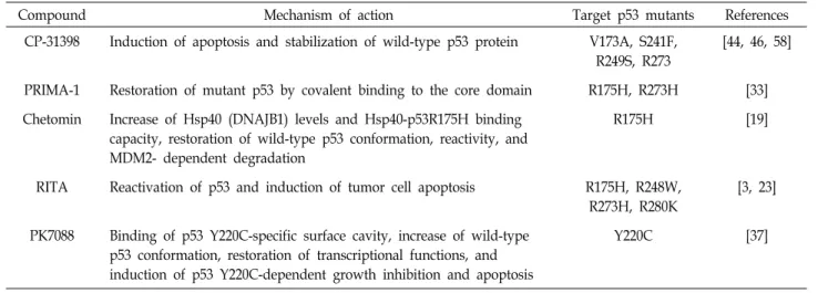
Table 1. Compounds to target mutant p53
Compound Mechanism of action Target p53 mutants References
CP-31398 Induction of apoptosis and stabilization of wild-type p53 protein V173A, S241F, R249S, R273
[44, 46, 58]
PRIMA-1 Restoration of mutant p53 by covalent binding to the core domain R175H, R273H [33]
Chetomin Increase of Hsp40 (DNAJB1) levels and Hsp40-p53R175H binding capacity, restoration of wild-type p53 conformation, reactivity, and MDM2- dependent degradation
R175H [19]
RITA Reactivation of p53 and induction of tumor cell apoptosis R175H, R248W, R273H, R280K
[3, 23]
PK7088 Binding of p53 Y220C-specific surface cavity, increase of wild-type p53 conformation, restoration of transcriptional functions, and induction of p53 Y220C-dependent growth inhibition and apoptosis
Y220C [37]
ramer formation of p53 is essential to DNA binding, post- translational modification, and protein-protein interactions.
The structure of TD has been solved by NMR and X-ray crystallography. Each monomer comprises a short β-strand, a sharp turn (Gly334), and an α-helix. Two monomers form a dimer via an anti-parallel interaction of the β–strands, and two dimers interact as a four-helix bundle to form a tetramer [25]. The CTD is subject to alternative splicing and post-translational modifications. It is known that the CTD affects non-specific DNA binding and transcriptional activity.
Additionally, the CTD is involved in binding to 14-3-3, GSK3 β, hGcn5, PARP-1, S100B (ββ), TAF, TAF1, TRRAP and many other proteins. These p53 domains interact with a vari- ety of partner proteins that create highly complex signaling pathways involving post-translational modifications and these pathways can be used therapeutically.
Therapeutic targets
Many cancer-associated p53 mutants result in the ex- pression of point-mutated p53 proteins that can exert neg- ative effects and acquire novel oncogenic functions [16].
Mutations are clustered within seven hot-spot amino acids (Arg175, Tyr220, Gly245, Arg248, Arg249, Arg273, and Arg282) in the DNA-binding domain of p53. Interestingly, these p53 mutations were found to be associated with new oncogenic functions (mutant p53 gain-of-function, GOF) that include cancer cell survival, invasion, and metastasis [39]. Recent studies have focused on the degradation of mutant p53 or the restoration of wild-type p53 function (Table 1) [60]. One of the mutant specific inhibitors reported, CP-31398, was tar- geted by either formation of wild-type or mutant p53. In
the wild-type human rhabdomyosarcoma (RMS) cell line, CP-31398 increased the expression of p53, downstream tar- get p21, and MDM2. In addition, CP-31398 induced mi- tochondrial translocation of mutant p53, resulting in cyto- chrome c release and ROS-dependent apoptosis [58]. In an- other study, interference with the interaction between mu- tant p53 and other proteins was targeted. The growth sup- pressive and pro-apoptotic activities of p53 require that its function be tightly regulated by the E3 ubiquitin protein li- gase MDM2, which has been shown to be involved in that regulation [15]. Therefore, drug-discovery groups have tar- geted the p53 pathway, with the interaction between p53 and MDM2 being the most attractive target (Table 2).
Attempts to regulate the p53-MDM2 interaction have in- cluded the generation of anti-MDM2 antisense oligonucleo- tides and scaffold-attached peptides [28, 54]. Additionally, the studies have revealed potent and selective small mole- cule inhibitors that block the MDM2–p53 interaction. From a structural standpoint, the p53-MDM2 interaction has been mapped to the N-terminal p53 binding domain (aa 18-101) of MDM2 and the N-terminal transactivation domain 1 (TAD1; aa 1-40) of p53 [7]. The first potent and selective small molecule p53-MDM2 binding inhibitors, the Nutlins (cis-imidazoline analogs), were reported to competitively bind to the p53-binding pockets of MDM2 [50]. Importantly, Nutlin-3α inhibits proliferation and promotes apoptosis in cancer cells in vivo and in vitro [53]. Therefore, these selective small-molecule inhibitors suggest that targeted inhibition of p53-MDM2 interaction in p53-mutant cancers inhibits onco- genic functions.
The p53 is reported to be involved in energy, inflamma- tion, proliferation and oxidative stress mechanisms which
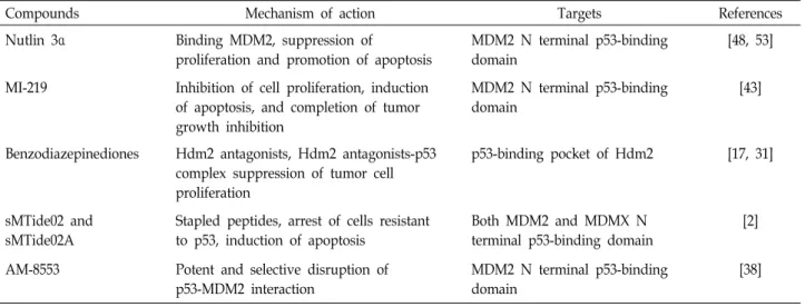
Table 2. p53-MDM2 interaction inhibitors
Compounds Mechanism of action Targets References
Nutlin 3α Binding MDM2, suppression of
proliferation and promotion of apoptosis
MDM2 N terminal p53-binding domain
[48, 53]
MI-219 Inhibition of cell proliferation, induction of apoptosis, and completion of tumor growth inhibition
MDM2 N terminal p53-binding domain
[43]
Benzodiazepinediones Hdm2 antagonists, Hdm2 antagonists-p53 complex suppression of tumor cell proliferation
p53-binding pocket of Hdm2 [17, 31]
sMTide02 and sMTide02A
Stapled peptides, arrest of cells resistant to p53, induction of apoptosis
Both MDM2 and MDMX N terminal p53-binding domain
[2]
AM-8553 Potent and selective disruption of p53-MDM2 interaction
MDM2 N terminal p53-binding domain
[38]
are also linked to tumor progression as well. Many cellular stimuli such as physical, physiological and oxidative stress result in the induction of crosstalk between NF-κB (Nuclear factor kappa B) and the p53 tumor suppressor [57]. In addi- tion, p53 and NF-κB crosstalk participates in regulation of tumor cell metabolism [24]. Therefore, the understanding of these processes could contribute towards the design of new therapy for cancer.
In summary, this review suggests the need for a better understanding of the structural function of p53 and the de- velopment of more effective anticancer therapy to facilitate treatment of various cancers.
Acknowledgment
This work was supported by a 2-Years Research Grant from Pusan National University (2017-2018).
References
1. Bochkareva, E., Kaustov, L., Ayed, A., Yi, G. S., Lu, Y., Pineda-Lucena, A., Liao, J. C., Okorokov, A. L., Milner, J., Arrowsmith, C. H. and Bochkarev, A. 2005. Single-stranded DNA mimicry in the p53 transactivation domain interaction with replication protein A. Proc. Natl. Acad. Sci. USA. 102, 15412-15417.
2. Brown, C. J., Quah, S. T., Jong, J., Goh, A. M., Chiam, P.
C., Khoo, K. H., Choong, M. L., Lee, M. A., Yurlova, L., Zolghadr, K., Joseph, T. L., Verma, C. S. and Lane, D. P.
2013. Stapled peptides with improved potency and specific- ity that activate p53. ACS Chem. Biol. 8, 506-512.
3. Burmakin, M., Shi, Y., Hedstrom, E., Kogner, P. and Seliva- nova, G. 2013. Dual targeting of wild-type and mutant p53 by small molecule RITA results in the inhibition of N-Myc
and key survival oncogenes and kills neuroblastoma cells in vivo and in vitro. Clin. Cancer Res. 19, 5092-5103.
4. Bykov, V. J. N., Eriksson, S. E., Bianchi, J. and Wiman, K.
G. 2018. Targeting mutant p53 for efficient cancer therapy.
Nat. Rev. Cancer 18, 89-102.
5. Canadillas, J. M. P., Tidow, H., Freund, S. M. V., Rutherford, T. J., Ang, H. C. and Fersht, A. R. 2006. Solution structure of p53 core domain: Structural basis for its instability. Proc.
Natl. Acad. Sci. USA. 103, 2109-2114.
6. Chi, S. W. 2014. Structural insights into the transcription- independent apoptotic pathway of p53. BMB Rep. 47, 167- 172.
7. Chi, S. W., Lee, S. H., Kim, D. H., Ahn, M. J., Kim, J. S., Woo, J. Y., Torizawa, T., Kainosho, M. and Han, K. H. 2005.
Structural details on mdm2-p53 interaction. J. Biol. Chem.
280, 38795-38802.
8. Collavin, L., Lunardi, A. and Del, Sal G. 2010. p53-family proteins and their regulators: hubs and spokes in tumor suppression. Cell Death Differ. 17, 901-911.
9. Demir, O., Ieong, P. U. and Amaro, R. E. 2017. Full-length p53 tetramer bound to DNA and its quaternary dynamics.
Oncogene 36, 1451-1460.
10. Di Lello, P., Jenkins, L. M. M., Jones, T. N., Nguyen, B. D., Hara, T., Yamaguchi, H., Dikeakos, J. D., Appella, E., Legault, P. and Omichinski, J. G. 2006. Structure of the Tfb1/p53 complex: Insights into the interaction between the p62/Tfb1 subunit of TFIIH and the activation domain of p53. Mol. Cell 22, 731-740.
11. Dornan, D., Shimizu, H., Burch, L., Smith, A. J. and Hupp, T. R. 2003. The proline repeat domain of p53 binds directly to the transcriptional coactivator p300 and allosterically con- trols DNA-dependent acetylation of p53. Mol. Cell. Biol. 23, 8846-8861.
12. Duan, J. X. and Nilsson, L. 2006. Effect of Zn2+ on DNA recognition and stability of the p53 DNA-binding domain.
Biochemistry 45, 7483-7492.
13. Espinoza-Fonseca, L. M. and Trujillo-Ferrara, J. G. 2006.
Transient stability of the helical pattern of region F19-L22
of the N-terminal domain of p53: a molecular dynamics sim- ulation study. Biochem. Biophys. Res. Commun. 343, 110-116.
14. Feng, H. Q., Jenkins, L. M. M., Durell, S. R., Hayashi, R., Mazur, S. J., Cherry, S., Tropea, J. E., Miller, M., Wlodawer, A., Appella, E. and Bai, Y. 2009. Structural basis for p300 Taz2-p53 TAD1 binding and modulation by phosphorylation.
Structure 17, 202-210.
15. Fotouhi, N. and Graves, B. 2005. Small molecule inhibitors of p53/MDM2 interaction. Curr. Top. Med. Chem. 5, 159-165.
16. Freed-Pastor, W. A. and Prives, C. 2012. Mutant p53: one name, many proteins. Genes Dev. 26, 1268-1286.
17. Grasberger, B. L., Lu, T., Schubert, C., Parks, D. J., Carver, T. E., Koblish, H. K., Cummings, M. D., LaFrance, L. V., Milkiewicz, K. L., Calvo, R. R., Maguire, D., Lattanze, J., Franks, C. F., Zhao, S., Ramachandren, K., Bylebyl, G. R., Zhang, M., Manthey, C. L., Petrella, E. C., Pantoliano, M. W., Deckman, I. C., Spurlino, J. C., Maroney, A. C., Tomczuk, B. E., Molloy, C. J. and Bone, R. F. 2005. Discovery and coc- rystal structure of benzodiazepinedione HDM2 antagonists that activate p53 in cells. J. Med. Chem. 48, 909-912.
18. Halazonetis, T. D., Gorgoulis, V. G. and Bartek, J. 2008. An oncogene-induced DNA damage model for cancer develop- ment. Science 319, 1352-1355.
19. Hiraki, M., Hwang, S. Y., Cao, S., Ramadhar, T. R., Byun, S., Yoon, K. W., Lee, J. H., Chu, K., Gurkar, A. U., Kolev, V., Zhang, J., Namba, T., Murphy, M. E., Newman, D. J., Mandinova, A., Clardy, J. and Lee, S. W. 2015. Small- mole- cule reactivation of mutant p53 to wild-type-like p53 through the p53-Hsp40 regulatory axis. Chem. Biol. 22, 1206-1216.
20. Ho, W. C., Luo, C., Zhao, K., Chai, X., Fitzgerald, M. X. and Marmorstein, R. 2006. High-resolution structure of the p53 core domain: implications for binding small-molecule stabi- lizing compounds. Acta Crystallogr. D Biol. Crystallogr. 62, 1484-1493.
21. Hollstein, M., Sidransky, D., Vogelstein, B. and Harris, C.
C. 1991. p53 mutations in human cancers. Science 253, 49-53.
22. Huang, L., Yan, Z., Liao, X. D., Li, Y., Yang, J., Wang, Z.
G., Zuo, Y., Kawai, H., Shadfan, M., Ganapathy, S. and Yuan, Z. M. 2011. The p53 inhibitors MDM2/MDMX com- plex is required for control of p53 activity in vivo. Proc. Natl.
Acad. Sci. USA. 108, 12001-12006.
23. Issaeva, N., Bozko, P., Enge, M., Protopopova, M., Verhoef, L. G., Masucci, M., Pramanik, A. and Selivanova, G. 2004.
Small molecule RITA binds to p53, blocks p53-HDM-2 inter- action and activates p53 function in tumors. Nat. Med. 10, 1321-1328.
24. Johnson, R. F. and Perkins, N. D. 2012. Nuclear factor-κB, p53, and mitochondria: regulation of cellular metabolism and the Warburg effect. Trends Biochem. Sci. 37, 317-324 25. Jeffrey, P. D., Gorina, S. and Pavletich, N. P. 1995. Crystal
structure of the tetramerization domain of the p53 tumor suppressor at 1.7 angstroms. Science 267, 1498-1502.
26. Joerger, A. C. and Fersht, A. R. 2010. The tumor suppressor p53: from structures to drug discovery. Cold Spring Harb.
Perspect. Biol. 2, a000919.
27. Kamada, R., Toguchi, Y., Nomura, T., Imagawa, T. and Sakaguchi, K. 2016. Tetramer formation of tumor suppressor protein p53: Structure, function, and applications. Biopol- ymers 106, 598-612.
28. Karlsson, G. B., Jensen, A., Stevenson, L. F., Woods, Y. L., Lane, D. P. and Sorensen, M. S. 2004. Activation of p53 by scaffold-stabilised expression of Mdm2-binding peptides:
visualisation of reporter gene induction at the single-cell level. Br. J. Cancer 91, 1488-1494.
29. Khoo, K. H., Andreeva, A. and Fersht, A. R. 2009. Adaptive evolution of p53 thermodynamic stability. J. Mol. Biol. 393, 161-175.
30. Khoo, K. H., Verma, C. S. and Lane, D. P. 2014. Drugging the p53 pathway: understanding the route to clinical efficacy.
Nat. Rev. Drug Discov. 13, 217-236.
31. Koblish, H. K, Zhao, S., Franks, C. F., Donatelli, R. R., Tominovich, R. M., LaFrance, L. V., Leonard, K. A., Gushue, J. M., Parks, D. J., Calvo, R. R., Milkiewicz, K. L., Marugan, J. J., Raboisson, P., Cummings, M. D., Grasberger, B. L., Johnson, D. L., Lu, T., Molloy, C. J. and Maroney, A. C.
2006. Benzodiazepinedione inhibitors of the Hdm2:p53 com- plex suppress human tumor cell proliferation in vitro and sensitize tumors to doxorubicin in vivo. Mol. Cancer Ther.
5, 160-169.
32. Kussie, P. H., Gorina, S., Marechal, V., Elenbaas, B., Moreau, J., Levine, A. J. and Pavletich, N. P. 1996. Structure of the MDM2 oncoprotein bound to the p53 tumor suppressor transactivation domain. Science 274, 948-953.
33. Lambert, J. M., Gorzov, P., Veprintsev, D. B., Soderqvist, M., Segerback, D., Bergman, J., Fersht, A. R., Hainaut, P., Wiman, K. G. and Bykov, V. J. 2009. PRIMA-1 reactivates mutant p53 by covalent binding to the core domain. Cancer Cell 15, 376-388.
34. Lane, D. P. and Verma, C. 2012. Mdm2 in evolution. Genes Cancer 3, 320-324.
35. Lee, H., Mok, K. H., Muhandiram, R., Park, K. H., Suk, J.
E., Kim, D. H., Chang, J., Sung, Y. C., Choi, K. Y. and Han, K. H. 2000. Local structural elements in the mostly un- structured transcriptional activation domain of human p53.
J. Biol. Chem. 275, 29426-29432.
36. Leroy, B., Anderson, M. and Soussi, T. 2014. TP53 mutations in human cancer: database reassessment and prospects for the next decade. Hum. Mutat. 35, 672-688.
37. Liu, X., Wilcken, R., Joerger, A. C., Chuckowree, I. S., Amin, J., Spencer, J. and Fersht, A. R. 2013. Small molecule induced reactivation of mutant p53 in cancer cells. Nucleic Acids Res.
41, 6034-6044.
38. Lucas, B. S., Fisher, B., McGee, L. R., Olson, S. H., Medina, J. C. and Cheung, E. 2012. An expeditious synthesis of the MDM2-p53 inhibitor AM-8553. J. Am. Chem. Soc. 134, 12855- 12860.
39. Muller, P. A., Vousden, K. H. and Norman, J. C. 2011. p53 and its mutants in tumor cell migration and invasion. J. Cell Biol. 192, 209-218.
40. Olivier, M., Hollstein, M. and Hainaut, P. 2010. TP53 muta- tions in human cancers: origins, consequences, and clinical
use. Cold Spring Harb. Perspect. Biol. 2, a001008.
41. Oren, M. and Rotter, V. 1999. Introduction: p53 - the first twenty years. Cell. Mol. Life Sci. 55, 9-11.
42. Saha, T., Kar, R. K. and Sa, G. 2015. Structural and sequen- tial context of p53: A review of experimental and theoretical evidence. Prog. Biophys. Mol. Biol. 117, 250-263.
43. Shangary, S., Qin, D., McEachern, D., Liu, M., Miller, R. S., Qiu, S., Nikolovska-Coleska, Z., Ding, K., Wang, G., Chen, J., Bernard, D., Zhang, J., Lu, Y., Gu, Q., Shah, R. B., Pienta, K. J., Ling, X., Kang, S., Guo, M., Sun, Y., Yang, D. and Wang, S. 2008. Temporal activation of p53 by a specific MDM2 inhibitor is selectively toxic to tumors and leads to complete tumor growth inhibition. Proc. Natl. Acad. Sci.
USA. 105, 3933-3938.
44. Takimoto, R., Wang, W., Dicker, D. T., Rastinejad, F., Lyssikatos, J. and el-Deiry, W. S. 2002. The mutant p53-con- formation modifying drug, CP-31398, can induce apoptosis of human cancer cells and can stabilize wild-type p53 protein. Cancer Biol. Ther. 1, 47-55.
45. Tan, B. X., Liew, H. P., Chua, J. S., Ghadessy, F. J., Tan, Y. S., Lane, D. P. and Coffill, C. R. 2017. Anatomy of Mdm2 and Mdm4 in evolution. J. Mol. Cell Biol. 9, 3-15.
46. Tang, X., Zhu, Y., Han, L., Kim, A. L., Kopelovich, L., Bickers, D. R. and Athar, M. 2007. CP-31398 restores mutant p53 tumor suppressor function and inhibits UVB-induced skin carcinogenesis in mice. J. Clin. Invest. 117, 3753-3764.
47. Tuncbag, N., Kar, G., Gursoy, A., Keskin, O. and Nussinov, R. 2009. Towards inferring time dimensionality in pro- tein-protein interaction networks by integrating structures:
the p53 example. Mol. Biosyst. 5, 1770-1778.
48. Uversky, V. N. 2016. p53 Proteoforms and intrinsic disorder:
an illustration of the protein structure-function continuum concept. Int. J. Mol. Sci. 17, 1874.
49. Valente, L. J., Gray, D. H., Michalak, E. M., Pinon-Hofbauer, J., Egle, A., Scott, C. L., Janic, A. and Strasser, A. 2013. p53 efficiently suppresses tumor development in the complete absence of its cell-cycle inhibitory and proapoptotic effectors p21, Puma, and Noxa. Cell Rep. 3, 1339-1345.
50. Vassilev, L. T., Vu, B. T., Graves, B., Carvajal, D., Podlaski, F., Filipovic, Z., Kong, N., Kammlott, U., Lukacs, C., Klein, C., Fotouhi, N. and Liu, E. A. 2004. In vivo activation of the p53 pathway by small-molecule antagonists of MDM2.
Science 303, 844-848.
51. Vogelstein, B., Lane, D. and Levine, A. J. 2000. Surfing the p53 network. Nature 408, 307-310.
52. Wallentine, B. D., Wang, Y., Tretyachenko-Ladokhina, V., Tan, M., Senear, D. F. and Luecke, H. 2013. Structures of oncogenic, suppressor and rescued p53 core-domain var- iants: mechanisms of mutant p53 rescue. Acta Crystallogr.
D Biol. Crystallogr. 69, 2146-2156.
53. Wang, B., Fang, L., Zhao, H., Xiang, T. and Wang, D. 2012.
MDM2 inhibitor Nutlin-3a suppresses proliferation and pro- motes apoptosis in osteosarcoma cells. Acta Biochim. Biophys.
Sin. (Shanghai). 44, 685-691.
54. Wang, H., Nan, L., Yu, D., Agrawal, S. and Zhang, R. 2001.
Antisense anti-MDM2 oligonucleotides as a novel ther- apeutic approach to human breast cancer: in vitro and in vivo activities and mechanisms. Clin. Cancer Res. 7, 3613- 3624.
55. Wang, S., Zhao, Y., Aguilar, A., Bernard, D. and Yang, C.
Y. 2017. Targeting the MDM2-p53 protein-protein interaction for new cancer therapy: progress and challenges. Cold Spring Harb. Perspect. Med. 7, a026245.
56. Wang, Y., Rosengarth, A. and Luecke, H. 2007. Structure of the human p53 core domain in the absence of DNA. Acta Crystallogr. D Biol. Crystallogr. 63, 276-281.
57. Webster, G. A. and Perkins, N. D. 1999. Transcriptional Cross Talk between NF-κB and p53. Mol. Cell. Biol. 19, 3485-3495.
58. Xu, J., Timares, L., Heilpern, C., Weng, Z., Li, C., Xu, H., Pressey, J. G., Elmets, C. A., Kopelovich, L. and Athar, M.
2010. Targeting wild-type and mutant p53 with small mole- cule CP-31398 blocks the growth of rhabdomyosarcoma by inducing reactive oxygen species-dependent apoptosis. Can- cer Res. 70, 6566-6576.
59. Zacchi, P., Gostissa, M., Uchida, T., Salvagno, C., Avolio, F., Volinia, S., Ronai, Z., Blandino, G., Schneider, C. and Del Sal, G. 2002. The prolyl isomerase Pin1 reveals a mechanism to control p53 functions after genotoxic insults. Nature 419, 853-857.
60. Zhao, D., Tahaney, W. M., Mazumdar, A., Savage, M. I. and Brown, P. H. 2017. Molecularly targeted therapies for p53- mutant cancers. Cell. Mol. Life Sci. 74, 4171-4187.
61. Zilfou, J. T. Hoffman, W. H., Sank, M., George, D. L. and Murphy, M. 2001. The corepressor mSin3a interacts with the proline-rich domain of p53 and protects p53 from protea- some-mediated degradation. Mol. Cell. Biol. 21, 3974-3985.
초록:암 치료 표적으로서 p53의 구조적 및 기능적 역할
한창우†․박소영†․정미숙․장세복*
(부산대학교 자연과학대학 분자생물학과)
p53 유전자는 스트레스, DNA 손상, 저산소증 및 종양 발생에 대한 세포 반응의 전사 조절에서 중요한 역할을 담당한다. 최근에 발견된 다양한 종류의 p53의 생리 활성을 생각한다면 p53이 암 조절에 관여한다는 것은 놀랄 만한 일이 아니다. 인간 암의 약 50%에는 p53 유전자의 돌연변이 또는 p53을 활성화시키는 기전의 결함을 통해 p53 단백질 기능의 불활성화가 나타난다. p53 기능의 이러한 장애는 p53 의존 반응으로부터 회피를 허용함으로써 종양의 진화에 결정적인 역할을 하게 된다. 최근의 많은 연구들은 p53의 돌연변이를 대폭 감소시키거나 p53의 종양 억제 기능을 복원하기 위하여 선택적인 저분자 화합물을 동정함으로써 p53의 돌연변이를 직접 표적하는 것 에 초점을 두고 있다. 이들 저분자는 좋은 약물과 유사한 특성을 유지하면서 다양한 상호작용을 효과적으로 조절 해야 한다. 이 중, p53의 음성조절인자 핵심인 MDM2의 발견은 p53과 MDM2 간의 상호작용을 차단하는 새로운 저분자 억제제의 설계를 제공하였다. 저분자 화합물 중 일부는 개념 증명 연구에서 임상 시험으로 옮겨졌으며 향후 맞춤형 항암제가 추가될 전망이다. 본 리뷰에서는 야생형 p53과 돌연변이 p53의 구조적 및 기능적 중요성과 p53을 직접 표적하는 치료제 개발, p53과 MDM2 간의 상호작용을 억제하는 화합물에 대하여 검토하였다.