Bone Metabolism and Estrogenic Effect of Phytochemicals
Bokyung Kim and Mihyang Kim*
Department of Food and Nutrition, Silla University, Busan 617-736, Korea Received July 13, 2018 /Revised July 24, 2018 /Accepted July 25, 2018
Osteoporosis is a disease that increases the risk of fracture by decreasing the mass and strength of bone. It is caused by imbalance of osteoclast bone formation and osteoclast bone resorption. Bone for- mation by osteoblast is activated via bone morphogenetic proteins and runt-related transcription factor 2. Wnt/β-catenin signaling and bone resorption by osteoclast are initiated by the binding of receptor activator of nuclear factor-κB ligand and receptor activator of nuclear factor-κB. Menopausal women are at risk for many diseases due to hormonal imbalances, and osteoporosis is the most common met- abolic disorder in 30% of postmenopausal women. When estrogen is deficient, bone resorption of os- teoclasts is promoted, and the risk of osteoporosis especially increases in postmenopausal women.
Hormone replacement therapy has been widely used to relieve or treat the symptoms of menopausal syndrome. However, long-term administration of hormone therapy has been associated with a high risk of side effects, such as breast cancer, ovarian cancer, and uterine cancer. Recently, phytochemicals have been actively studied as a phytoestrogen, which has an estrogen-like activity to cope with symp- toms of menopausal syndrome. Therefore, in this review, we investigated the differentiation mecha- nism of osteoblast and osteoclast and the role of estrogen and phytoestrogen in bone metabolism in relation to previous studies.
Key words : Bone metabolism, differentiation, estrogenic effect, osteoblast, osteoclast
*Corresponding author
*Tel : +82-51-999-5620, Fax : +82-51-999-6957
*E-mail : [email protected]
This is an Open-Access article distributed under the terms of the Creative Commons Attribution Non-Commercial License (http://creativecommons.org/licenses/by-nc/3.0) which permits unrestricted non-commercial use, distribution, and reproduction in any medium, provided the original work is properly cited.
Journal of Life Science 2018 Vol. 28. No. 7. 874~883 DOI : https://doi.org/10.5352/JLS.2018.28.7.874
서 론
인구고령화 현상은 소득수준의 증가 및 의료기술의 발전에 따른 현상으로 평균수명이 증가됨에 따라 노년기에서 다양한 건강문제가 발생하고 있어 이에 따른 대처가 필요한 실정이다 [34]. 우리나라 여성의 평균 수명은 약 85.48세(통계청, 2014)이 며, 평균 폐경 연령은 49.7세(통계청, 2010)로 일생의 1/3 이상 을 폐경 상태로 지내게 됨에 따라 폐경 이후의 여성의 삶은 매우 중요하다. 폐경의 주된 원인으로는 여성의 생리주기에 직접적인 영향을 주는 estrogen 분비 감소에 의한 것으로 알려 져 있으며, 이러한 것은 정상적인 노화과정에서 나타나는 현 상으로 다양한 신체적, 정신적 증상을 동반하게 된다[77]. 대표 적인 폐경 증상으로는 무월경, 홍조, 수면장애, 심혈관계질환 및 골다공증 등이 있으며, 한 사례연구에서는 자연 폐경을 한 여성 중 89%가 폐경과 연관된 증상을 적어도 하나 이상 경험 한 것으로 나타났다[8]. 또한, 대부분의 여성은 증상의 완화가 필요하다고 생각하였으며, 이와 관련하여 폐경 증상의 정도에 따라 폐경기 여성의 삶의 질에 영향을 준다는 보고도 있다[12,
33]. 이와 같이 폐경기 여성은 호르몬 불균형과 체내 산화적 스트레스 증가로 여러 질병의 위험에 처해 있으며, 폐경기 여 성의 약 30%에서 관찰되는 골다공증은 폐경기 여성에게서 발 생되는 가장 흔한 대사성 질환이기도 하다[46, 50]. 또한 폐경 기 이후 estrogen의 결핍 때문에 골다공증이 급격히 증가하고, 전 세계적으로 여성의 약 30~40%가 평생 동안 한번 이상의 골절 경험을 하는 것으로 알려져 있다[9].
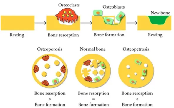
골은 조골전구제포(osteoprogenitor cell), 조골세포(osteo- blast) 골세포(osteocyte), 파골세포(osteoblasr) 등 다양한 종류 의 세포들로 구성되어 있으며, 이러한 여려 세포의 작용에 의 해 골량의 생성과 흡수가 이루어지고 있다[59]. 골 재형성 과정 (bone remodeling cycle)은 골의 구조 유지에 필수적이며, 주 기적으로 손상되거나 오래된 뼈는 흡수되고 그 자리에 새로운 뼈가 신생되는 과정으로 골 재흡수(bone response)와 골 형성 (bone formation)을 반복하는 것에 의해 정상적인 골 조직이 유지된다[47]. 골 흡수율이 증가하면 골 형성율도 따라서 증가 되어야 하나 균형이 이루어지지 않을 경우 골질량(bone mass) 또는 골밀도(bone density) 감소와 골격기능 손상 등으로 인하 여 골다공증(osteoporosis) 발병에 의해 골절이 쉽게 유발될 수 있다[16](Fig. 1). 골다공증의 임상적인 중요성은 골절과 그 결과로 초래되는 합병증으로 인하여 이환률과 사망률을 야기 시키며 더 나아가 경제적인 측면에 있어서 재활 치료비용을 증가시킬 뿐만 아니라, 골다공증 예방 및 치료법은 대부분 골 흡수를 억제하는 작용을 할 뿐 진행된 골 소실을 완전히 회복 할 수 없다[49].
- Review -
Fig. 1. The schematic outlines of the bone remodeling cycle and the balances of bone resorption and bone formation.
호르몬대체요법(hormone replacement therapy, HRT)은 폐경기 증후군의 증상을 경감시키거나 치료하기 위해 널리 사용되어 왔으나[6, 19], 호르몬 치료를 장기간 실시할 경우 유방암, 난소암, 자궁암 등의 부작용 위험성이 매우 높은 것으 로 알려져 있다[4-7]. 따라서 최근 들어 여러 부작용을 보완하 기 위해 폐경기 증후군 증상에 대처할 수 있는 estrogen과 유 사한 활성을 지닌 식물성 estrogen인 phytoestrogen에 대한 연구가 활발히 진행되고 있다[6, 48]. 약용식물 및 한약재의 약용부위 물질들의 생리활성 효능을 이용한 건강 증진용 식품 이나 건강기능성 식품 소재로 개발함은 자원의 효율적인 이용 이라는 측면과 식품 신소재 개발의 측면에서 매우 의미 있는 일이라 사료된다. 그러므로 본 총설에서는 폐경기 증후군 증 상 중 골 대사와 관련하여 골의 형성 및 흡수 경로에 대한 고찰과 더불어 골 대사에 효능을 가지는 천연소재와 식물성 estrogen에 초점을 맞추어 이에 대한 다양한 연구결과를 소개 하고자 한다.
조골세포의 분화 과정과 phytoestrogen의 효과
골 형성에 있어 조골세포의 분화는 여러 용해성 단백질에 의해 조절되며 초기 증식단계(4~10일)에서 골 형성 및 성숙단 계(10~16일)를 거쳐 석회화(무기질화)의 최종 단계(16일~30 일)로 순차적으로 진행된다[4, 70]. 초기 증식단계의 대표적인 바이오마커인 alkaline phosphatase (ALP)는 뼈 및 신장 등의 거의 모든 조직에서 발견되는 염기성 인산분해 효소로 골 성
장이 활발하게 일어날 때 그 활성이 높아져 초기 분화지표로 주로 사용된다[23]. ALP는 세포 외 β-glycerol phosphate를 가수분해하고 국소적으로 PO4-2 농도를 증가시켜 후기 석회화 의 촉발제 역할을 한다[79]. 이외에도 조골세포의 초기분화에 관여하는 인자로는 runt-related transcription factor 2 (Runx2) 가 있는데, RUNX2는 runt domain 유전자의 종류로 다분화능 세포 및 전조골세포에서 ALP, osteocalcin 및 bone sialopro- tein과 같은 유전자를 조절하여 조골세포 분화를 촉진하는 역 할을 하며, inhibitors of DNA binding/ differentiation (ID1) 및 distal-less homeobox 5 (Dlx5) 또한 조골세포의 초기 분화 에 관여하는 것으로 알려져 있다[20, 38, 42, 56](Fig. 2).
Bone morphogenetic proteins (BMPs)는 조골세포의 성장 및 분화에 관여하는 인자로 BMP sub family로는 BMP2, RUNX2, osterix 등이 있으며, BMP2는 BMP수용체와 결합하 여 Smad1/5/8를 인산화시켜 활성화하는 인자로 Smad4와 결 합된 복합체는 전사활성 인자로 작용하여 5Dlx5의 발현을 촉 진한다. Dlx5는 조골세포 분화의 핵심 전사 인자로 작용하는 Runx2의 발현을 유도하며, 이때 유도된 Runx2는 osteocalcin (OCN), osteopontin (OPN), collagen type Ⅰ과 같은 조골세포 분화 마커 유전자의 프로모터에 결합하여 이러한 유전자들의 발현을 증가시킴으로써 조골세포의 분화를 촉진한다[53, 68].
조골세포 분화의 중기단계에 해당하는 골 형성 및 성숙기 (10일~16일)에는 collagen type I의 발현 및 생합성이 이루어 지고, fibronectin, TGF-β1 및 osteonectin의 발현 또한 높아진 다. 성숙기의 collagen type I과 TGF-β1는 최종 단계인 석회화
Fig. 2. Runx2 and Wnt/β-catenin pathway for bone remodeling regulation.
기간에서보다 높은 함량을 나타내며, fibronectin과 osteo- nectin은 성숙기 및 석회화 기간에서 초기 증식단계보다 높은 발현을 나타낸다는 보고가 있다[13]. 석회화 형성은 칼슘 흡착 력이 높은 식물성 염료인 alizarin을 처리하여 염색 정도에 따 라 판단할 수 있으며, 이러한 원리를 이용하여 조골세포의 결 절 정도를 알 수 있다[10]. OCN은 석회화 단계(16~30일)에서 발현이 높아지는 것으로 알려져 있는데[11], 무기질인 수산화 인회석(hydroyapatite)과 결합하는 골 및 치아에 발견되는 특 이적 단백질로 조골세포에서 생산되어 골의 세포 외 기질에 축적되며, 새로 합성된 OCN의 약 30%는 혈중으로 배출되므 로 혈액 중의 함량 분석을 통해 무기화 정도를 확인할 수 있는 지표로써 분화 28일까지 증가된다는 보고가 있다[31]. 특히, 조골세포의 후기 분화과정인 석회화는 앞서 기술한 것처럼 조골세포의 초기 증식단계의 지표가 되는 ALP 활성과도 관련 이 깊은데, ALP는 석회화 과정에서 기능이 정확히 규명되지 는 않았으나 organic phosphatate를 가수분해하고 국소적으 로 PO4의 농도를 증가시켜 석회화를 유도한다는 보고가 있다 [61, 74].
조골세포 분화에 관여하는 인자인 ALP, OPN 및 OCN의 mRNA 발현에 영향을 미치는 물질에 대한 연구로는 관절염 등 항염증 치료제로 사용되는 천수근의 뿌리에서 분리한 har- pagide를 이용한 사례가 있다[14]. 그 결과에 따르면 harpa- gide는 조골세포주인 MC3T3-E1 cell의 분화에 관여하는 인자
인 ALP, OPN 및 OCN의 mRNA 발현을 증가시키는 것으로 보고되고 있다. 난소절제 후 estrogen 결핍을 유도한 실험동물 을 이용한 연구에서도 harpagide 투여는 bone morphometric parameters including trabecular bone volume (BV/ TV), tra- becular thickness (Tb.Th), trabecular number (Tb.N), tra- becular separation (Tb.Sp), structure model index (SMI) 및 bone mineral density (BMD)에 있어서 대조군에 비해 유의적 인 증가를 나타내었으며, 3D-mCT 촬영결과에서도 harpagide 를 투여한 군에서 골 감소가 억제되었음을 알 수 있다[8]. 미나 리과에 속하는 벌사상자(Cnidium monnieri)에서 분리된 천연 쿠마린 성분인 osthole을 조골세포에 처리한 연구에서도 MC3T3-E1의 ALP 활성이 증가되었을 뿐 아니라 분화를 유도 하는 인자인 ALP, collagen I, RUNX2 및 OCN의 mRNA 발현 량이 증가되었다는 결과가 있다[84]. 또한, 강향(Dalbergia odorifera)에서 발견되는 2,4,5-trimethoxyldalbergiquinol도 앞 선 연구결과와 유사하게 ALP 활성을 증가시키며 석회화 염색 실험을 통해 석회화 정도가 증가된 것으로 나타났으며, 이는 조골세포의 분화를 유도한 결과로 이러한 분화는 BMP/Smad 경로의 조절에 의한 결과라는 연구보고가 있다[81]. 이외에도 조골세포 분화와 관련된 바이오마커 활성에 미치는 천연물에 대한 연구로는 미역취(Solidago virga - aurea var. gigantea Miq.) 뿌리 추출물[61], 마가목 열매에서 추출한 cryptochlorogenic Acid[39], 유향추출물[25, 38], 홍화 및 홍화씨 추출물[27], 감국
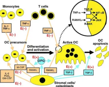
Fig. 3. Molecular mechanism of osteoclast differentiation and activation in- volving the RANKL/RANK/OPG system.
추출물[82], 복분자 추출물[45], 머위 및 여주 추출물[32], 톳 분획물[58] 등이 있으며, 조골세포의 증식단계, 골 형성 및 성 숙단계 및 석회화(무기질화)의 최종 단계에 이르기까지 식물 성 유래 물질에 의해 분화가 촉진되는 사례는 다수 연구되어 있다.
한편, Wnt 신호전달과정에 관여하는 LRP5/6 및 β-catenin 등의 인자들이 조골 세포의 분화 및 골 형성에 관여한다는 보고가 있으며, BMP 경로의 활성화는 Wnt/β-catenin 경로를 활성화시켜 상호적 신호전달 과정에 의해 조골세포의 분화 가 조절되는 것으로 알려져 있다[40, 83]. BMP 및 Wnt 경로는 복합적인 메커니즘을 가지는데 sclerostin, CtGF, cerberus 및 sFRP와 같은 일부 인자는 BMP, Wnt 경로의 리간드, antago- nists 또는 수용체에 결합하는 것으로 알려져 있으며, 세포내 의 Smads는 disheveled-1, Axin, GSK3 및 β-catenin과 같은 Wnt 신호 분자와 복합체를 형성하며, 이러한 복합체는 Smads 와 β-catenin의 인산화와 활성을 조절한다[15, 17, 18, 21, 55, 72]. BMP 및 Wnt 경로에서 Smad, Tcf / Lef response ele- ments (TREs)와 같은 표적 유전자의 전사 조절과정은 매우 중요하며, 특히, Smads는 β-catenin/Tcf/Lef와 전사 복합체를 형성 할 수 있고 BMP와 Wnt 신호 전달에 반응하여 이들 결합 요소를 통해 많은 표적 유전자의 전사를 활성화시킬 수 있는 역할을 한다[11, 26, 28, 43, 51, 66, 83].
Herba siegesbeckia에 함유된 kirenol을 이용한 연구에서 kir- enol은 ALP, collagen type Ⅰ, OPN, BMP2, RUNX2, osterix 와 같은 조골세포 분화 표지인자의 mRNA 수준을 증가시켜 골 형성을 촉진하였으며, 이러한 효과는 Wnt/β-catenin 경로 의 주요 조절인자인 LRP5, DVL2, β-catenin, CCND1, p-GSK3 β의 발현 수준 증가에 의한 것으로 보고되었다[38]. 또한, 동 연구에서 β-catenin을 knock down 시킨 조골세포의 CCND1, ALP, collagen type Ⅰ의 발현이 감소된 결과로부터 Wnt/β-
catenin 경로를 통해 골 형성이 조절된다는 것을 알 수 있다 [40]. 또한, phytoestrogen으로 알려진 대두 이소플라본을 이 용한 연구에서도 조골세포의 ALP 활성 및 Wnt3a, Wnt7b, β -catenin의 유전자 발현 수준이 증가된 것으로 보아 Wnt/β- catenin 경로를 매개하여 조골세포의 분화가 촉진된 것으로 나타났다[80]. 최근 Wnt/ β-catenin 활성화에 의한 조골세포 의 분화 효과는 estradiol을 이용한 연구에서 ERβ/GSK-3β-를 매개한 Wnt/β-catenin 신호 전달 경로를 통해 관련 유전자 발현을 유도함으로써 증식 및 분화를 촉진한다는 연구 결과가 있다[78]. 이외에도 geraniin [54], 구아바(guava) 추출물[63], 은행잎(Ginkgo biloba) 추출물[24] 및 음양각(Epimedium pu- bescens)의 플라보노이드 성분인 icariin 등을 이용한 연구가 있다.
파골세포의 분화 과정과 phytoestrogen의 효과
파골세포는 조골세포보다 세포 크기가 크고 골수 mono- cyte-macrophage 계통의 조혈모세포로부터 유래하며 핵의 전 구세포가 다핵성 거대 세포화되면서 세포로써 기능을 담당한 다. 파골세포 분화는 조골세포에서 발현되는 tumor necrosis factor (TNF) 계열의 cytokine인 RANKL (receptor activator of nuclear factor-κB ligand)과 파골전구세포에서 발현되는 RANK (receptor activator of nuclear factor-κB)의 결합으로 신호전달체계가 활성화되어 시작된다[3, 37, 65](Fig. 3). 이때 조골세포에서 분비되는 다양한 국소인자인 macrophage col- ony-stimulating factor (M-CSF), tumor necrosis factor (TNF- α), IL-1, IL-2은 파골세포의 분화 및 분화 조절에 관여하게 된 다[62]. 활성화된 파골세포는 세포막과 골기질 사이에 수많은 actin ring을 형성하고, 단백질 분해효소와 proton을 분비하며, 골기질을 흡수하여 뼈의 밀도를 감소시킨다[44, 57]. 파골세포 분화에는 골수의 mesenchymal cells (MSC)에 의해 만들어지
는 M-CSF와 RANKL이 가장 중요한 역할을 하는데, 파골세포 전구체에서 M-CSF와 그 수용체가 결합함에 따라 파골세포의 생존 및 분화에 관여하는 신호전달이 활성화되며, tumor ne- crosis factor super-family인 RANKL은 가용성 막과 결합한 형태로 존재하며 파골세포의 분화 및 전구체의 융합에 중요한 역할을 한다. 즉, M-CSF는주로 파골 세포 전구 세포의 생존과 증식에 기여하는 반면, RANKL은 수용체(RANK)를 통한 세 포간의 융합을 포함한 역동적인 분화 과정을 가능하게 한다 [22]. 파골세포를 생성하는 경로에는 이외에도 MAPKs 및 NF-κB가 활성화가 관여하는데, 활성화된 T세포의 nuclear factor인 NFATc1에 의해 파골세포의 분화가 가속화되고, 파 골세포 형성 및 분화 후기 단계에서 NFATc1은 자체 프로모터 에 결합하여 활성화를 시키는 NFATc1 autoamplification 현 상이 일어난다[2]. 또한, NFATc1은 파골세포 형성을 위한 마 커인 Acap5, Atp6v0d와 같은 파골세포 생성 유전자를 활성화 시키는 것으로 알려져 있다[2, 22, 76].
TRAP (tartrate-resistant acid phsphatase)은 파골세포가 골 흡수 작용을 할 때 분비되는 인자로 ATP, nitrophenyl phos- phate 존재 하에 활성을 나타내며, 골 조직 내 다른 세포와 구별하는 파골세포의 골 분해 표지인자로 파골세포의 분화 정도 측정에 활용되는 지표효소이다[35, 75]. Type IV collage- nase인 MMP-9은 파골세포에서 과 발현될 경우 extracellular matrix를 분해하는 것으로 알려져 있으며, protease인 cathe- psin K는 골기질, collagen, osteonectin 등을 분해할 수 있고 calcitonin receptor는 calcium의 항상성에 관여하므로 파골세 포형성 및 분화과정의 특이적으로 작용하는 마커로 사용되고 있다[29]. 조골세포에서 분비되는 OPG는 RANKL과 특이적으 로 결합하는 decoy receptor로 OPG와 RANKL이 결합함에 따라 RANKL이 RANK와 결합하여 파골세포로 분화하는 것 을 차단함으로써 파골세포의 분화를 간접적으로 조절하는 인 자로 알려져 있다[1]. OPG 유전자가 결여된 실험동물에서 파 골세포의 형성이 촉진되어 골 흡수가 이루어짐에 따라 골다공 증 현상이 일어난다는 보고가 있으며, 조골세포에서 분비되는 OPG와 RANKL의 비율은 파골세포 분화에 중요한 영향을 끼 친다[1, 8, 47].
앵초과에 속하는 아시아 약용식물인 Labisia pumila 을 이 용한 연구에서 난소절제를 실시한 실험동물의 혈중 OPG가 감소하고 RANKL의 함량은 증가하였으나, Labisia pumila 추 출물을 8주 동안 투여하였을 때 대조군에 비해 혈중 OPG 함량 은 증가하고 RANKL의 함량은 감소하여 골 흡수가 저해되었 다는 보고가 있다[60]. 중동 식물인 육종용(Cistanche tubulosa) 을 이용한 연구에서 육종용 추출물이 조골세포의 분화를 촉진 하는 ALP 활성 및 collagen I과 OCN의 함량을 증가시킬 뿐 아니라, OPG의 함량은 증가시키고 RANKL의 함량은 감소시 켜 파골세포의 분화를 억제한다는 보고가 있다[52].
이 이외에도 골형성 및 골다공증에 관한 연구로는 황금
(Scutellaria radix) 추출물, 한약재 복합 추출물[30], 아보카도 추출물[41], 플라보노이드 물질인 quercitrin과 taxifolin [67]
및 석류씨 오일(pomegranate seed oil) [69] 등을 이용하여 조 골세포의 분화 활성뿐만 아니라 파골세포의 분화억제 연구를 동시에 수행하여 bone remodeling cycle이 정상적으로 유지 될 수 있도록 하는 연구가 진행되고 있다.
골 흡수와 phytoestrogen의 효과
Estrogen은 골밀도 유지에 매우 중요한 역할을 하는 호르몬 으로 폐경에 의해 estrogen 수준이 감소할 때 고관절 및 척추 의 골 밀도 감소로 골절의 위험이 증가하는 것으로 알려져 있다[10]. Estrogen은 남성과 여성 모두에 존재하며 생리적 기 능을 가지는 호르몬으로 17β-estradiol (E2)과 같은 여성호르 몬은 estrogen 수용체(estrogen receptor, ER)인 ERα과 ERβ가 결합하여 세포의 성장 및 분화를 촉진한다[36]. 또한 이 두 수 용체는 조골 및 파골세포에서도 존재하여 세포의 성장 및 퇴 화에 관여하며, 파골세포에서 estrogen은 수용체와 결합하여 IL-1과 TNF-α의 생산을 감소시키고 estrogen 결핍 시에는 증 가시키는 것으로 알려져 있다[5, 7]. 난소 절제로 인해 estrogen 이 결핍된 동물모델에서 T세포는 종양괴사인자(TNF) 및 RANKL을 증가시켜 골 흡수를 촉진하지만 TNF가 결핍된 동 물모델에서는 골 손실이 억제된다는 보고가 있다[64]. 또한 MG-63 세포를 이용한 연구에서도 E2가 OPG의 발현을 증가 시키고 RANKL 및 IL-6의 발현을 감소시켜 골 손실을 예방한 다는 연구결과[71]가 있어, 폐경 이후 estrogen의 감소는 골 흡수를 촉진하고 이에 따라 골다공증이 진행될 수 있음을 알 수 있다(Fig. 4).
이소플라본 유도체인 daidzein의 영향을 검토한 연구에서, 조골세포인 MG-63 세포의 OPG를 증가시킨 반면 RANKL과 IL-6를 감소시켜 분화를 억제하였음을 확인하였으며, OPG와 RANKL의 발현에는 ERα와 ERβ 모두 매개하나 IL-6의 발현은 ERα에 매개하여 조절된다는 연구 결과가 있다[71]. 갈근에 존 재하는 phytoestrogen으로 알려진 puerarin 및 daidzein을 이 용한 연구에서도 유사한 결과를 보였는데, RANKL의 발현 양 을 감소시킨 반면 OPG의 발현을 증가시켜 파골세포의 분화를 간접적으로 조절하였으며, puerarin을 처리하였을 때 ALP의 mRNA 발현 양이 2배 이상 증가하였으나 ER antagonist (ICI 182780)를 처리하였을 때 ALP의 증가가 관찰되지 않는 것으 로 보아 puerarin의 골 형성 효과는 ER에 의존한 결과라는 보고가 있다[73].
이러한 연구결과를 바탕으로 phytoestroen은 파골세포의 분화억제인자인 OPG와 RANKl의 발현변화를 조절하며, 파골 세포의 분화촉인진자인 IL-6은 ERα를 매개하여 조절한다는 결론을 얻을 수 있다.
Fig. 4. Major cytokines in the bone microenvir- onment that regulate OC function.
Stimulatory factors are shown in orange and inhibitory factors are shown in blue. Positive (+) or negative (–) effects of E on these regulatory factors are shown in red. The blow-up circle shows that TNF-α and RANKL act through separate receptors, but both activate the NF-κB and JNK intracellular signaling pathways. GM-CSF, granulocyte macro- phage-colony-stimulating factor.
결 론
골 항상성은 조골세포의 골 형성 및 파골세포의 골 흡수가 반복적으로 진행되면서 골량이 유지되는데 골 형성에 비해 골 흡수가 과도하게 진행될 경우 골다공증이 발생하게 된다.
골다공증의 임상적인 중요성은 골절과 그 결과로 초래되는 합병증으로 인하여 이환률과 사망률을 야기시키며 더 나아가 경제적인 측면에 있어서 재활 치료비용을 증가시킨다. 또한, 골다공증 예방 및 치료법은 대부분 골 흡수를 억제하는 작용 을 할 뿐 진행된 골 소실을 완전히 회복할 수 없다. 한편, 폐경 기 여성은 호르몬 불균형과 체내 산화적 스트레스 증가로 여 러 질병의 위험에 처해 있으며, 폐경기 여성의 약 30%에서 관찰되는 골다공증은 폐경기 여성에게서 발생되는 가장 흔한 대사성 질환이기도 하다. 호르몬대체요법(hormone replace- ment therapy, HRT)은 폐경기 증후군의 증상을 경감시키거나 치료하기 위해 널리 사용되어 왔으나, 호르몬 치료를 장기간 실시할 경우 유방암, 난소암, 자궁암 등의 부작용 위험성이 매우 높은 것으로 알려져 있다. 따라서 최근 들어 여러 부작용 을 보완하기 위해 폐경기 증후군 증상에 대처할 수 있는 estro- gen겐과 유사한 활성을 지닌 식물성 에스트로겐인 phytoes- trogen에 대한 연구가 활발히 진행되고 있다.
골 형성과 관련된 천연소재의 연구에서 osthole, trimethox- yldalber, giquinol, cryptochorogenic acid , kirenol, geraniin, icariin 등이 ALP, collagen type Ⅰ, OPN, BMP2, RUNX2, os- terix와 같은 조골세포 분화 표지인자의 mRNA 수준을 증가시 키고, 이러한 효과는 Wnt/β-catenin 경로의 주요 조절인자인 LRP5, DVL2, β-catenin, CCND1, p-GSK3 β의 발현 수준 증가 로 이어지며 그 결과 골 형성을 촉진하는 것으로 나타났다.
파골세포의 분화는 RANKL과 RANK의 결합에 의해서 시작
되고, MAPKs, NK-κB, NFATc1 등에 의하여 분화가 가속화되 며, 관련 연구로는 quercitrin, taxifolin 등에 의해 억제되는 것으로 보고되고 있다.
앞서 살펴본 선행 연구들에서 언급하고 있는 골 형성 및 골 흡수에 영향을 미치는 천연소재들은 주로 벌사상자, 강향, 마가목 열매, 대두, 육종용, 황금 등 육상 식물 중의 phytoes- trogen이 대부분으로 해양식물에 대한 연구는 그리 많지 않은 수준으로 해양소재에 대한 연구가 더 이루어질 필요가 있다.
또한, 현재 phytoestrogen의 분자생물학적인 기작에 미치는 영향에 대한 연구는 다양하게 수행되고 있는 실정이나 estre- gon receptor와의 결합 및 estrogen 유사활성을 나타내는 과정 등에 관한 심도 있는 연구가 필요하며, phytoestrogen 활성을 나타내는 천연 소재의 활성 compound가 규명된다면 phy- toestrogen을 이용한 산업적 적용도 활발해 질 것으로 사료된 다.
References
1. Akune, T., Ohba, S., Kamekura, S., Yamaguchi, M., Chung, U. I, Kubota, N., Terauchi, Y., Harada, Y., Azuma, Y., Naka- mura, K., Kadowaki, T. and Kawaguchi, T. 2004. PPARgam- ma insufficiency enhances osteogenesis through osteoblast formation from bone marrow progenitors. J. Clin. Invest. 113, 846-855.
2. Asagiri, M., Sato, K., Usami, T., Ochi, S., Nishina, H., Yoshida, H., Morita, I., Wagner, E. F., Mak, T. W. and Serfling, E. 2005. Autoamplification of NFATc1 expression determines its essential role in bone homeostasis. J. Exp.
Med. 202, 1261-1269.
3. Asagiri, M. and Takayanagi, H. 2007. The molecular under- standing of osteoclast differentiation. Bone 40, 251-264.
4. Aubin, J. E., Liu, F., Malaval, L. and Gupta, A. K. 1995.
Osteoblast and chondroblast differentiation. Bone 17, S77- S83.
5. Bae, S. J. 2012. Estrogen deficiency stimulates sclerostin ex- pression by TNF-α. PhD Thesis, Ulsan University.
6. Belchetz, P. E. 1994. Hormonal treatment of postmenopausal women. N. Engl. J. Med. 330, 1062-1071.
7. Boyle, W. J., Simonet, W. S. and Lacey, D. L. 2003. Osteoclast differentiation and activation. Nature 423, 337.
8. Bucay, N., Sarosi, I., Dunstan, C. R., Morony, S., Tarpley, J., Capparelli, C., Scully, S.,. Tan, H. L., Xu, W., Lacey, D. L., Boyle, W. J. and Simonet, W. S. 1998. Osteoprotegerin- Deficient Mice Develop Early Onset Osteoporosis and Arterial Calcification. Genes Dev. 12, 1260-1268.
9. Byun, J. S., Rho, S. N., Park, J. S. and H. Park, H. M. 2005.
Effect of isoflavone supplementation on bone metabolism in ovariectomized rats at different ages. Kor. J. Food Nutr.
34, 1350-1356.
10. Cauley, J. A. 2015. Estrogen and bone health in men and women. Steroids 99, 11-15.
11. Chakladar, A., Dubeykovskiy, A., Wojtukiewicz, L. J., Pratap, J., Lei, S. and Wang, T. C. 2005. Synergistic activation of the murine gastrin promoter by oncogenic Ras and β-cat- enin involves SMAD recruitment. Biochem. Biophys. Res.
Commun. 336, 190-196.
12. Cheon, J. S. 2003. Reproductive psychiatry: perimenopause and menopause. J. Kor. Neuropsychiatr Assoc. 42, 46-53.
13. Choi, J. Y., Lee, B. H., Song, K. B., Park, R. W., Kim, I. S., Sohn, K. Y., Jo, J. S. and Ryoo, H. M. 1996. Expression pat- terns of bone-related proteins during osteoblastic differ- entiation in MC3T3-E1 cells. J. Cell. Biochem. 61, 609-618.
14. Chung, H. J., Kim, W. K., Park, H. J., Cho, L., Kim, M. R., Kim, M. J., Shin, J. H. Lee, J. H., Ha, I. H. and Lee, S. K.
2016. Anti-osteoporotic activity of harpagide by regulation of bone formation in osteoblast cell culture and ovar- iectomy-induced bone loss mouse models. J. Ethnopharmacol.
179, 66-75.
15. Edlund, S., Lee, S. Y., Grimsby, S., Zhang, S., Aspenstrom, P., Heldin, C. H. and Landstrom, M. 2005. Interaction be- tween Smad7 and beta-catenin: importance for transforming growth factor beta-induced apoptosis. Nat. Rev. Mol. Cell Biol. 25, 1475-1488.
16. Eisman, J. A. 1999. Genetics of osteoporosis. Endocr. Rev.
20, 788-804.
17. Eivers, E., Demagny, H. and De Robertis, E. M. 2009.
Integration of BMP and Wnt signaling via vertebrate Smad1/
5/8 and Drosophila Mad. Cytokine Growth Factor Rev. 20, 357-365.
18. Eivers, E., Demagny, H., Choi, R. H. and De Robertis, E.
M. 2011. Phosphorylation of Mad controls competition be- tween wingless and BMP signaling. Sci. Signal. 4, ra68.
19. Elfituri, A., Sherif, F., Elmahaishi, M. and Chrystyn, H. 2005.
Two hormone replacement therapy (HRT) regimens for middle-eastern postmenopausal women. Maturitas 52, 52-59.
20. Franceschi, R. T., Ge, C., Xiao, G., Roca, H. and Jiang, D.
2007. Transcriptional regulation of osteoblasts. Ann. N. Y.
Acad. Sci. 1116, 196-207.
21. Fuentealba, L. C., Eivers, E., Ikeda, A., Hurtado, V., Kuroda, H., Pera, E. M. and De Robertis, E. M. 2007. Integrating pat- terning signals: Wnt/GSK3 regulates the duration of the BMP/Smad1 signal. Cell 131, 980-993.
22. Fuji, H., Ohmae, S., Noma, N., Takeiri, M., Yasutomi, H., Izumi, K., Ito, M., Toyomoto, K., Iwaki, S., Takemoto, K., Seo, S., Taura, K., Hida, S., Aoyama, M., Ishihama, Y., Hagiwara, M., Takeda, N., Hatano, E., Iwaisako, K., Uemoto, S. and Asagiri, M. 2018. Necrostatin-7 suppresses RANK- NFATc1 signaling and attenuates macrophage to osteoclast differentiation. Biochem. Biophys. Res. Commun. doi: 10.1016/
j.bbrc.2018.05.153.
23. Golub, E. E. and Boesze-Battaglia, K. 2007. The role of alka- line phosphatase in mineralization. Curr. Orthop. Pract. 18, 444-448.
24. Gu, Q., Chen, C., Zhang, Z., Wu, Z., Fan, X., Zhang, Z., Di, W. and Shi, L. 2015. Ginkgo biloba extract promotes osteo- genic differentiation of human bone marrow mesenchymal stem cells in a pathway involving Wnt/β-catenin signaling.
Pharmacol. Res. 97, 70-78.
25. Han, S. H., Kim, M. D., You, S. H., You, Y. O., You, H.
K. and Shin, H. S. 2001. Effects of olibanum extracts on the activity and differentiation of MC3T3-E1 cells. J. Kor. Acad.
Periodontol. 31, 287-298.
26. Hu, M. C. and Rosenblum, N. D. 2005. Smad1, beta-catenin and Tcf4 associate in a molecular complex with the Myc promoter in dysplastic renal tissue and cooperate to control Myc transcription. Development 132, 215-225.
27. Huh, J. S., Kang, J. H., Yoo, Y. J., Kim, C. S., Cho, K. S.
and Choi, S. H. 2001. The effect of safflower seed fraction extract on periodontal ligament fibroblast and MC3T3-E1 cell in vitro. J. Kor. Acad. Periodontol. 31, 833-846.
28. Hussein, S. M., Duff, E. K. and Sirard, C. 2003. Smad4 and beta-catenin co-activators functionally interact with lym- phoid-enhancing factor to regulate graded expression of Msx2. J. Biol. Chem. 278, 48805-48814.
29. Hwang, J. H., Lee, M. R., Kang, C. H. and Bu, H. J. 2016.
Effects of sulraphane on osteoclastogenesis in RAW 264.7.
J. Agric. Life Sci. 50, 151-160.
30. Im, N. K., Kim, H. J., Kim, M. J., Lee, E. J., Kim, H. I. and Lee, I. S. 2010. Effects of medicinal herb extracts on osteo- blast differentiation and osteoclast formation. Kor. J. Food Nutr. 42, 637-642.
31. Jeon, S. K. 2008. Effects of Rosmarinus officinalis L. on the Activity and Differentiation Osteoblast cell. MS Thesis, Keimyung University.
32. Ji, S. H, Ahn, D. H. and Jun, M. R. 2010. Effects of petasites japonicus and Momordica charantia L. extracts on MC3T3- E1 osteoblastic cells. Kor. J. Food Nutr. 39, 203-209.
33. Jun, Y. J., Lee, T. Y., Kong, M. H., Joo, N. S. and Park, S.
B. 2007. Effect of osteoporosis therapy & bone marker change in peri menopousal women. J. Osteoporos. 5, 27-36.
34. Jung, C. 2012. Aging society and labor market. J. Digit.
Converg. 10, 185-194.
35. Karst, M., Gorny, G., Galvin, R. J. S and Oursler, M. J. 2004.
Roles of stromal cell RANKL, OPG, and M-CSF expression in biphasic TGF-β regulation of osteoclast differentiation.
J. Cell. Physiol. 200, 99-106.
36. Khalid, A. B. and. Krum, S. A. 2016. Estrogen receptors al- pha and beta in bone. Bone 87, 130-135.
37. Kim, D. Y. 2014. The effects of collagen and collagen-con- tainig foods on bone metabolism. MS Thesis, Sookmyung Women’s University.
38. Kim, K. M., Kim, T. H. and Jang, W. G. 2017. Effect of cryp- tochlorogenic acid extracted from fruits of sorbus commixta on osteoblast differentiation. Kor. J. Food Nutr. 46, 314-319.
39. Kim, K. M., Kim, T. H. and Jang, W. G. 2017. Effect of cryp- tochlorogenic acid extracted from fruits of sorbus commixta on osteoblast differentiation. Kor. J. Food Nutr. 46, 314-319.
40. Kim, M. B., Song, Y and Hwang, J. 2014. Kirenol stimulates osteoblast differentiation through activation of the BMP and Wnt/β-catenin signaling pathways in MC3T3-E1 cells. Fito- terapia 98, 59-65.
41. Kim, M. J., Im, N. K., Yu, M. H., Kim, H. J and Lee, L.
S. 2011. Effects of extracts from sarcocarp, peels, and seeds of avocado on osteoblast differentiation and osteoclast formation. Kor. J. Food Nutr. 40, 919-927.
42. Komori, T. 2005. Regulation of skeletal development by the Runx family of transcription factors. J. Cell. Biochem. 95, 445-453.
43. Labbe, E., Letamendia, A. and Attisano, L. 2000. Association of Smads with lymphoid enhancer binding factor 1/T cell-specific factor mediates cooperative signaling by the transforming growth factor-beta and wnt pathways. Proc.
Natl. Acad. Sci. USA. 97, 8358-8363.
44. Lacey, D. L., Tan, H. L., Lu, J., Kaufman, S., Van, G., Qiu, W., Rattan, A., Scully, S., Fletcher, F. and Juan, T. 2000.
Osteoprotegerin ligand modulates murine osteoclast surviv- al in vitro and in vivo. Am. J. Pathol. 157, 435-448.
45. Lee, J. and Lee, I. 2004. Effects of Rubus coreanus Miquel extracts on the activity and differentiation of MC3T3-E1 os- teoblastic cell. J. Life Sci. 14, 967-974.
46. Lee, J. W., Kim, H. J., Jhee, O. H., Won, H. D., Yu, J. Y., Lee, M. H., Kim, T. W., Om, A. S. and Kang, J. S. 2005.
Effects of alternative medicine extract on bone mineral den- sity, bone strength and biochemical markers of bone metab- olism in ovariectomized rats. Kor. J. Food Nutr. 18, 72-80.
47. Lee, M. J., Lee, S. Y., Park, S. H and Ahn, D. W. 2010.
Differential expression of RANKL and OPG by the PPAR gamma agonist rosiglitazone in osteoblasts. J. Kor. Soc.
Osteoporos. 8, 47-55.
48. Lee, S. H., Jung, B. H., Kim, S. Y and Chung, B. C. 2004.
Determination of phytoestrogens in traditional medicinal herbs using gas chromatography–mass spectrometry. J.
Nutr. Biochem. 15, 452-460.
49. Lee, W. J and Kim, N. A. 2005. A statistical compare analysis of bone mineral density of lumbar spine and femur, risk factors for osteoporosis in the women of pre, postmeno- pausal. J. Radiol. Sci. Technol. 28, 227-234.
50. Lee, Y. B., Lee, H. J., Kim, K. S., Lee, J. Y., Nam, S. Y., Cheon, S. H and Sohn, H. S. 2004. Evaluation of the preventive ef-
fect of isoflavone extract on bone loss in ovariectomized rats. Biosci. Biotechnol. Biochem. 68, 1040-1045.
51. Lei, S., Dubeykovskiy, A., Chakladar, A., Wojtukiewicz, L and Wang, T. C. 2004. The murine gastrin promoter is syn- ergistically activated by transforming growth factor-be- ta/Smad and Wnt signaling pathways. J. Biol. Chem. 279, 42492-42502.
52. Li, F., Yang, Y., Zhu, P., Chen, W., Qi, D., Shi, X., Zhang, C., Yang, Z. and Li, P. 2012. Echinacoside promotes bone regeneration by increasing OPG/RANKL ratio in MC3T3-E1 cells. Fitoterapia 83, 1443-1450.
53. Li, J., Hao, L., Wu, J., Zhang, J. and Su, J. 2016. Linarin pro- motes osteogenic differentiation by activating the BMP-2/
RUNX2 pathway via protein kinase A signaling. Int. J. Mol.
Med. 37, 901-910.
54. Li, K., Zhang, X., He, B., Yang, R., Zhang, Y., Shen, Z., Chen, P. and Du, W. 2018. Geraniin promotes osteoblast proliferation and differentiation via the activation of Wnt/β- catenin pathway. Biomed. Pharmacother. 99, 319-324.
55. Liu, Z., Tang, Y., Qiu, T., Cao, X. and Clemens, T. L. 2006.
A dishevelled-1/Smad1 interaction couples WNT and bone morphogenetic protein signaling pathways in uncommitted bone marrow stromal cells. J. Biol. Chem. 281, 17156-17163.
56. Marie, P. J. 2008. Transcription factors controlling osteo- blastogenesis. Arch. Biochem. Biophys. 473, 98-105.
57. Martin, P. M., Horwitz, K. B., Ryan, D. S and Mcguire, W.
L. 1978. Phytoestrogen interaction with estrogen receptors in human breast cancer cells. Endocrinology 103, 1860-1867.
58. Jeon, M. H. and Kim, M. H. 2011. Effect of Hijikia fusiforme fractions on proliferation and differentiation in osteoblastic MC3T3-E1 cells. J. Life Sci. 21, 300-308.
59. Mok, S. K., You, H. K. and Shin, H. S. 1996. The effects of prostaglandin and dibutyryl cAMP on osteoblastic cell activity and osteoclast generation. J. Periodontal Implant Sci.
26, 448-468.
60. Nurdiana, N., Mariati, N., Noorhamdani, N., Setiawan, B.
Budhiparama, N. and Noor, Z. 2018. Effects of Labisia pum- ila on bone turnover markers and OPG/RANKL system in a rat model of post-menopausal osteoporosis. Clin. Nutr.
Exp. 20, 41-47.
61. Park, J., Lee, J., Kim, H. and Lee, I. 2005. Effects of Solidago virga-aurea var. gigantea Miq. root extracts on the activity and differentiation of MC3T3-E1 osteoblastic cell. Kor. J. Food Nutr. 34, 929-936.
62. Pols, H. A., Felsenberg, D., Hanley, D. A., Štepán, J., Munoz-Torres, M., Wilkin, T. J., Qin-Sheng, G., Galich, A.
M., Vandormael, K. and Yates, A. 1999. Multinational, place- bo-controlled, randomized trial of the effects of alendronate on bone density and fracture risk in postmenopausal wom- en with low bone mass: results of the FOSIT study.
Osteoporosis Int. 9, 461-468.
63. Porwal, K., Pal, S., Dev, K., China, S. P., Kumar, Y., Singh, C., Barbhuyan, T., Sinha, N., Sanyal, S., Trivedi, A. K., Maurya, R. and Chattopadhyay, N. 2017. Guava fruit extract and its triterpene constituents have osteoanabolic effect:
Stimulation of osteoblast differentiation by activation of mi-
tochondrial respiration via the Wnt/β-catenin signaling. J.
Nutr. Biochem. 44, 22-34.
64. Roggia, C., Gao, Y., Cenci, S., Weitzmann, M. N., Toraldo, G., Isaia, G. and Pacifici, R. 2001. Up-regulation of TNF-pro- ducing T cells in the bone marrow: a key mechanism by which estrogen deficiency induces bone loss in vivo. Proc.
Natl. Acad. Sci. USA. 98, 13960-13965.
65. Roodman, G. D. 2006. Regulation of osteoclast differentiation.
Ann. N. Y. Acad. Sci. 1068, 100-109.
66. Sakai, D., Tanaka, Y., Endo, Y., Osumi, N., Okamoto, H. and Wakamatsu, Y. 2005. Regulation of Slug transcription in em- bryonic ectoderm by β-catenin-Lef/Tcf and BMP-Smad signaling. Dev. Growth Differ. 47, 471-482.
67. Satué, M., del Mar Arriero, M., Monjo, M. and Ramis, J.
M. 2013. Quercitrin and taxifolin stimulate osteoblast differ- entiation in MC3T3-E1 cells and inhibit osteoclastogenesis in RAW 264.7 cells. Biochem. Pharmacol. 86, 1476-1486.
68. Son, B. S., Do, H. H., Kim, E. G., Youn, B. H. and Kim, W. Y. 2017. Circadian clock genes, PER1 and PER2, as tumor suppressors. J. Life Sci. 27, 1225-1231.
69. Spilmont, M., Léotoing, L., Davicco, M. J., Lebecque, P., Mercier, S., Miot-Noirault, E., Pilet, P., Rios, L., Wittrant, Y. and Coxam, V. 2013. Pomegranate seed oil prevents bone loss in a mice model of osteoporosis, through osteoblastic stimulation, osteoclastic inhibition and decreased inflamma- tory status. J. Nutr. Biochem. 24, 1840-1848.
70. Stein, G. S., Lian, J. B. and Owen, T. A. 1990. Relationship of cell growth to the regulation of tissue-specific gene ex- pression during osteoblast differentiation. FASEB J. 4, 3111- 3123.
71. Sun, J., Sun, W. J., Li, Z. Y., Li, L., Wang, Y., Zhao, Y., Wang, C., Yu, L. R., Li, L. Z. and Zhang, Y. L. 2016. Daidzein in- creases OPG/RANKL ratio and suppresses IL-6 in MG-63 osteoblast cells. Int. Immunopharmacol. 40, 32-40.
72. Tang, Y., Liu, Z., Zhao, L., Clemens, T. L. and Cao, X. 2008.
Smad7 stabilizes beta-catenin binding to E-cadherin com- plex and promotes cell-cell adhesion. J. Biol. Chem. 283, 23956-23963.
73. Tiyasatkulkovit, W., Charoenphandhu, N., Wongdee, K., Thongbunchoo, J., Krishnamra, N. and Malaivijitnond, S.
2012. Upregulation of osteoblastic differentiation marker mRNA expression in osteoblast-like UMR106 cells by
puerarin and phytoestrogens from Pueraria mirifica.
Phytomedicine 19, 1147-1155.
74. Torii, Y., Hotomi, K., Yamagishi, Y. and Tsukagoshi, N.
1996. Demonstration of alkaline phosphatase participation in the mineralization of osteoblasts by antisense RNA approach. Cell Biol. Int. 20, 459-464.
75. Väänänen, H. K. and Horton, M. 1995. The osteoclast clear zone is a specialized cell-extracellular matrix adhesion structure. J. Cell. Sci. 108, 2729-2732.
76. Walsh, M. C. and Choi, Y. 2014. Biology of the RANKL–
RANK–OPG system in immunity, bone, and beyond. Front Immunol. 5, 511.
77. Whittington, R. and Faulds, D. 1994. Hormone replacement therapy. Pharmacoeconomics 5, 419-445.
78. Yin, X., Wang, X., Hu ,X., Chen, Y., Zeng, K. and Zhang, H. 2015. ERβ induces the differentiation of cultured osteo- blasts by both Wnt/β-catenin signaling pathway and estro- gen signaling pathways. Exp. Cell Res. 335, 107-114.
79. Yoon, Y. N. 2007. The study of Bio-material Effects on the Osteoblast and Osteoclast Differentiation. MS Thesis, Sook- myung Women’s University.
80. Yu, F., Liu, Z. Tong, Z., Zhao, Z. and Liang, H. 2015. Soybean isoflavone treatment induces osteoblast differentiation and proliferation by regulating analysis of Wnt/β-catenin path- way. Gene 573, 273-277.
81. Yun, H. M., Park, K. R., Quang, T. H., Oh, H., Hong, J. T., Kim, Y. C and Kim, E. C. 2015. 2, 4, 5-Trimethoxyldalbergi- quinol promotes osteoblastic differentiation and mineraliza- tion via the BMP and Wnt/β-catenin pathway. Cell Death Dis. 6, e1819.
82. Yun, J. H., Hwang, E. S. and Kim, G. H. 2011. Effects of Chrysanthemum indicum L. extract on the growth and dif- ferentiation of osteoblastic MC3T3-E1 Cells. Kor. J. Food Nutr. 40, 1384-1390.
83. Zhang, R., Oyajobi, B. O., Harris, S. E., Chen, D., Tsao, C., Deng, H. W. and Zhao, M. 2013. Wnt/β-catenin signaling activates bone morphogenetic protein 2 expression in osteo- blasts. Bone 52, 145-156.
84. Zhang, Z. R., Leung, W. N., Li, G., Kong, S. K., Lu, X., Wong, Y . M. and Chan, C. W. 2017. Osthole enhances osteogenesis in osteoblasts by elevating transcription factor osterix via cAMP/CREB signaling in vitro and in vivo. Nutrients 9, 588.
초록:골 대사 및 phytochemicals의 estrogen 효과
김보경․김미향*
(신라대학교 식품영양학과)
전 세계적인 인구 고령화 현상으로 인하여 골다공증은 주요한 질병으로 대두되고 있다. 골다공증은 뼈의 질량 과 강도가 감소하여 골절의 위험이 증가하는 질환으로 조골세포의 골 형성 및 파골세포의 골 흡수의 불균형으로 인해 발생하는 질환이다. 조골세포에 의한 골 형성은 BMP, RUNX2, Wnt/β-catenin 경로 등을 통하여 활성화 되 며, 파골세포에 의한 골 흡수는 RANKL과 RANK의 결합에 의해서 시작된다. 폐경기 여성은 호르몬 불균형에 의 해 여러 질병의 위험에 처해 있으며, 폐경기 여성의 약 30%에서 관찰되는 골다공증은 폐경기 여성에게서 발생되 는 가장 흔한 대사성 질환이기도 하다. Estrogen이 부족할 때 파골세포의 골 흡수가 촉진되므로, 특히 폐경 여성에 서 골다공증의 발생위험이 증가하게 된다. 호르몬대체요법은 폐경기 증후군의 증상을 경감시키거나 치료하기 위 해 널리 사용되어 왔으나, 호르몬 치료를 장기간 실시할 경우 유방암, 난소암, 자궁암 등의 부작용 위험성이 매우 높은 것으로 알려져 있다. 따라서 최근 들어 여러 부작용을 보완하기 위해 폐경기 증후군 증상에 대처할 수 있는 estrogen과 유사한 활성을 지닌 식물성 estrogen인 phytoestrogen에 대한 연구가 활발히 진행되고 있다. 따라서, 본 총설에서는 조골세포 및 파골세포의 분화 기전에 대한 선행연구를 알아보고 골 대사에서의 estrogen의 역할 및 phytoestrogen과 관련한 연구들에 대해서도 살펴보았다.
![Fig. 2. Runx2 and Wnt/β-catenin pathway for bone remodeling regulation. 기간에서보다 높은 함량을 나타내며, fibronectin과 osteo-nectin은 성숙기 및 석회화 기간에서 초기 증식단계보다 높은 발현을 나타낸다는 보고가 있다[13]](https://thumb-ap.123doks.com/thumbv2/123dokinfo/5000536.548028/3.892.146.746.159.598/remodeling-regulation-기간에서보다-나타내며-fibronectin과-기간에서-증식단계보다-나타낸다는.webp)
![Fig. 3. Molecular mechanism of osteoclast differentiation and activation in-volving the RANKL/RANK/OPG system.추출물[82], 복분자 추출물[45], 머위 및 여주 추출물[32], 톳 분획물[58] 등이 있으며, 조골세포의 증식단계, 골 형성 및 성숙단계 및 석회화(무기질화)의 최종 단계에 이르기까지 식물성 유래 물질에 의해 분화가 촉진](https://thumb-ap.123doks.com/thumbv2/123dokinfo/5000536.548028/4.892.83.572.826.1125/molecular-osteoclast-differentiation-조골세포의-증식단계-성숙단계-무기질화-이르기까지.webp)