Lipolysis Effect of Daucosterol Isolated from Mulberry (Morus alba) Leaves
Ke Li1, Mi Lim Lee2, Lu Que1, Mae Li1, Jum Soon Kang1, Yung Hyun Choi3, Kyung Mi Kim4, Jae-Chul Jung4, Dae Youn Hwang2* and Young Whan Choi1*
1Department of Horticultural Bioscience, College of Natural Resources & Life Science/Life and Industry Convergence Research Institute, Pusan National University, Miryang 50463, Korea
2Department of Biomaterials Science, College of Natural Resources & Life Science/Life and Industry Convergence Research Institute, Pusan National University, Miryang 50463, Korea
3Department of Biochemistry, College of Oriental Medicine, Dongeui University, Busan 47340, Korea
4NOVAREX Co., Ltd. Life Science Institute, Chungbuk 363-885, Korea
Received October 23, 2017 /Revised November 15, 2017 /Accepted December 13, 2017
Plants are reservoirs of naturally occurring chemical compounds and of structurally diverse bioactive molecules. The aim of this investigation was to screen for the presence of phytochemicals responsible for the lipolysis activity in mulberry (Morus alba) leaves, which are important in traditional Asian me- dicinal plants. Powdered mulberry leaves were extracted with hexane, ethyl acetate, and methanol.
Daucosterol was isolated from the EtOAc extract of mulberry leaves, and its structure was elucidated by NMR spectral analyses. The NMR assignments for the compound were determined using 1H, 13C, DEPT, COSY, HSQC, and HMBC NMR spectral data. Daucosterol showed a concentration-dependent lipolysis activity that may impart medicinal properties that can be exploited by medical practitioners for the treatment of various diseases. However, further studies should be conducted to elucidate addi- tional mechanisms of daucosterol.
Key words : Column chromatography, daucosterol, lipolysis, mulberry, NMR
*Corresponding authors
*Tel : +82-55-350-5388, Fax : +82-55-350-5389
*E-mail : [email protected] (Dae Youn Hwang)
*Tel : +82-55-350-5522, Fax : +82-55-350-5529
*E-mail : [email protected] (Young Whan Choi)
This is an Open-Access article distributed under the terms of the Creative Commons Attribution Non-Commercial License (http://creativecommons.org/licenses/by-nc/3.0) which permits unrestricted non-commercial use, distribution, and reproduction in any medium, provided the original work is properly cited.
ISSN (Online) 2287-3406 Journal of Life Science 2017 Vol. 27. No. 12. 1500~1506 DOI : https://doi.org/10.5352/JLS.2017.27.12.1500
Introduction
Moraceae is a family of flowering plants that comprises about 40 genera and over 1,000 species. Belonging to this family, Morus is a genus of 10–16 species of deciduous trees native to warm, temperate, and subtropical regions of Asia, Africa, North America, and southern Europe [29]. The leaves are used to feed silkworms (Bombyx mori L.), and it plays a key factor in the economic and ecological mulberry-grow- ing countries such as silk, silkworms, and mulberry trees [28].
Mulberry plants used in traditional Chinese medicine, health foods and while almost all of the parts of the tree are used for pharmacological actions all over the world [12, 20, 21, 36]. The benefits of mulberry leaf functional compo-
nents have been reported in various countries [4, 7, 19, 26]
and to show diuretic, hypoglycemic, antidiabetic, and hypo- tensive activities [14, 21]. The secondary metabolites in fruits contained lignans, anthocyanins, flavonoids, benzofuran de- rivatives, and phenolic constituents [6, 33, 35], which show anti-inflammatory, tonic, sedative, laxative, odontalgic, an- thelmintic, expectorant, emetic, antioxidant, and neuro- protective effects [21, 33]. Previous phytochemical studies on the mulberry root bark contained 2-arylbenzofurans, cou- marins, Diels−Alder type adducts, dihydrofuran de- rivatives, stilbenes, terpenes [22, 32] and 1-deoxynojirimycin as an alkaloids [1, 30], and moracin C, mulberrofuran Y, and mulberrofuran H, kuwanon E, kuwanon C, sanggenon H, cudraflavone B, morusinol, and sanggenon E [33], which tra- ditionally used as an antiinflammatory, antipyretic, anti- microbial, antioxidant, antiviral, cytotoxic, hypoglycemic, hypolipidemic, and neuroprotective, antitussive, diuretic, and expectorant activities [8, 21, 27].
Plant sterols generally known as phytosterols are integral component of the membrane lipid bilayer involved in regu- lators of membrane fluidity and as precursors of plant hor- mones [17]. Phytosterols have also been connected with health promoting, inhibition of cancer-cell growth, angio- genesis and apoptosis of cancer cells in lung, stomach, pros- - Note -
tate, ovarian and breast cancers, but also with cytotoxicity for aorta endothelial cells [3, 23, 31]. Phytosterols are present as free sterols, esterified to fatty or phenolic acids, or as glu- cosides [17]. Over 40 different sterols have been identified in plants [25]. Three most common and abundant phytoster- ol are β-sitosterol, compesterol and stigmasterol [2, 13, 24], which are 4-desmethyl sterols [24]. β-Sitosterol was reported to include in 500 plants [25] and predominantly supplied by food products, especially in plant oils, nuts, seeds, cereals, fruits and vegetables [17]. Especially, β-sitosterol-β-D-gluco- side also possess anti-inflammatory activity [5], im- munomodulatory [18], inhibitory platelet aggregation effect [34]. increases fasting plasma insulin level [11].
There are many people who suffer from life-style related diseases such as obesity, diabetes, hyperlipidemia, and hy- pertension as they grow older. Among these diseases, obe- sity (diabetes) continues to increase worldwide and is well recognized as a major global health problem. Current scien- tific evidence demonstrates that isolation and structure eluci- dation of pure compounds from mulberry can be eliminated by aggressive treatment with pharmacological approaches to achieve better control of lipolysis. Its original isolation was prompted by the knowledge that mulberry extracts were able to promote the lipolysis.
However, despite intensive chemical investigations of mulberry leaves, to the best of our knowledge, there have been rarely reports on lipolysis bioactive compounds. The aim of the present work was to screening for the first time the functional components in mulberry leaves, with screen- ing lipolysis active compound and to contribute to the im- provement of the potential value of these fruits as food.
Materials and Methods
General experimental procedure
NMR spectra were obtained on a Agilent FT-NMR Spectrometer System (600 MHz) in pyridine-d5 at room tem- perature (25℃). The spectrometer was operated at 600 MHz for 1H and 150 MHz for 13C. A 30 mg sample was dissolved in 0.7 ml of pyridine-d5 with TMS (δH 0.00 ppm, δC 0.00 ppm) and placed in the NMR tube. Sephadex LH-20 pur- chased from Amersham Bio-sciences AB (Uppsala, Sweden) and TLC Silica gel 60 F254 SiO2 from Merk (Darmstadt, Germany). Column chromatography was conducted with silica gel (SiO2, 40 μm flash, J.C. Baker, Jamaica, YW, USA) and other materials were purchased from Sigma-Aldrich (St.
Louis, MO, USA).
Plant sample
The plant sample was collected from Sangju, Gyeongbuk province, South Korea. The botanical identification was made by Dr. Young Whan Choi (College of Natural Resources and Bioscience, Pusan National University, Korea), and a voucher specimen (No. ML20160001) was deposited at the laboratory of the Natural Products Research Lab., College of Natural Resources and Bioscience, Pusan National University, Korea.
Extraction, isolation and characterize properties of isolated compound
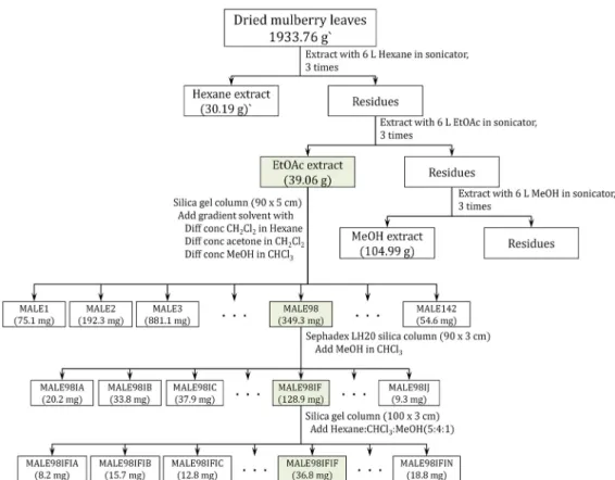
The powdered mulberry leaves (1,933.76 g) successively extracted with hexane, ethylacetate (EtOAc) and methanol (MeOH) in sonicator for one hour at room temperature and repeated the extraction three times. After evaporation of the solvent with evaporator, the dried EtOAc extract (39.06 g) was imbedded in silica gel and first chromatographed on the silica gel column (80×6 cm) eluting with gradient mix- ture of 75% CH2Cl2 in hexane, 100% CH2Cl2, 5% and 15%
acetone in CH2Cl2, and 5%, 15%, 25% and 50% MeOH in CHCl3 to give 142 fractions (MLE1-142). Fraction 98 (MLE98, 349.3 mg) was separated by Sephadex LH-20 (100x3 cm) elut- ing with 50% MeOH in CHCl3 to give 10 fractions (MLE98IA-IJ). Fraction 98IF (128.9 mg) was rechromato- graphed by silica gel column (100×3 cm) eluting with hexane : CHCl3 : MeOH (50 : 40 : 10) to give 36.8 mg of pure com- pound (MLE98IFIF, later structure elucidated daucosterol (β -sitosterol-glucoside) (Fig. 1). The purity of compound ex- ceeded 98%, as checked via NMR.
The MLE98IFIF was subjected to TLC using several sol- vent systems and it showed to be homogenous compound.
The white powder was further subjected to Proton NMR (600 MHz), Carbon NMR (150 MHz) and 2D NMR (COSY, HSQC, HMBC).
Measurement of free glycerol release
Briefly, the intra-abdominal adipose tissues were dis- sected from eight-week-old male SD rats after sacrifice using CO2 gas. These tissues (30 g) were then minced in 5 ml of DMEM supplemented with 1 mg/ml type I collagenase (Worthington Biochemical Co., Freehold, NJ, USA) and 1%
BSA (MP Biomedicals, Illkirch, France) and subsequently in- cubated at 37℃ for 30 min in a shaking incubator (JSR,
Fig. 1. Extraction and isolation scheme of the daucosterol from mulberry leaves (Morus alba).
Gongju-City, Korea). The homogenate of adipose tissue was filtered through a 100 μm nylon mesh and washed three times in KRH (Krebs ringer/HEPES solution: 25 mM NaHCO3, 125 mM NaCl, 5 mM glucose, 2.5 mM KCl, 1.25 mM NaH2PO4, 2 mM CaCl2, 1 mM MgCl2, 25 mM HEPES) containing 1% BSA. Finally, the pellets of adipocytes har- vested with centrifugation were diluted in KRH containing 3% BSA to generate a cell suspension. Primary adipocytes were grown in a humidified incubator at 37℃ under 5% CO2
and 95% air in KRH containing 3% BSA. Thereafter, these adipocytes were seeded onto 24-wellplates for each ex- perimental purpose and incubated with different concen- tration of EMfCs to measure the release of free glycerol and cell viability.
Free glycerol release was measured using free glycerol reagent (Sigma-Aldrich Co., St. Louis, MO, USA) according to the manufacturer’s protocols [9]. To measure the glycerol level, adipocytes were seeded at a density of 1×10⁵cells/ml of KRH and grown in a 37℃ incubator. After 24 hr, they were either untreated (No group), treated with vehicle (dH2O or DMSO), or pretreated with 10 μM of isoproterenol hydrochloride (Sigma-Aldrich Co., St. Louis, MO, USA), or 25, 50, 100 and 200 μg/ml of daucosterols. Following in-
cubation for 24 hr, the culture medium was collected from primary adipocytes treated with daucosterols and heated at 65℃ for 15 min to inactivate any enzymes released by the adipocytes. The inactivated medium (10 μl) was then mixed with 200 μl of glycerol detection reagent, after which the absorbance was read at 570 nm directly in the wells using a Vmax plate reader (Molecular Devices, Sunnyvale, CA, USA).
Results
Identification of daucosterol
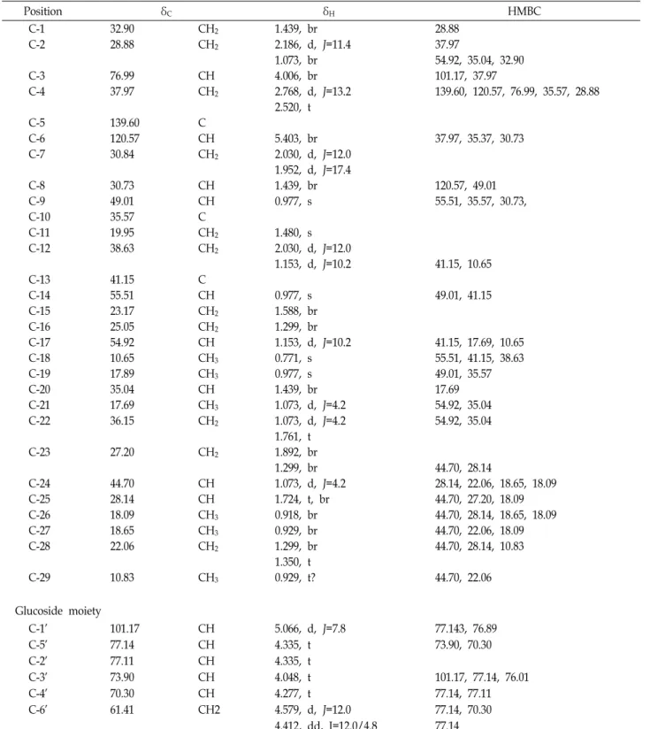
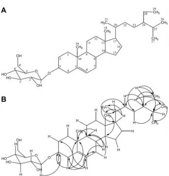
Chemical investigation of the EtOAc extracts of mulberry leaves resulted in the identification of a daucosterol (β -sitosterol-glucoside) (Fig. 2). Data obtained from 1D NMR including 1H-NMR, 13C-NMR, DEPT, 1H–1H COSY, and 2D NMR including 1H–13C HSQC, 1H–13C HMBC (Table 1 and Fig. 2).
Daucosterol (isolated as MLE98IFIF) was obtained as a colorless sticky compound. The structure of daucosterol was elucidated previously reported NMR spectral data [10, 15, 16]. Briefly, 1H and 13C NMR in conjunction with DEPT ex- periments indicated the presence of 35 carbon atoms, con-
Table 1. 1H NMR (600 MHz in pyridine), 13C NMR (150 MHz in pyridine) and HMBC spectroscopic data of daucosterol
Position δC δH HMBC
C-1 32.90 CH2 1.439, br 28.88
C-2 28.88 CH2 2.186, d, J=11.4 37.97
1.073, br 54.92, 35.04, 32.90
C-3 76.99 CH 4.006, br 101.17, 37.97
C-4 37.97 CH2 2.768, d, J=13.2 139.60, 120.57, 76.99, 35.57, 28.88
2.520, t
C-5 139.60 C
C-6 120.57 CH 5.403, br 37.97, 35.37, 30.73
C-7 30.84 CH2 2.030, d, J=12.0
1.952, d, J=17.4
C-8 30.73 CH 1.439, br 120.57, 49.01
C-9 49.01 CH 0.977, s 55.51, 35.57, 30.73,
C-10 35.57 C
C-11 19.95 CH2 1.480, s
C-12 38.63 CH2 2.030, d, J=12.0
1.153, d, J=10.2 41.15, 10.65
C-13 41.15 C
C-14 55.51 CH 0.977, s 49.01, 41.15
C-15 23.17 CH2 1.588, br
C-16 25.05 CH2 1.299, br
C-17 54.92 CH 1.153, d, J=10.2 41.15, 17.69, 10.65
C-18 10.65 CH3 0.771, s 55.51, 41.15, 38.63
C-19 17.89 CH3 0.977, s 49.01, 35.57
C-20 35.04 CH 1.439, br 17.69
C-21 17.69 CH3 1.073, d, J=4.2 54.92, 35.04
C-22 36.15 CH2 1.073, d, J=4.2 54.92, 35.04
1.761, t
C-23 27.20 CH2 1.892, br
1.299, br 44.70, 28.14
C-24 44.70 CH 1.073, d, J=4.2 28.14, 22.06, 18.65, 18.09
C-25 28.14 CH 1.724, t, br 44.70, 27.20, 18.09
C-26 18.09 CH3 0.918, br 44.70, 28.14, 18.65, 18.09
C-27 18.65 CH3 0.929, br 44.70, 22.06, 18.09
C-28 22.06 CH2 1.299, br 44.70, 28.14, 10.83
1.350, t
C-29 10.83 CH3 0.929, t? 44.70, 22.06
Glucoside moiety
C-1' 101.17 CH 5.066, d, J=7.8 77.143, 76.89
C-5' 77.14 CH 4.335, t 73.90, 70.30
C-2' 77.11 CH 4.335, t
C-3' 73.90 CH 4.048, t 101.17, 77.14, 76.01
C-4' 70.30 CH 4.277, t 77.14, 77.11
C-6' 61.41 CH2 4.579, d, J=12.0 77.14, 70.30
4.412, dd, J=12.0/4.8 77.14
sisting of the following functional groups: six methyl carbon (-CH3), twelve methylene (-CH2-) carbon, fourteen methine (=CH-) carbon and three quaternary (=C=) carbon. The 1H and 13C NMR values for all the protons and carbons were assigned on the basis of COSY, HMQC and HMBC correla-
tions and were given in Table 1. The HMBC correlations of H-1’ in glucoside to the oxygenated carbon (δC 76.99, C-3) of β-sitosterol. H-18 correlated to the δC 55. 51 (C-14), 41.15 (C-13) and 38.63 (C-12) of C ring, revealed that the methyl group (H-18) was attached at C-13. H-19 correlated to the
A
B
Fig. 2. Structure (A) and key HMBC (B) of daucosterol (β-sitos- terol-glucoside).
Fig. 3. Dose dependent response of daucosterol on the lipolytic activity. The cell viability was measured in 3T3-L1 cells treated with 25, 100, 200 and 400 μg/ml of daucosterol.
The free glycerol release was measured in the super- natant of primary adipocytes treated with daucosterol.
The data shown represent the means ± SD of three replicates. *, p<0.05 relative to the Vehicle treated group.
ISOP; isoproterenol.
δC 49.01 (C-9) and 35.57 (C-10) of B ring, revealed that the methyl group (H-19) was attached at C-10. H-3 connected to the δC 101.17 (C-6) and 37.97 (C-4); H-6 to the δC 37.97 (C-4), 35.37 (C-10 and 30.73 (C-8); H-17 to the δC 41.15 (C-13), 17.69 (C-21) and 10.65 (C-18); H-24 to 28.14 (C-25), 22.06 (C-28), 18.65 (C-27) and 18.09 (C-26). From the further analy- sis of HMQC and HMBC spectra confirmed the presence of the β-sitosterol and glucoside groups, the structure were
unambiguously assigned. So, the compound MLE98IFIF has been identified as daucosterol (sitosterol-β-D-glucoside).
Lipolytic activity of daucosterol
To identify novel extracts with lipolytic activity of daucos- terol, the free glycerol levels were analyzed in the culture medium of primary adipocytes derived from SD rats follow- ing treatment with 25, 50, 100 and 200 μg/ml of daucosterol.
The glycerol level increased significantly in a dose depend- ent manner in the culture medium of primary adipocytes although increase rate was very low between 50 and 100 ug/ml treated group (Fig. 3). Therefore, these results in- dicate that daucosterol derived from fermented mulberry leaves have very strong lipolytic activity.
Discussion
Mulberry plants contain natural products with a large number of pharmacologically active compounds. In previous studies, crude extracts of mulberry plants showed diuretic, hypoglycemic, antidiabetic, and hypotensive activities [14, 21]. However, there is limit information on the purification and identification of the compounds responsible for the obe- sity activity. In this study, the ethyl acetate extracts was frac- tionated on an open column chromatography, and then puri- fied the daucosterol. This study demonstrated that daucos- terol from the leaves of mulberry possessed good lipolysis activity. To the best of our knowledge, this study is the first report to demonstrate the lipolysis of daucosterol. This in- vestigation elucidates that the daucosterol has a great medic- inal importance. However, further detailed experiment need to elucidate the exact mechanism of action and any potential impact on lipolysis of daucosterol.
Acknowledgement
This work was supported by Korea Institute of Planning and Evaluation for Technology in Food, Agriculture, Fores- try and Fisheries (IPET) through High Value-added Food Technology Development Program, funded by Ministry of Agriculture, Food and Rural Affairs (MAFRA) (116027-03- 1-HD020 and 314043032HD020).
References
1. Asano, N., Nash, R. J., Molyneux, R. J. and Fleet, G. W.
J. 2000. Sugar-mimic glycosidase inhibitors: Natural occur-
rence, biological activity and prospects for therapeutic application. Tetrahedron Asymmetry 11, 1645-1680.
2. Awad, A. B., Gan, Y. and Fink, C. S. 2000. Effect of β-sitos- terol, a plant sterol, on growth, protein phosphatase 2A, and phospholipase D in LNCaP cells. Nutr. Cancer 36, 74-78.
3. Bradford, P. G. and Awad, A. B. 2007. Phytosterols as anti- cancer compounds. Mol. Nutr. Food Res. 51, 161-170.
4. Chauhan, S., Devi, U., Kumar, V. R., Kumar, V., Anwar, F. and Kaithwas, G. 2015. Dual inhibition of arachidonic acid pathway by mulberry leaf extract. Inflammopharmacology 23, 65-70.
5. Choi, J. N., Choi, Y. H., Lee, J. M., Noh, I. C., Park, J. W., Choi, W. S. and Choi, J. H. 2012. Anti-inflammatory effects of β-sitosterol-β-D-glucoside from Trachelospermum jasmi- noides (Apocynaceae) in lipopolysaccharide-stimulated RAW 264.7 murine macrophages. Nat. Prod. Res. 26, 2340-2343.
6. Choi, S. J., Jeon, H., Lee, C. U., Yoon, S. H., Bae, S. K., Chin, Y. W. and Yoon, K. D. 2015. Isolation and development of quantification method for cyanidin-3-glucoside and cyani- din-3-rutinoside in mulberry fruit by high-performance countercurrent chromatography and high-performance liq- uid chromatography. Nat. Prod. Sci. 21, 20-24.
7. Evans, S. V., Fellows, L. E., Shing, T. K. M. and Fleet, G.
W. J. 1985. Glycosidase inhibition by plant alkaloids which are structural analogues of monosaccharides. Phytochemistry 24, 1953-1955.
8. Fukai, T., Satoh, K., Nomura, T. and Sakagami, H. 2003.
Antinephritis and radical scavenging activity of prenyl- flavonoids. Fitoterapia 74, 720-724.
9. Ho, J. N., Kim, O. K., Nam, D. E., Jun, W. and Lee, J. 2014.
Pycnogenol supplementation promotes lipolysis via activa- tion of cAMP-dependent PKA in ob/ob mice and pri- mary-cultured adipocytes. J. Nutr. Sci. Vitaminol. 60, 429- 435.
10. Hui, W. H., Li, M. M. and Wong, K. M. 1976. A New com- pound, 21-a-Hydroxy friedel-4,23-En-3-one and other tri- terpenoids from Phyllanthus reticulantus. Phytochemistry 15, 797-798.
11. Ivorra, M. D., D’Ocon, M. P., Paya, M. and Villar, A. 1988.
Antihyperglycemic and insulin-releasing effects of β-sitos- terol-3-β-D-glucoside and its aglycone, β-sitosterol. Arch.
Int. Pharmacodyn. Ther. 296, 224-231.
12. Jeong, J. Y., Jo, Y. H., Kim, S. B., Liu, Q., Lee, J. W., Mo, E. J., Lee, K. Y., Hwang, B. Y. and Lee, M. K. 2015. Pancreatic lipase inhibitory constituents from Morus alba leaves and op- timization for extraction conditions. Bioorg. Med. Chem. Lett.
25, 2269-2274.
13. Jimenez-Escrig, A., Santos-Hidalgo, A. B. and Saura-Calixto, F. 2006. Common sources and estimated intake of plant ster- ols in the Spanish diet. J. Agric. Food Chem. 54, 3462-3471.
14. Kelkar, S. M., Bapat, V. A., Ganapathi, T. R., Kaklig, G. S., Rao, P. S. and Heble, M. R. 1996. Determination of hypo- glycemic activity in Morus indica L. (mulberry) shoot cul- tures. Curr. Sci. 71, 71-72.
15. Khan, M. U. N. M. and Hossain, S. Md. 2015. Scopoletin and β-sitosterol glucoside from roots of Ipomoea digitata. J.
Pharmacog. Phytochem. 4, 5-7.
16. Kobayashi, M., Tsuru, R., Todo, K. and Mitsuhashi, H. 1973.
Marine sterols-II : Asterosterol, a new C26 sterol from Asterias amurensis lütken. Tetrahedron 29, 1193-1194.
17. Moreau, R. A., Whitaker, B. D. and Hicks, K. B. 2002. Phy- tosterols, phytostanols, and their conjugates in foods:
Structural diversity, quantitative analysis, and health pro- moting uses. Prog. Lipid Res. 41, 457-500.
18. Nair, P. P., Turjman, N., Kessie, G., Calkins, B., Goodman, G. T., Davidovitz, H. and Nimmagadda, G. 1984. Diet, nu- trition intake, and metabolism in populations at high and low risk for colon cancer. Dietary cholesterol, β-sitosterol, and stigmasterol. Am. J. Clin. Nutr. 40(4 Suppl), 927-930.
19. Nakagawa, K. 2013. Studies targeting α-glucosidase in- hibition, antiangiogenic effects, and lipid modification regu- lation: background, evaluation, and challenges in the devel- opment of food ingredients for therapeutic purposes. Biosci.
Biotechnol. Biochem. 77, 900-908.
20. Nomura, T. 1999. The chemistry and biosynthesis of iso- prenylated flavonoids from moraceous plants. Pure Appl.
Chem. 71, 1115-1118.
21. Pawlowska, A. M., Oleszek, W. and Braca, A. 2008. Quali- quantitative analyses of flavonoids of Morus nigra L. and Morus alba L. (Moraceae) fruits. J. Agric. Food Chem. 56, 3377- 3380.
22. Piao, S. J., Chen, L. X., Kang, N. and Qiu, F. 2011. Simultane- ous determination of five characteristic stilbene glycosides in root bark of Morus albus L. (Cortex Mori) using high-per- formance liquid chromatography. Phytochem. Anal. 22, 230- 235.
23. Rubis, B., Paszel, A., Kaczmarek, M., Rudzinska, M., Jelen, H. and Rybczynska, M. 2008. Beneficial or harmful influence of phytosterols on human cells? Br. J. Nutr. 100, 1183-1191.
24. Ryan, E., Galvin, K., O’Connor, T. P., Maguire, A. R. and O’Brien, N. M. 2006. Fatty acid profile, tocopherol, squalene and phytosterol content of Brazil, pecan, pine, pistachio and cashew nuts. Int. J. Food Sci. Nutr. 57, 219-228.
25. Shirishkumar, D. A., Ashwini, V. M. and Prashant, D. A.
2014. Pharmacological, nutritional, and analytical aspects of β-sitosterol: a review. Orient. Pharm. Exp. Med. 14, 193-211.
26. Tsuduki, T., Kikuchi, I., Kimura, T., Nakagawa, K. and Miyazawa, T. 2013. Intake of mulberry 1-deoxynojirimycin prevents diet-induced obesity through increases in adipo- nectin in mice. Food Chem. 139, 16-23.
27. Venkatesh, K. P. and Chauhan, S. J. 2008. Mulberry: Life enhancer. J. Med. Plants Res. 2, 271-278.
28. Vijayan, K., Chauhan, S., Das, N. K., Chakraborti, S. P. and Roy, B. N. 1997. Leaf yield component combining abilities in mulberry (Morus s pp). Euphytica 98, 47-52.
29. Watson, L. and Dallwitz, M. J. 2007. In The Families of Flowering Plants: Descriptions, Illustrations, Identification, and Information Retrieval, 1992 onward; June 1, 2007, ver- sion; http://delta-intkey.com.
30. Winchester, B. and Fleet, G. W. J. 1992. Amino-sugar glyco- sidase inhibitors: Versatile tools for glycobiologists. Glycobiology 2, 199-210.
초록:뽕잎으로부터 순수분리한 daucosterol의 lipolysis 효과
이커1․이미림2․루쿼1․이매1․강점순1․최영현3․김경미4․정재철4․황대연2*․최영환1*
(1부산대학교 원예생명과학과/생명산업융합연구원, 2부산대학교 바이오소재과학과, 3동의대학교 생화학과, 4(주)
노바렉스)
뽕나무는 약 40여 속과 1,000여 종이 있는 것으로 알려져 있으며, 항염증, 항진정, 지사작용, 노화억제 및 신경보 호작용이 있는 것으로 알려져 있다. 본 연구에서는 아시아 지역에서 전통 한약재로 사용되는 뽕 나무 잎의 지방분 해 활성에 관여하고 있는 물질을 스크린하기 위하여 뽕잎 분말을 헥산, 에틸 아세테이트 및 메탄올로 순차 추출하 였다. 뽕 잎의 EtOAc 추출물로부터 daucosterol이 순수분리 되었으며, 그 구조는 1H, 13C, DEPT, COSY, HSQC 및 HMBC 등의 NMR스펙트럼 분석에 의해 밝혀졌다. Daucosterol은 농도 의존적으로 지질분해 효과를 나타내었 는데, 본 연구의 결과로부터 뽕나무 잎으로부터 순수분리한 daucosterol의 지분분해 활성은 다양한 질병을 치료하 기 위한 천연물 소재 또는 지표성분으로서 활용이 가능할 것으로 생각된다. 그러나 보다 효율적으로 이용하기 위해서는 daucosterol의 비만에 관한 생리활성 기작에 대한 추가적인 연구가 필요할 것이다.
31. Woyengo, T. A., Ramprasath, V. R. and Jones, P. J. H. 2009.
Anticancer effects of phytosterols. Eur. J. Clin. Nutr. 63, 813- 820.
32. Yang, Z. G., Matsuzaki, K., Takamatsu, S. and Kitanaka, S.
2011. Inhibitory effects of constituents from Morus alba var.
multicaulis on differentiation of 3T3-L1 cells and nitric oxide production in RAW264.7 cells. Molecules 16, 6010-6022.
33. Zelová, H., Hanáková, Z., Čermáková, Z., Šmejkal, K., Dalĺ Acqua, S., Babula, P., Cvačka, J. and Hošek, J. 2014. Evalu- ation of anti-inflammatory activity of prenylated substances isolated from Morus alba and Morus nigra. J. Nat. Prod. 77, 1297-1303.
34. Zhao, J., Zhang, C. Y., Xu, D. M., Huang, G. Q., Xu, Y. L., Wang, Z. Y., Fang, S. D., Chen, Y. and Gu, Y. L. 1990. The antiatherogenic effects of components isolated from pollen typhae. Thromb. Res. 57, 957-966.
35. Zhao, M., Chen, C. C., Yang, S. Q. and Zhang, S. J. 2012.
Chemical constituents from leaves of Morus alba Linn.
Zhongchengyao 34, 1126-1131.
36. Zhao, S., Park, C. H., Li, X., Kim, Y. B., Yang, J., Sung, G.
B., Park, N. I., Kim, S. and Park, S. U. 2015. Accumulation of rutin and betulinic acid and expression of phenyl- propanoid and triterpenoid biosynthetic genes in mulberry (Morus alba L.). J. Agric. Food Chem. 63, 8622-8630.